Astigmatism Correction in Children with Cataracts
M. Edward Wilson
Muralidhar Ramappa
Rupal H. Trivedi
Corneal astigmatism is often documented during preoperative biometry measurements in children and adults. In infancy, there is a high prevalence (≥42%) of astigmatism (≥1.00 D).1,2,3,4,5,6,7,8,9,10,11,12,13A reduction in this infantile astigmatism occurs in the first years of life because of a natural decrease in toricity of the cornea and the anterior lens, along with a decrease in the variation of the cornea and lenticular surfaces.14 Hispanic and African-American children are more likely to have astigmatism than are non-Hispanic white children, and this association remains even after correcting for the presence of spherical equivalent refractive error (myopia or hyperopia).15 In adults, residual postoperative corneal astigmatism, if uncorrected, has been noted to reduce uncorrected visual acuity and induce symptomatic blur, ghosting of images, and halos.16 Children with astigmatism >2.00 D in 3-year-olds or >1.50 D in 4-year-olds have been shown to have visual motor integration deficits.17 Amblyopia treatment seems to be less favorable in patients with either hyperopic or myopic against-the-rule (ATR) astigmatism.18
To meet parents’ expectations for good visual acuity after cataract surgery and to help amblyopic management, it is important to address both sphere and cylinder errors that may affect final refractive outcomes. Parents want to know how clear the vision will be when glasses are removed, even if it has been explained that the child’s best visual acuity will always be with bifocal glasses. Preoperative assessment for cataract surgery should be followed by comprehensive counseling that focuses on a patient’s individual vision needs. The refractive part of the discussion should include a discussion about the refractive aim for immediately after surgery and also what the refraction is expected to be years later. In a young child, this may include a discussion of far-sighted bifocal glasses after surgery with a gradual reduction in the strength of the glasses as the eye grows. If the child is older than 3 years and has preoperative astigmatism, parents are told that the astigmatism will likely be unchanged after surgery and will be corrected with glasses. For older children, a review of surgical options for reducing astigmatism is becoming more common. These options include astigmatism management with peripheral corneal-relaxing incisions (PCRIs), toric intraocular lens (IOL) (Fig. 30.1), and the possibility of postoperative laser refractive surgery. It is important to estimate the likelihood of less spectacle dependence based on the age that can realistically be achieved with a given surgical option. Videos or computer- based illustrations may be a useful and effective way of explaining the different approaches. Spherical refractive errors are managed by accurate biometry and IOL power calculations. The target refraction should be based on age at surgery and other factors (see Chapter 7). Relying solely on manual keratometry to characterize a patient’s corneal curvature is commonly done during standard biometry for IOL power calculations. However, if the surgeon desires to surgically manage astigmatism, manual keratometry may be insufficient. Significant cylindrical errors may exist inside and outside the central 3.2-mm optical zone measured by keratometry. In addition, meridian changes over the entrance pupil and irregular astigmatism may be missed. Using the IOL Master® (Carl Zeiss Meditec, CA, USA) for corneal curvature measurements can result in similar mistakes.19 For these reasons, computerized corneal topography is the current standard of care when astigmatism correction is being considered. Topography accurately measures the global corneal astigmatism, quantifies the nature of the astigmatism (i.e., symmetric bow tie versus asymmetric or irregular astigmatism), and identifies the steep meridian of astigmatism. Patients implanted with monofocal IOLs usually achieve better-uncorrected visual acuity if the spherical error approaches emmetropia and residual manifest astigmatism is less than 1 D. For patients implanted with multifocal IOLs, the postoperative refractive goal is to achieve <0.75 D of residual corneal astigmatism. However, as mentioned above, children have a targeted refraction that depends on age at surgery. Final results are also dependent on the pupil size and the amount of residual spherical error. Overcorrections and large rotations in the axis of residual astigmatism should be avoided.
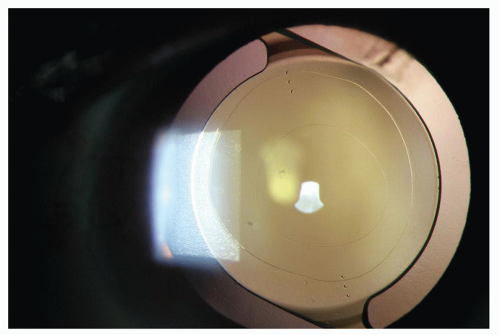 Figure 30.1. Six-month follow-up of toric IOL implantation in a child. |
Astigmatism correction at the time of cataract surgery or in aphakic/pseudophakic eyes of children is still uncommon and is only now beginning to be reported.20,21 However, witnessing impressive outcomes in adults, pediatric cataract surgeons have started discussing correction of astigmatism in children. In the following sections of this chapter, we review the literature on astigmatism correction in adult eyes. We anticipate a more conservative approach in the growing eyes of children.
STEPLADDER APPROACH TO ASTIGMATISM MANAGEMENT
Subjects with symmetrical corneal astigmatism can be placed into one of four categories.22 These categories are as shown in Table 30.1. Cases with <1 D of corneal astigmatism can be neutralized by designing the surgical incision on the steep corneal meridian. For 1 to 3 D of corneal astigmatism, single or paired PCRIs may be fashioned. Toric IOLs may be implanted in eyes with 1 to 4 D of corneal astigmatism. Toric IOLs and PCRIs may be combined for eyes with 4.5 to 7 D of corneal astigmatism. Finally, astigmatism of >4.5 to 6 D may warrant the use of high-power toric IOLs that are available in Europe and elsewhere.
Table 30.1 STEPLADDER APPROACH TO THE MANAGEMENT OF CORNEAL ASTIGMATISM AT THE TIME OF ADULT CATARACT SURGERY | ||||||||||||
|---|---|---|---|---|---|---|---|---|---|---|---|---|
|
Miller PCRI Nomogram
For symmetrical bow tie corneal astigmatism on corneal topography, clear corneal-paired incisions are placed in the steeper axis, just inside the limbus. The incisions are 500 to 550 microns in depth and as long in clock hours as the cornea is steep in diopters by simulated keratometry. For asymmetric bow tie astigmatism, in which the axis of astigmatism is constant across the cornea, longer incisions are made on the side of the cornea that is steeper.
The astigmatic correction in diopters equals the sum of the lengths of the incisions in clock hours divided by 2. For asymmetric bow tie astigmatism in which the axis of astigmatism changes across the cornea, the incisions are rotated to coincide with the location where the steep axis intersects the limbus. Surgical management options in cases of very high postoperative residual astigmatism include piggyback IOL implantation, IOL exchange, toric IOL rotation, photorefractive keratectomy (PRK), and laser-assisted in situ keratomileusis (LASIK).
Concepts in Applying the Stepladder Approach
Surgical incisions placed on the steep meridian and PCRIs do not alter the spherical equivalent power of the cornea enough to alter IOL power calculations. The location and architecture of the corneal incision are the most important surgical variables. Any clear corneal incision (CCI) flattens the meridian in which it is placed and steepens the perpendicular meridian with a coupling effect.23 For a 3.2-mm wide incision, the resultant surgically induced astigmatism (SIA) is approximately 0.5 D (95% CI: 0.4-0.6 D).24 Incisions <2.4 mm wide may induce smaller degrees of SIA, but not in a linear logarithmic manner and not significantly <0.5 D, possibly due to surgical manipulation while IOL implantation. Consequently, when placing a cataract incision on the steep meridian, the surgeon can predict an approximate 0.5-D correction of preexisting corneal astigmatism, which is ideal for patients with <1 D of preexisting corneal astigmatism. To accurately calculate surgeon-specific SIA, software is available.25 Preferably, it is better to place a CCI inside a convenient PCRI if a relaxing incision approach to astigmatism management is planned. If a toric IOL is planned, the surgeon must determine the vector sum of the preexisting corneal astigmatism and SIA before selecting the appropriate IOL to be implanted.23,26 Most of the toric IOL manufacturers
(Alcon Laboratories, STAAR Surgical, Rayner, and Carl Zeiss Meditec) have toric IOL calculator software that performs this function (Table 30.2). These calculators use subject’s corneal topography measurement and SIA to select an appropriate toric IOL model and determine orientation IOL in the capsular bag.
(Alcon Laboratories, STAAR Surgical, Rayner, and Carl Zeiss Meditec) have toric IOL calculator software that performs this function (Table 30.2). These calculators use subject’s corneal topography measurement and SIA to select an appropriate toric IOL model and determine orientation IOL in the capsular bag.
Table 30.2 ONLINE TORIC IOL CALCULATORS | ||||||||
|---|---|---|---|---|---|---|---|---|
|


