In this article the author describes in detail the adaptation of adult vestibular testing techniques to the pediatric population. Assessment tools discussed include video-oculography (VOG), computerized rotary chair (CRC), computerized dynamic posturography (CDP), and vestibular evoked myogenic potentials (VEMPs). As with hearing impairment, the earlier a vestibular disorder is identified, the earlier remediation strategies may begin. Among the most crucial aspects of vestibular testing in pediatric patients are obtaining of pediatric normative data and adapting adult techniques so that younger children may be evaluated.
The area of pediatric vestibular evaluation has become popular in audiology and otolaryngology clinics in recent years. As early identification of hearing impairment has unfolded, so too has early identification of vestibular disorders. With regard to both audiology and otolaryngology, the earlier the identification, the earlier remediation strategies can be implemented. Researchers and clinicians have contributed valuable information related to vestibular disorders in the pediatric population. There has been a paucity of both clinical and research work related to vestibular evaluation techniques useful for young children. This review focuses on adaptation of adult vestibular evaluation techniques for use with pediatric patients, beginning with the medical/physical examination and progressing through major tests of vestibular function. An important concept recurring throughout is that the use of pediatric normative data is crucial, so that results obtained after testing a pediatric patient are not compared with adult normative data.
History and the medical evaluation
Many clinicians who perform vestibular evaluations have remarked that the case history is one of the most important diagnostic tools available when evaluating a dizzy patient. Although the author firmly believes that this premise also holds true with pediatric patients, a need exists within the profession for development of a reliable case history instrument. On searching the literature regarding vestibular evaluation in children, one may view several different bodies of work. On one hand, Physical Therapy and Occupational Therapy studies document evaluation and remediation techniques utilized with such disorders as autism, motor delay, learning disability, and behavioral disorders. While fascinating, these evaluative tools are very different from those discovered within the Audiology and Otolaryngology bodies of literature. Many studies within this latter group link vestibular disorders to such entities as migraine syndrome, benign recurrent vertigo of childhood, otitis media, and sensorineural hearing loss. Additional research is needed regarding other syndromes associated with childhood dizziness including CHARGE association, Cogan syndrome, and Usher syndrome, among others. As evaluative tools and remediation strategies are also variable among disciplines, greater understanding of other disciplines and greater degree of collaboration are worthy goals.
Many challenges exist with regard to vestibular evaluation of children. For example, pediatric patients may not be able to thoroughly describe dizziness and other symptoms, necessitating the development of appropriate checklists and parent questionnaires. Another challenge related to vestibular evaluation is that the cost of many pieces of technologically advanced equipment is prohibitive, regardless of patient age. This article highlights adaptation of adult screening techniques for pediatric patients, and describes the relative ease of performance of these tests in an office setting with minimal equipment.
Physical examination
Within an office setting, the clinician may begin by checking for any spontaneous and/or gaze-evoked nystagmus that may be present. One simple method is to have the child look straight ahead, if able to follow such verbal commands, and then to follow an examiner’s finger as it moves to the right and left and in upward and downward directions. Mental alerting is important with regard to many vestibular measures, and must be geared to the child’s age and capabilities. In this case and especially in viewing spontaneous nystagmus, informal conversation and establishing rapport should suffice. It may be possible to use Frenzel lenses for magnification and alleviation of visual fixation suppression, depending on the child’s age. At this point the clinician may also briefly check to determine that the eyes are moving conjugately.
As the clinician approaches a dynamic evaluation, consideration may be given to testing “head-shake nystagmus” (HSN). According to Fife and colleagues, the vestibulo-ocular reflex (VOR) should be readily observable by the age of 9 to 12 months in typically developing infants. Visualization may be enhanced via Frenzel lenses, electro-oculography (EOG), or infrared video. As with adults, the child’s head may be gently rotated back and forth in a rhythmic, horizontal manner. Theoretically, symmetric vestibular function will not induce symptoms, although some children may not be in a developmental position to verbalize the presence of such symptoms. HSN will not be seen in patients with symmetric peripheral vestibular function, although it may be observed in the presence of a vestibular asymmetry. HSN screening may be advantageous in situations where bithermal caloric irrigations and/or rotary chair testing may not be feasible. If the child is able to perform these tests, HSN screening may provide complementary information by incorporating higher frequencies of head motion.
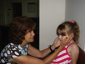
Head-thrust testing (HTT) may also be performed with young children as part of a medical/physical workup. This procedure is based on the VOR, which as previously mentioned should be observable by the age of 9 to12 months in typically developing children. The examiner gently rotates the child’s head by approximately 30° in the yaw plane, while asking the child to focus on the examiner’s nose. One may creatively devise placement of colorful stickers or cartoon characters to facilitate such testing. The examiner incorporates a brief and rapid head thrust back to center, making certain that visual fixation is maintained. Any “catch-up” saccade may indicate a disorder of the ipsilateral semicircular canal. Figs. 1 and 2 display the successful demonstration of HTT with a 4-year-old child.
Children may also be evaluated via dynamic visual acuity (DVA) techniques, with modification of the typical Snellen Eye Chart for this population. Familiar to most, this chart is highly utilized during eye examinations as the patient reads smaller and smaller lines of various letter “E” configurations. For example, child-friendly characters could be easily substituted for the more traditional ones. With this procedure, a baseline is obtained in the form of the smallest line of characters that the child can discern from a calibrated distance. The child again attempts to read the smallest line of figures during back and forth rotation of the head, approximating 1 to 2 Hz. On reading, a loss of one line may be considered insignificant whereas a loss of 3 lines or more may indicate a VOR deficiency. Advances in computerized technology may facilitate this evaluation. The clinician may also perform rudimentary oculomotor tasks, such as checking rapid saccadic eye movement and smoothness of pursuit, by following an examiner’s finger or other sinusoidal maneuver.
Although children rarely demonstrate the true benign paroxysmal positional vertigo (BPPV) that is prevalent in adults (see article by Cho and White in this publication), they may experience positional vertigo and/or sudden vertiginous episodes. Positioning and positional testing can be successfully performed with children. With all procedures, establishing rapport with the child is critical, and the child’s comfort level may be increased by being seated on a parent’s lap and/or having a parent present. The traditional Dix-Hallpike maneuver involves rapidly taking the patient from a seated to a lying-back position. The clinician assists the patient in turning the head to the right or left and lying back, and then the procedure is repeated as the patient lies back while turning the head to the opposite side. As Hallpike testing is very time-efficient and noninvasive, it may be performed with children along with more traditional positional testing. It is important for the child to be able to understand instructions and to receive continual reassurance, in that the room environment may be darkened. Typical positions may be similar to those performed with adults: supine, head right, head left, right lateral, and left lateral. As the clinician searches for the presence of nystagmus, it is also important to supplement this information with any subjective symptoms that the child may report.
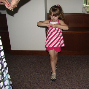
Finally, during the physical examination, the clinician may ask the child to perform various types of standing balance tasks for screening postural stability. An example is the Romberg maneuver, for which the child stands with feet together and sway is noted with eyes both open and closed. A variation is the tandem Romberg, whereby such stability is evaluated in a heel-to-toe format. Fig. 3 depicts the author as she performs the tandem Romberg maneuver with a 4-year-old patient.
Examiners may ask the child to march in place with eyes closed to determine straightness versus deviation of walking path and/or may also encourage the child to walk forward in a heel-to-toe pattern. With the latter, cerebellar dysfunction may be suspected if difficulties arise. Other cerebellar signs may include slurred speech, upper or lower extremity dysfunction, ataxia, muscle incoordination, tremors, and abnormalities noted on oculomotor tasks.
Physical examination
Within an office setting, the clinician may begin by checking for any spontaneous and/or gaze-evoked nystagmus that may be present. One simple method is to have the child look straight ahead, if able to follow such verbal commands, and then to follow an examiner’s finger as it moves to the right and left and in upward and downward directions. Mental alerting is important with regard to many vestibular measures, and must be geared to the child’s age and capabilities. In this case and especially in viewing spontaneous nystagmus, informal conversation and establishing rapport should suffice. It may be possible to use Frenzel lenses for magnification and alleviation of visual fixation suppression, depending on the child’s age. At this point the clinician may also briefly check to determine that the eyes are moving conjugately.
As the clinician approaches a dynamic evaluation, consideration may be given to testing “head-shake nystagmus” (HSN). According to Fife and colleagues, the vestibulo-ocular reflex (VOR) should be readily observable by the age of 9 to 12 months in typically developing infants. Visualization may be enhanced via Frenzel lenses, electro-oculography (EOG), or infrared video. As with adults, the child’s head may be gently rotated back and forth in a rhythmic, horizontal manner. Theoretically, symmetric vestibular function will not induce symptoms, although some children may not be in a developmental position to verbalize the presence of such symptoms. HSN will not be seen in patients with symmetric peripheral vestibular function, although it may be observed in the presence of a vestibular asymmetry. HSN screening may be advantageous in situations where bithermal caloric irrigations and/or rotary chair testing may not be feasible. If the child is able to perform these tests, HSN screening may provide complementary information by incorporating higher frequencies of head motion.
Head-thrust testing (HTT) may also be performed with young children as part of a medical/physical workup. This procedure is based on the VOR, which as previously mentioned should be observable by the age of 9 to12 months in typically developing children. The examiner gently rotates the child’s head by approximately 30° in the yaw plane, while asking the child to focus on the examiner’s nose. One may creatively devise placement of colorful stickers or cartoon characters to facilitate such testing. The examiner incorporates a brief and rapid head thrust back to center, making certain that visual fixation is maintained. Any “catch-up” saccade may indicate a disorder of the ipsilateral semicircular canal. Figs. 1 and 2 display the successful demonstration of HTT with a 4-year-old child.
Children may also be evaluated via dynamic visual acuity (DVA) techniques, with modification of the typical Snellen Eye Chart for this population. Familiar to most, this chart is highly utilized during eye examinations as the patient reads smaller and smaller lines of various letter “E” configurations. For example, child-friendly characters could be easily substituted for the more traditional ones. With this procedure, a baseline is obtained in the form of the smallest line of characters that the child can discern from a calibrated distance. The child again attempts to read the smallest line of figures during back and forth rotation of the head, approximating 1 to 2 Hz. On reading, a loss of one line may be considered insignificant whereas a loss of 3 lines or more may indicate a VOR deficiency. Advances in computerized technology may facilitate this evaluation. The clinician may also perform rudimentary oculomotor tasks, such as checking rapid saccadic eye movement and smoothness of pursuit, by following an examiner’s finger or other sinusoidal maneuver.
Although children rarely demonstrate the true benign paroxysmal positional vertigo (BPPV) that is prevalent in adults (see article by Cho and White in this publication), they may experience positional vertigo and/or sudden vertiginous episodes. Positioning and positional testing can be successfully performed with children. With all procedures, establishing rapport with the child is critical, and the child’s comfort level may be increased by being seated on a parent’s lap and/or having a parent present. The traditional Dix-Hallpike maneuver involves rapidly taking the patient from a seated to a lying-back position. The clinician assists the patient in turning the head to the right or left and lying back, and then the procedure is repeated as the patient lies back while turning the head to the opposite side. As Hallpike testing is very time-efficient and noninvasive, it may be performed with children along with more traditional positional testing. It is important for the child to be able to understand instructions and to receive continual reassurance, in that the room environment may be darkened. Typical positions may be similar to those performed with adults: supine, head right, head left, right lateral, and left lateral. As the clinician searches for the presence of nystagmus, it is also important to supplement this information with any subjective symptoms that the child may report.
Finally, during the physical examination, the clinician may ask the child to perform various types of standing balance tasks for screening postural stability. An example is the Romberg maneuver, for which the child stands with feet together and sway is noted with eyes both open and closed. A variation is the tandem Romberg, whereby such stability is evaluated in a heel-to-toe format. Fig. 3 depicts the author as she performs the tandem Romberg maneuver with a 4-year-old patient.
Examiners may ask the child to march in place with eyes closed to determine straightness versus deviation of walking path and/or may also encourage the child to walk forward in a heel-to-toe pattern. With the latter, cerebellar dysfunction may be suspected if difficulties arise. Other cerebellar signs may include slurred speech, upper or lower extremity dysfunction, ataxia, muscle incoordination, tremors, and abnormalities noted on oculomotor tasks.
Video-oculography
Video-oculography (VOG) has replaced EOG in most clinics, with the advent of infrared cameras mounted in eye goggles to replace electrodes for recording eye movements. An example of such eye goggles is seen in Fig. 4 , with the clinician making use of the smallest size possible for evaluation of the pediatric patient.
This vestibular evaluation procedure is actually a battery of tests that assists the clinician in determining whether dizziness is of central or peripheral origin. If peripheral, components of the test battery may help determine the affected side of the deficit. Components of this test battery include testing for spontaneous nystagmus and gaze nystagmus, performing various oculomotor tests, measuring positioning and positional nystagmus, and performing bithermal caloric irrigations. Several investigators have effectively described some of the adaptations of adult VOG techniques that may be used with children.
As the parent makes certain that the child is optimally prepared for vestibular evaluation, it is important that certain medications (such as those for cough or cold) be withheld for 24 to 48 hours. As some aspects of the test battery may induce nausea, it is important that the child eat only lightly prior to the appointment. VOG may be performed in a darkened room so that visual fixation of nystagmus does not occur; therefore, children may feel more comfortable when a parent is present in the examining room. During calibration, a light bar is utilized with adult patients, and the patient is asked to look at targets appearing to the left and right of center gaze. Children’s attention may be maximized if asked to focus on interesting cartoon characters that illuminate in place of the lights. Various oculomotor tests may also be performed using the light bar: measurement of saccades or rapid eye movements, optokinetic testing (following lights that induce the slow and then fast phase of nystagmic eye movements), and sinusoidal tracking that should result in smooth pursuit eye movements. With all of these oculomotor tests, interesting cartoon characters may replace the traditional light bar used with adults. Just as with audiometry, the clinician may wish to employ an assortment of novel stimuli (cartoon characters in this case) to regain attention, should habituation take place.
Measurement of spontaneous nystagmus, addressed in a previous section, may be more formally measured as an initial component of the VOG test battery. The child is asked to sit quietly as the clinician records eye movements while the child looks at a target and while vision is compromised (ie, in complete darkness). Nystagmus is not elicited in any way, and the audiologist exercises care in making certain that mental alerting measures appropriate for the individual child are carried out. Gaze nystagmus may also be measured with a light bar displaying interesting cartoons, or the clinician may ask the child to “look at the lightning bugs” if a traditional light bar is utilized. In this manner, it is feasible to measure gaze nystagmus in right, left, upward, and downward positions. Standard positioning and positional tests previously described also may be performed in the pediatric vestibular laboratory. The clinician may use discretion regarding expanding the test battery to include right and left lateral, as well as other positions. As with the office examination, it is important to check for the presence of nystagmus and to recognize subjective report of symptoms.
Adaptations of positioning and positional testing with children include establishing excellent rapport, having a parent present, and ensuring that the child is comfortable at all times. The clinician should be conscious of allaying fears at all times, especially if the testing session progresses within a darkened room. Hearing-impaired children may need special modifications, such as making certain hearing devices are in place and/or providing signs with written instructions when necessary. Children who communicate via sign language may be effectively tasked by asking them to sign stories or words to familiar children’s songs. The clinician may think of additional creative modifications that may be implemented with children demonstrating other types of disability. Bithermal caloric irrigation, considered a “gold standard” in evaluating the status of each ear independently, is arguably the most challenging to implement with a pediatric patient. In this author’s experience, this section of the test battery may efficiently be performed by the developmental age of 5 to 6 years (although much variability exists among children). The clinician irrigates each ear with warm and cool water or air, stimulating the vestibular system and recording slow-phase eye movement velocity of each subsequent response. Small children may not tolerate this stimulating irrigation or lying still for several minutes as the post-irrigation nystagmic response is recorded and measured. Children may be fearful of the irrigation equipment and procedure, and also of becoming dizzy or experiencing the sensation of movement following stimulation. Fig. 5 displays state-of-the art VOG equipment that may be easily adapted for children, including the goggles, light bar, and computerized recording equipment.


