Purpose
To compare the in vitro effect of rose bengal and riboflavin as photosensitizing agents for photodynamic therapy (PDT) on fungal isolates that are common causes of fungal keratitis.
Design
Experimental study.
Methods
Three isolates ( Fusarium solani , Aspergillus fumigatus , Candida albicans ) recovered from patients with confirmed fungal keratitis were used in the experiments. Isolates were grown on Sabouraud-Dextrose agar, swabbed, and prepared in suspension, and 1 mL aliquots were inoculated onto test plates in triplicate. Test plates were separated into 5 groups: Group 1, no treatment; Group 2, 0.1% rose bengal alone; Group 3, 518 nm irradiation alone; Group 4, riboflavin PDT (riboflavin + 375 nm irradiation); and Group 5, rose bengal PDT (rose bengal + 518 nm irradiation). Irradiation was performed over a circular area using either a green light-emitting diode (LED) array (peak wavelength: 518 nm) or an ultraviolet-A LED array (peak wavelength: 375 nm). Test plates were irradiated with an energy density of 5.4 J/cm 2 . Later, plates were placed in a 30 C incubator and observed for growth.
Results
Rose bengal–mediated PDT successfully inhibited the growth of all 3 fungal isolates in the irradiated area. All other groups exhibited unrestricted growth throughout the plate.
Conclusions
Rose bengal–mediated PDT successfully inhibited the growth of 3 types of fungi. No other experimental groups, including riboflavin-mediated PDT, had any inhibitory effect on the isolates. The results might be useful for the treatment of patients suffering from corneal infection.
Fungal keratitis is a potentially blinding disease. Its incidence has been reported to be between 1% and 44% of all microbial keratitis cases depending on the geographic location. It is more common in tropical and subtropical geographic locations. Fusarium is the most common isolate found globally in patients with fungal keratitis, followed by Aspergillus in tropical climates and Candida in temperate areas. No standard treatment has been established for these infections, and treatment remains limited by scarcity of effective antifungal agents and poor penetration of existing medications. Resolution of the infections thus tends to be slow, usually lasting several months, with many cases requiring a therapeutic penetrating keratoplasty.
Photodynamic therapy (PDT) involves the activation of a photosensitizing agent by light ranging from ultraviolet-A (UV-A) to near-infrared wavelengths. The photosensitizer reaches an excited state that undergoes a reaction with ambient oxygen to create a reactive oxygen species (ROS). These ROS then react with intracellular components and produces cell inactivation and death. In the field of ophthalmology, PDT has been used for numerous applications including choroidal neovascularization in age-related macular degeneration, corneal neovascularization, and experimentally, using photosensitizer dihematoporphyrin ether (DHE; Photofin II) for tumor treatment (Chan YC, et al., IOVS 1990;31:Abstract 421), acanthamoeba keratitis, and to prevent lens epithelial cell proliferation (Takesue Y, et al., SPIE 1993;Proc 1877), and corneal neovascularization.
Recently, PDT has been proposed as an alternative approach for localized corneal infections. The studies regarding PDT for keratitis thus far have followed the collagen cross-linking (CXL) protocol using riboflavin and UV-A irradiation for patients having keratoconus. In vitro studies of CXL have found this treatment to be effective against certain common bacteria strains but ineffective against Candida, Fusarium , and Acanthamoeba .
Rose bengal is a dye routinely used in ophthalmology clinics to stain for defects and degeneration of the ocular surface epithelium. Recent studies have attempted to use rose bengal and green light for CXL and in other medical fields this technology has been used for PDT on Candida albicans in biofilms. Therefore, our study was designed to assess the in vitro efficacy of PDT using rose bengal and riboflavin as photosensitizing agents on 3 types of fungi: Fusarium solani , Aspergillus fumigatus , and Candida albicans .
Materials and Methods
All work was performed at the Ophthalmic Biophysics Center & Ocular Microbiology Laboratory, Bascom Palmer Eye Institute, University of Miami Miller School of Medicine, Miami, Florida, USA.
Preparation of Fungal Isolates
All fungal isolates ( Fusarium solani , Aspergillus fumigatus , Candida albicans ) were isolated from the corneal scrapings of patients diagnosed with a fungal keratitis at Bascom Palmer Eye Institute. All isolates were confirmed using traditional and phenotypic microbiology techniques. Spore inoculum was prepared as previously described by Aberkane and associates with modification ; spore suspensions were prepared by gently scraping 3-day-old colonies grown on Sabouraud-Dextrose Emmons (Sab-Dex) agar plates (W20; Hardy Diagnostics, Santa Maria, California, USA) at 30 C in a non-CO 2 incubator. Colonies were covered with 5 mL of sterile water and spores collected by rubbing with a sterile cotton swab and transferring the suspension to a sterile 15 mL conical tube. Spore concentration and presence of hyphae or clumps were checked with an initial hematocytometer screen (Neubauer chamber). Suspensions were vortexed, diluted, and placed in racks for 5 minutes to let hyphae and clumps settle. Ten microliters of the supernatant were loaded into a clean hemacytometer and all of the spores in each of the 4 0.1 mm 2 corner squares were counted. Spores touching the top, bottom, left, or right borders were not counted. Spore counts were determined by the equation cfu/mL = (n) × 10 4 , where n = the average cell count per square of the 4 corner squares counted. Isolates were suspended in a sterile saline solution and the concentration adjusted to 10 3 cfu/mL with each photosensitizing agent. Sterile water was used to dilute the organisms to 10 3 in the control run.
The final concentrations used in the experiments for each of the organisms were: Fusarium solani , 5.7 × 10 3 cfu/mL, Aspergillus fumigatus , 5.4 × 10 3 cfu/mL, Candida albicans , 3.8 × 10 3 cfu/mL.
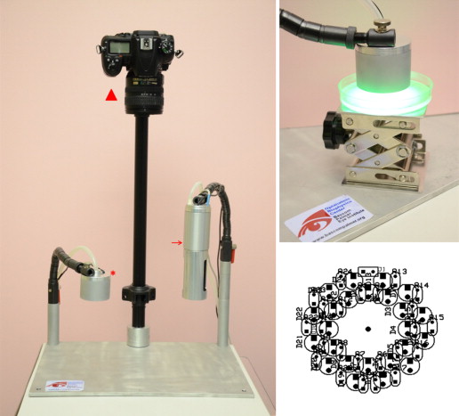
Light Source and Irradiation
A custom-built LED source was fabricated with 2 irradiation heads: green (518 nm) and UV-A (375 nm) ( Figure 1 ). Each irradiation head was assembled using an array of 24 LEDs. The green LED source (L1-0-G5TH45-1; LEDSupply, Randolph, Vermont, USA) had a 518 nm peak irradiance (I 40% : 500–541 nm) and produced 2.2mW/cm 2 over a surface of 28.3 cm 2 . The UV-A source used LEDs with a peak wavelength of 375 nm (I 40% : 370–383 nm) producing an irradiance of 2.91 mW/cm 2 on a surface of 13.8 cm 2 . The spectra were measured using a spectrometer (SM442; Spectral Products, Putnam, Connecticut, USA) and the irradiances were measured with an optical power meter (Model 1916C; Newport, Irvine, California, USA).
Preparation of the Photosensitizing Agents
The 0.1% rose bengal solution was produced by dissolving 100 mg of rose bengal (198250; Sigma-Aldrich, St Louis, Missouri, USA) in 100 mL of sterile water. Similarly, the 0.1% riboflavin solution was made by dissolving 100 mg of riboflavin (R7774; Sigma-Aldrich) in 100 mL of sterile water. Solutions were made at room temperature immediately before experimentation and kept in the dark until irradiation to ensure that photobleaching of the solutions did not occur.
Experimental Protocol
The plates were divided into 5 groups according to the treatment group: Group 1, no treatment; Group 2, 0.1% rose bengal only; Group 3, 518 nm irradiation only; Group 4, riboflavin PDT (0.1% riboflavin + 375 nm irradiation); and Group 5, rose bengal PDT (0.1% rose bengal + 518 nm irradiation). Separate groups testing 0.1% riboflavin and 375 nm against the fungi were not included in the experimental design.
Groups 1 and 2 received no irradiation. For the irradiated experiments, the Ophthalmic Biophysics Center’s (OBC) irradiating source prototypes were used; Groups 3 and 5 were irradiated with the 518 nm source and Group 4 was irradiated using the 375 nm irradiating source. The study was conducted in triplicate under aseptic conditions. One milliliter of each fungal suspension was inoculated onto a 100-mm-diameter Sabouraud-Dextrose agar plate and allowed to diffuse evenly over the entire plate. The agar plates were placed at 1 cm from the irradiator heads ( Figure 1 ) and subjected to an energy density of 5.4 J/cm 2 (30 minutes of irradiation for the 375 nm source and 40 minutes for the 518 nm sources using the irradiator prototypes), corresponding to the value defined by the Dresden protocol for clinical cornea cross-linking. Using a digital 2K thermocouple (DM6802A+; MN Measurement Instruments LLC, St Paul, Minnesota, USA), the temperature of the plate was measured for each source. The rise in temperature after a full 5.4 J/cm 2 exposure was only 9 C. After the irradiation treatment the last step of the protocol was to seal the plates and to put them into an incubator (ThermoFisher Scientific, Waltham, Massachusetts, USA) at 30 C.
LabVIEW Program for Fungal Viability Assessment
Images of the agar plates were taken at each 24 hour checkpoint with a digital camera (Nikon D7000; Nikon Inc, Tokyo, Japan) and processed using a custom-made program written in LabVIEW 6 (National Instruments, Austin, Texas, USA) to determine the percent growth of the fungus on the irradiated area of each plate. Each image has a resolution of 13.5 (±0.5) pixels/mm and was calibrated using the standard dimensions of the 100-mm-diameter agar plate. Following calibration, the irradiation zone of each image was extracted and segmented by application of a threshold based on hue, saturation, and luminance. Inner diameters of 50 mm for the 518 nm and 42 mm for the 375 nm for UV-A of each plate were analyzed because they correspond to the irradiation zone of the OBC sources. The total area of the growth inhibition was then divided by the area of the irradiation zone to determine the percent inhibition.
Statistics
A 1-tailed 2-sample z test for proportions was performed to compare the percent growth of each experimental group with respect to the control for each organism. Statistical significance was set at P < .05.
Results
For F solani , the growth of the organisms was visible beginning at day 2. At day 3 they reached the peak of growth. Group 5 was the only group exhibiting statistically significant inhibition ( P < .049). Beginning at day 3, the organism started growing from the area outside of the irradiation zone into the region of interest for Group 5. However, for all other groups, there was uniform growth throughout the entire surface of the agar plates. The findings for day 0 and day 3 are shown in Figure 2 .
Similarly, A fumigatus organisms were visible beginning at day 2. At day 3 they reached the peak of growth. Group 5 was the only group exhibiting statistically significant inhibition ( P < .049). Beginning at day 3, the organism started growing from the area outside of the irradiation zone into the region of interest for Group 5. For all other groups, there was uniform growth throughout the entire surface of the agar plates. The findings for day 0 and day 3 are shown in Figure 3 .
Stay updated, free articles. Join our Telegram channel

Full access? Get Clinical Tree


