Purpose
To evaluate and report the results of a 3-year study investigating argon laser photoablation of superficial conjunctival nevus.
Design
Retrospective, interventional case series.
Methods
The medical charts of patients referred to our clinic for the removal of a conjunctival nevus were reviewed. Argon laser photoablation of superficial conjunctival nevi was performed for 230 patients during the period from March 2006 to February 2009. The clinical course, complications, and recurrence rates were evaluated.
Results
The mean follow-up period was 71.29 ± 19.51 months (range, 36-100 months). The horizontal and vertical diameters of conjunctival nevi were 4.53 ± 1.85 mm and 3.41 ± 1.62 mm, respectively. Most nevi were faintly pigmented and located on the nasal bulbar conjunctiva. The argon laser spot size was 200 μm; its duration was 0.1 second; and the power was 321.50 ± 9.23 mW. One session was required to completely remove the nevus in 212 eyes (81%). The total number of laser spots that were required to remove 1 nevus was 107.83 ± 33.68 (range, 53-172) for cases in which the horizontal and vertical diameters were <4 mm. In all cases, the laser procedure resulted in complete removal of the conjunctival nevus. There were no significant complications, such as infection, corneal damage, or permanent injection.
Conclusions
Argon laser photoablation could be a safe and effective treatment for benign superficial conjunctival pigmentation. This method is especially helpful for large conjunctival nevi, for which surgical excision would be impossible.
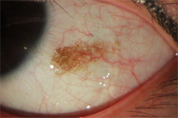
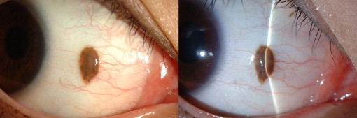
The conjunctival nevus is the most common melanocytic conjunctival tumor. Conjunctival nevi can be divided into 2 categories according to their clinical manifestation: superficial nevi and deep-vascularized nevi. A superficial nevus has a flat brownish color, is not connected to surrounding vessels, is free to move over the sclera, and does not contain a cyst ( Figure 1 ). On the other hand, a deep-vascularized nevus is elevated, can contain a cyst, and has feeder vessels that originate from the conjunctiva ( Figure 2 ).
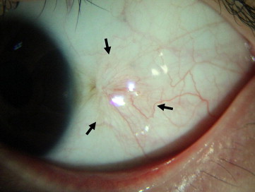
The removal of a conjunctival nevus is not always necessary. However, it may cause a cosmetic problem and result in an undue psychiatric burden, especially if it is large and noticeable because of its location. A conjunctival nevus is removed using 1 of 2 methods: surgical excision or argon laser photoablation. However, the surgical excision of a nevus often results in the dragging of tissue and neovascularization ( Figure 3 ). Surgical excision is therefore not recommended for a large, superficial, pigmented nevus. We previously reported that argon laser photoablation of a conjunctival pigmented nevus is a safe and viable alternative to surgical excision. This technique has repeatedly yielded good outcomes for patients.
In this study, we report the results of a 3-year follow-up study of patients who have undergone argon laser photoablation of superficial conjunctival pigmented nevus. We have evaluated the long-term efficacy and possible complications of this technique.
Methods
We retrospectively reviewed the medical records of 265 patients (83 men and 182 women) and analyzed 230 patients who had undergone argon laser photoablation for clinically benign superficial conjunctival pigmented nevus. The procedures were performed during the period from March 7, 2006 to February 17, 2009 at Seoul National University Hospital, and patients were followed up for at least 3 years at Seoul National University Hospital and Myongji Hospital, Kwandong University. Approval for the retrospective collection of these data was obtained from the Institutional Review Board of Seoul National University Hospital and Myongji Hospital, Kwandong University. The study was performed in accordance with the tenets of the Declaration of Helsinki, and written informed consent was obtained from each study participant. Only patients who had provided prior consent for their medical records to be used in research were included. The procedures and possible complications were explained to the patients before the initiation of laser treatment.
Clinical manifestations were examined by slit-lamp examination, and each conjunctival nevus was classified as either a superficial nevus or a deep-vascularized nevus ( Figures 1 and 2 ). Laser photoablation was conducted for superficial nevi that met the following inclusion criteria: (1) simple superficial pigmented conjunctival nevus; (2) freely movable over the underlying sclera; (3) no change in size and color for the previous 5 years; (4) no feeder vessels connected; (5) no cystic spaces within the nevus; (6) no involvement in cornea; and (7) nevus confined to bulbar conjunctiva. Surgical excision and biopsy were conducted for deep-vascularized nevi.
The pigmentation intensity, location, and size of each superficial conjunctival nevus to be subjected to laser photoablation were recorded. The intensity of pigmentation was classified as dark for cases in which the conjunctival vessels and sclera were not visible because they were covered by nevi, and faint if the vessels and sclera were visible. The location was classified as nasal, temporal, superior, or inferior bulbar conjunctiva using the cornea as the point of reference. With respect to size, the maximum horizontal and vertical diameters were measured by slit-lamp examination.
A maximum 4 × 4-mm section was removed by a single photoablation treatment. If the nevus was larger than 4 × 4 mm, the laser treatment was performed 2 or more times at 2-week intervals. When a patient had nevi in both eyes, the laser procedure was performed on 1 eye first, and on the second eye after an interval of 2 weeks.
Laser Procedure
The same ophthalmologist (J.W.K.) performed all procedures using an argon green laser (wavelength, 514 nm; Viridis; Quantel Medical, Cournon d’Auvergne, France). After administration of proparacaine hydrochloride 0.5% (Alcaine; Alcon Laboratories, Fort Worth, Texas, USA), the laser was focused directly on an area of the nevus. The laser spots did not overlap and were 200 μm in size. The duration of the laser pulse was 0.1 seconds; the energy delivered ranged from 300 to 340 mW.
After laser photoablation, we removed the nevus by gently rubbing it with a cotton-tipped swab. All nevi were easily removed from the conjunctival tissue.
Postoperative Care
Patients received levofloxacin (Cravit; Santen Pharmaceutical Company, Osaka, Japan) and 0.1% fluorometholone acetate (Flarex; Alcon Laboratories) 4 times a day for 1 week. Patients were followed up on postoperative days 7 and 14, and at 1, 3, 6, 12, 24, and 36 months after the procedure.
The best-corrected visual acuity was measured for each patient. Anterior segment photography and slit-lamp examinations were used to document the occurrence and duration of conjunctival injection, subconjunctival hemorrhage, scarring, and tissue dragging. The patient’s satisfaction level was determined 3 months after the photoablation. The answers were classified as “satisfactory” or “unsatisfactory.”
Results
This study included 299 eyes from 265 patients. Among the 265 patients presenting with conjunctival nevi, 230 (87%) had superficial nevi and 35 (13%) had deep-vascularized nevi. Among the patients presenting with superficial nevi, 68 (30%) were men and 162 (70%) were women. The mean age of these patients was 30.75 ± 10.09 years (range, 7-72 years). The mean duration of follow-up was 71.29 ± 19.51 months (range, 36-100 months). Argon laser photoablation was used to remove superficial nevi from 263 eyes belonging to 230 patients ( Table 1 ).
| Total | Superficial Nevus | Deep-vascularized Nevus | |
|---|---|---|---|
| Number of patients (%) | 265 | 230 (87) | 35 (13) |
| Sex (M/F), n (%) | 83 (31)/182 (69) | 68 (30)/162 (70) | 15 (43)/20 (57) |
| Age (y) | |||
| Mean ± SD | 29.40 ± 10.59 | 30.75 ± 10.09 | 20.54 ± 9.65 |
| Range | 6-72 | 7-72 | 6-52 |
| Period of follow-up (mo) | |||
| Mean ± SD | 70.23 ± 19.38 | 71.29 ± 19.51 | 58.36 ± 13.30 |
| Range | 36-100 | 36-100 | 37-76 |
| Number of eyes | 299 | 263 | 36 |
| Both | 34 | 33 | 1 |
| Right eyes | 121 | 98 | 23 |
| Left eyes | 110 | 99 | 11 |
With respect to the intensity of pigmentation, the vast majority of conjunctival nevi were classified in the faint group (243/263 eyes; 92%). The nevi were most likely to be found on the nasal bulbar conjunctiva (183/263 eyes; 70%), followed by the temporal, superior, and inferior bulbar conjunctiva. When the nevi were classified according to size based on a 4-mm standard, 198 of 263 eyes (75%) were less than 4 mm in both horizontal and vertical diameter ( Table 2 ).
| Total number of eyes | 263 |
| Intensity of pigmentation, no. of eyes (%) | |
| Faint | 243 (92) |
| Dark | 20 (8) |
| Location, no. of eyes (%) | |
| Nasal bulbar conjunctiva | 183 (70) |
| Temporal bulbar conjunctiva | 72 (27) |
| Superior bulbar conjunctiva | 5 (2) |
| Inferior bulbar conjunctiva | 3 (1) |
| Size (mm), mean ± SD (range) | |
| Horizontal diameter | 4.53 ± 1.85 (0.50-12.0) |
| Vertical diameter | 3.41 ± 1.62 (0.50-11.5) |
| Classification of size, no. of eyes (%) | |
| Horizontal and vertical diameter: <4 mm | 198 (75) |
| Horizontal or vertical diameter: >4 mm and <8 mm | 58 (22) |
| Horizontal and vertical diameter: >8 mm | 7 (3) |
Stay updated, free articles. Join our Telegram channel

Full access? Get Clinical Tree


