Anterior Ethmoid Artery Ligation
Metin Onerci
INTRODUCTION
With the advances in technology, endoscopic sinus surgery is now performed for the surgical management of epistaxis, and pathologies involving the skull base and orbit. Complex vascular malformations and tumors of the anterior cranial base can receive significant blood supply from the anterior ethmoidal artery (AEA). Proximal control of these feeding vessels is essential to minimize blood loss. Embolization through the ophthalmic artery puts vision at risk. A detailed knowledge of the anatomy can help the surgeon find the AEA and the best points for proximal control of the blood supply to these lesions.
The AEA originates from the ophthalmic artery, crosses to the medial orbital wall inferior to the superior oblique muscle, reaches the anterior ethmoid foramen where it leaves the orbit, and combines with the homonymous vein and nerves to form the neurovascular bundle. This bundle traverses above the ethmoid air cells, or enters and passes through the anterior ethmoid canal, and then turns superiorly at the cribriform plate to form the anterior falx artery. However, the course of the AEA may show variations, primarily related to the pneumatization of the ethmoid cells. If the supraorbital cells are well pneumatized, the AEC (anterior ethmoid canal) is often identified within a separate canal (Fig. 9.1A and B); if the ethmoid cells are poorly pneumatized, the canal is usually embedded in the roof of the ethmoid (Fig. 9.1C).
The anterior ethmoid artery canal may present a partial dehiscence in 11.4% to 66.7% according to previously published literature. These data show that the protection of the artery by the inferior bony wall is weak, and the artery is more accessible when distant from the roof of the ethmoid. If not handled carefully during surgery, the artery is vulnerable to injury.
The artery crosses the ethmoid sinus from the orbit in a diagonally anteromedial course to reach the cribriform plate at an angle ranging from 30 to 45 degrees. The distance between the artery and the axilla formed by the anterior attachment of the middle turbinate and the lateral nasal wall has a mean value of 20 mm, ranging from 17 to 25 mm, and is considered the most reliable landmark. The average distance between the AEA and the posterior wall of the frontal recess has a mean value of 11 mm ranging from 6 to 15 mm. The anterior ethmoid canal is between the second and third lamellae of the ethmoid sinus in the majority of patients, and the second and third lamellae can be used as anatomical references to locate the artery during endoscopic sinus surgery.
HISTORY
One cannot overestimate the importance of taking a detailed history. The severity of epistaxis and estimated amount of blood loss should be determined first. Although major epistaxis quickly comes from both nares, the side of bleeding should be localized to one side, if possible. Important associated signs prior to onset (easy bruising, bloody or tarry stools, hemoptysis, blood in urine, excess bleeding following tooth brushing),
any contributing or inciting factors such as trauma, and the frequency and duration of bleeding should be established, as well as any trigger factors (e.g., sneezing, nose blowing, nose picking). Any known bleeding disorders (including a family history) and conditions associated with defects in platelets or coagulation, particularly cancer, cirrhosis, HIV, and pregnancy, should be noted. The use of drugs that may promote bleeding, including aspirin and other NSAIDs, other antiplatelet drugs, heparin, and warfarin, should be asked about. Ethanol, vitamin E, and alternative medicines such as the “3 Gs” (garlic, ginseng, and gingko) also have antiplatelet effects and should be discussed with the patient.
any contributing or inciting factors such as trauma, and the frequency and duration of bleeding should be established, as well as any trigger factors (e.g., sneezing, nose blowing, nose picking). Any known bleeding disorders (including a family history) and conditions associated with defects in platelets or coagulation, particularly cancer, cirrhosis, HIV, and pregnancy, should be noted. The use of drugs that may promote bleeding, including aspirin and other NSAIDs, other antiplatelet drugs, heparin, and warfarin, should be asked about. Ethanol, vitamin E, and alternative medicines such as the “3 Gs” (garlic, ginseng, and gingko) also have antiplatelet effects and should be discussed with the patient.
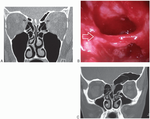 FIGURE 9.1 A: Coronal paranasal sinus CT scan, AEAs are located below the skull base and identified as a separate canal (arrows). There is an incidental septal spur toward the right side. B: Endoscopic view of AEA (arrow). C: Asymmetric frontal sinus pneumatization; on the right side, the AEA canal is embedded in the ethmoid roof; on the left side, AEA is seen as a separate canal (Courtesy of TESAV). |
PHYSICAL EXAMINATION
Vital signs should be assessed. High blood pressure should be controlled. Persistent tachycardia may be an early indicator of significant blood loss requiring intravenous (IV) fluid replacement and, potentially, transfusion. The skin should be examined for evidence of bruises or petechiae that may indicate an underlying hematologic abnormality. Associated signs such as facial hypesthesia and diplopia may indicate a sinonasal neoplasm.
Localization of the Bleeding Site
A thorough and methodical examination of the nasal cavity should be performed. The mucosa of the nasal cavity can be decongested by applying a vasoconstrictor (e.g., 0.05% oxymetazoline) with a topical anesthetic (e.g., 4% aqueous lidocaine). Blood clots should then be suctioned out to permit a thorough examination. The examination should start with inspection of the anterior aspect of the nasal cavity. Approximately 90% of bleeding vessels can be visualized in the anterior aspect of the nasal cavity. Nasal guide packs can also help the physician to identify the possible site of bleeding. In patients without a visible anterior source of bleeding, if the hemorrhage is from both nares, if the pack becomes bloody at the posterior part, or if constant dripping of blood is seen at the posterior pharynx with the anterior pack in place, the bleeding is more likely from a posterior site. Massive epistaxis may be confused with hemoptysis or hematemesis. Blood seen dripping from the nasopharynx confirms a nasal source.
Intranasal endoscopy may be performed with a rigid or flexible endoscope. The rigid endoscope is preferred because of its superior optics and its ability to allow endoscopic suction and cauterization. Endoscopy may show blood coming from superior and lateral to the middle turbinate. Visual evaluation is critical in patients with trauma to the AEA with retraction of the artery into the orbit.
INDICATIONS
Control of refractory bleeding coming from the superior aspect of the nasal cavity in the distribution of the AEA
Continued bleeding despite nasal packing
Continued bleeding despite medical treatment
Patients requiring transfusion due to a hematocrit below 38%
Problems with packing
Nasal anomaly precluding packing
Patients who are at risk for complications from nasal packing, for example, chronic pulmonary, cardiovascular, or central nervous system disease
Patient refusal/intolerance of packing
Trauma
Stay updated, free articles. Join our Telegram channel

Full access? Get Clinical Tree


