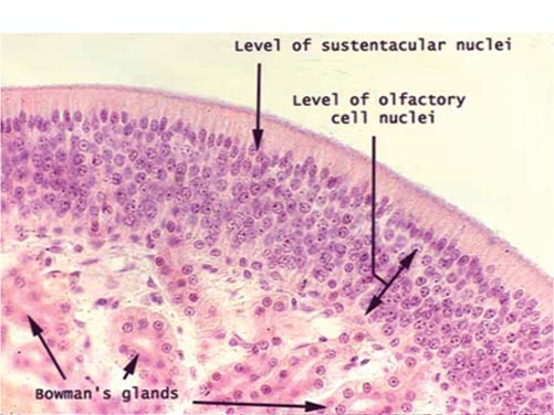
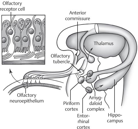
31 Anosmia and Olfactory Disturbance Olfaction can be affected by many different disease processes. Olfactory disorders may be organized into categories: The olfactory neuroepithelium is a complex network of primary and secondary neurons that make up the most ancient of the senses in humans as well as other species. This basic sensory input is more developed in other mammalian species but is critical to vertebrates and invertebrates alike. Fig. 31.1 Nerve bundles of the bipolar receptor cell course through foramina in the cribriform plate and form the olfactory bulb. They then branch out and form synaptic connections with dendrites of secondary neurons. The bipolar cells are the likely transit route by which viral and other inflammatory processes progress into the central nervous system. Fig. 31.2 Olfactory neuroepithelium—histology.
 Anosmia is the loss of the ability to smell.
 Hyposmia is a diminished ability to smell.
 Parosmia is an alteration in an olfactory cue resulting in a specific smell perceived differently.
 Phantosmia is a perception of a smell when there is no external olfactory stimulation.
 Presbyosmia pertains to the smell impairment associated with age. The loss of smell is commonly the true cause of taste disturbance complaints.
 Review of Anatomy
 The primary olfactory neuroepithelium is a pseudostratified columnar epithelium lying upon the cribriform plate. Olfactory neuroepithelium is made up of several different components.
 Olfactory receptor cells lie on the cribriform plate as well as on the superior septum and middle and superior turbinates.
 Nerve bundles of the bipolar receptor cell then course through foramina in the cribriform plate and form the olfactory bulb, then branch out and form synaptic connections with dendrites of secondary neurons. The bipolar cells are the likely transit route by which viral and other inflammatory processes progress into the central nervous system (Fig. 31.1).
 A secondary cell or sustentacular cell lies with the bipolar cells in the olfactory neuroepithelium and helps to regulate mucus production and odorant physiology.
 Additional cells include microvillar cells, supporting cells of the secretory Bowman glands, and dark horizontal and globose basal cells, which are the primordial cells from which all the others arise (Fig. 31.2).
 Olfactory neuroepithelium possesses a regenerative capacity. The degree of regeneration of olfactory neuroepithelium is dependent on the severity of the inciting event.
 The basal globose cells serve as the pluripotential cells, which regenerate the supporting and sensory bipolar cells. If the environmental insult is slight, then regeneration may occur. If the damage is severe, respiratory-like epithelium infiltrates the area of damage.
 This regeneration may take many years as realized in cohorts of smokers who stop their habit and realize an improvement in their sense of smell over several years.
 Clinical Factors to Consider when Differentiating Causes of Small Disturbance
 The onset of disturbed smell is important to differentiating the cause.
 A gradual onset is most often associated with the aging process; about 50% of patients 65 to 80 years of age have diminished olfactory function. The gradual loss is associated with additive environmental exposures, viral and bacterial damage, closure and stenosis of the cribriform foramina through which the sensory bipolar cells travel, as well as associations with dementia and neurodegenerative conditions associated with aging.
 Sudden loss of smell is more associated with either viral inflammatory damage of the olfactory neuroepithelium or shear injury of the axonal nerve fibers at the level of the cribriform plate occurring with closed head injury.
 Unilateral versus bilateral
 Associated symptoms, such as memory loss, allergic rhinitis, headache, and seizure disorder
 Diagnostic Testing
 Smell identification tests
 UPSIT (University of Pennsylvania Smell Identification Test, Sensonics, Inc., Haddon Heights, NJ). Unilateral testing can be aided by obstructing the nontest nares with a piece of tape such that cross stimulation via the nasopharynx (retronasal stimulation) does not occur. The UPSIT is a psychophysical test that is well validated and has excellent reliability compared with other testing instruments. Four booklets containing 10 odorants each are embedded on a scratch and sniff strip. Patients are asked to choose from one of four responses for each side of the nose. This test can distinguish from malingering because a 10 out of 40 response is statistically the chance performance. Lower scores suggest malingering. The UPSIT is scored on norms based on age and sex.
 Electrophysiological tests are available, including odor event-related potentials (OERPs), which involve the electroencephalographic (EEG) measurement of brain activity with surface scalp electrodes while the patient is administered odorants.
 The electroolfactogram (EOG) involves electrode application onto the surface of the olfactory neuroepithelium, which measures generator potentials of the sensory neuronal activity. This testing is experimental and not widely available.
 Radiographic testing
 Computed tomography
 Magnetic resonance imaging
 Useful Clinical Classification
 Conductive versus sensorineural
![]()
Stay updated, free articles. Join our Telegram channel

Full access? Get Clinical Tree


