Disease
Clinical parameters
Signs/symptoms
Differential diagnosis
Seasonal allergic conjunctivitis (SAC)
Sensitized individuals
Ocular itching
Infective conjunctivitis
Both females and males
Tearing (watery discharge)
Preservative toxicity
Bilateral involvement
Chemosis, redness
Medicamentosa
Seasonal allergens
Often associated with rhinitis
Dry eye
Self-limiting
Not sight threatening
PAC/AKC/VKC
Perennial allergic conjunctivitis (PAC)
Sensitized individuals
Ocular itching
Infective conjunctivitis
Both females and males
Tearing (watery discharge)
Preservative toxicity
Bilateral involvement
Chemosis, redness
Medicamentosa
Year-round allergens
Often associated with rhinitis
Dry eye
Self-limiting
Not sight threatening
SAC/AKC/VKC
Atopic keratoconjunctivitis (AKC)
Sensitized individuals
Severe ocular itching
Contact dermatitis
Peak incidence 20–50 years of age
Red flaking periocular skin
Infective conjunctivitis blepharitis
Both females and males
Mucoid discharge, photophobia
Pemphigoid
Bilateral involvement
Corneal erosions
VKC/SAC/PAC/GPC
Seasonal/perennial allergens Atopic dermatitis
Scaring of conjunctiva
Chronic symptoms
Cataract (anterior subcapsular)
A. Sight threatening
Vernal keratoconjunctivitis (VKC)
Some sensitized individuals
Severe ocular itching
Infective conjunctivitis blepharitis
Peak incidence 3–20 years of age
Severe photophobia
Males predominate 3:1
Thick, ropy discharge
Bilateral involvement
Cobblestone papillae
AKC/SAC/PAC/GPC
Warm, dry climate Seasonal/perennial allergens
Corneal ulceration and scaring
Chronic symptoms
Sight threatening
Giant papillary conjunctivitis (GPC)
Sensitization not necessary
Mild ocular itching
Infective conjunctivitis
Both females and males
Mild mucoid discharge
Bilateral involvement
Giant papillae
Preservative toxicity
Prosthetic exposure
Contact lens intolerance Foreign body sensation
Occurs anytime
Protein buildup on contact lens
SAC/PAC/AKC/VKC
Chronic symptoms
Not sight threatening
Table 3.2
Histopathologic and laboratory manifestations of allergic ocular disease
Disease | Histopathology | Laboratory manifestations |
|---|---|---|
Seasonal/perennial allergic conjunctivitis | Mast cell/eosinophil infiltration in conjunctival epithelium and substantia propria | Increased tear |
Mast cell activation | Specific IgE antibody | |
Upregulation of ICAM-1 on epithelial cells | Histamine | |
Tryptase | ||
TNFα | ||
Atopic keratoconjunctivitis | Increased mast cells, eosinophils in conjunctival epithelium, and substantia propria | Increased specific IgE antibody in tears |
Epithelial cell/goblet cell hypertrophy | Depressed cell-mediated immunity | |
Increased CD4/CD8 ratio in conjunctival epithelium and substantia propria | Increased IgE antibody and eosinophils in blood | |
Increased collagen | Eosinophils found in conjunctival scrapings | |
Vernal keratoconjunctivitis | Increased mast cells, eosinophils in conjunctival epithelium and substantia propria | Increased specific IgE/IgG antibody in tears |
Eosinophil major basic protein deposition in conjunctiva | Elevated histamine and tryptase in tears | |
CD4+ clones from conjunctiva found to have helper function for local production of IgE antibody | Reduced serum histaminase activity | |
Increased collagen | Increased serum levels of nerve growth factor and substance P | |
Increased ICAM-1 on corneal epithelium | ||
Giant papillary conjunctivitis | Giant papillae | No increased histamine in tears |
Conjunctival thickening | Increased tryptase in tears | |
Mast cells in epithelium |
3.2 Allergic Conjunctivitis: Seasonal/Perennial
Allergic conjunctivitis (AC) is a bilateral, self-limiting conjunctival inflammatory process. It occurs in sensitized individuals (no gender difference) and is initiated by allergen binding to IgE antibody on resident mast cells. The importance of this process is related more to its frequency rather than its severity of symptoms. The two forms of AC are defined by whether the inflammation and symptoms occur seasonally (spring, fall) or perennially (year-round). While the inflammatory signs and symptoms are similar for both entities, seasonal allergic conjunctivitis (“hay fever conjunctivitis”) is more common. It accounts for the majority of cases of AC and is related to pollens (e.g., grass, trees, ragweed) that appear during specific seasons. Perennial allergic conjunctivitis is often related to animal dander, dust mites, or other allergens that are present in the environment year-round. Both SAC and PAC must be differentiated from the sight-threatening allergic diseases of the eye, namely, AKC and VKC.
3.2.1 Historical Perspective
Hypersensitivity reaction description of the ocular surface dates to the earliest descriptions of hay fever or rhinitis. Some of the earliest allergy provocation testing was performed in the conjunctiva.
3.2.2 Epidemiology
Prevalence estimates for allergic conjunctivitis are difficult because allergies in general tend to be considerably underreported. As high as 40 % of the population may suffer from symptoms of allergic conjunctivitis (Rosario and Bielory 2011). Importantly, 46 % of all allergic conjunctivitis sufferers have associated allergic rhinitis (Palmares et al. 2010). The distribution of SAC depends largely on the climate. For example, in the United States grass pollen-induced SAC generally occurs in the Gulf Coast and southwestern areas of the country from March to October and from May to August, in most of the rest of the country. Conversely, ragweed pollen-induced SAC occurs in most of the country during August through October, but in the southernmost states, it can begin as early as July and stretch out through November. Tree pollens can become a problem as early as January in the south and March in the north. Race and gender predilection follows that of rhinitis sufferers.
3.2.3 Clinical Features
The dominant symptom reported in allergic conjunctivitis is ocular itching (Table 3.1). Itching can range from mild to severe. Other symptoms include tearing (watery discharge), redness, swelling, burning, a sensation of fullness in the eyes or eyelids, an urge to rub the eyes, sensitivity to light, and occasionally blurred vision. As stated previously, allergic conjunctivitis is often associated with symptoms of allergic rhinitis. Conjunctival hyperemia and chemosis with palpebral edema are typical (Fig. 3.1). A rapid test for tear IgE level has correlated the objective sign of giant papillae with the total IgE tear level (Mimura et al. 2012). Hyperemia is the result of vascular dilatation, while edema (chemosis) occurs because of altered permeability of postcapillary venules. “Allergic shiners” (periorbital darkening), due to an increase of periorbital pigmentation resulting from the decreased venous return in the skin and subcutaneous tissue, are also common.
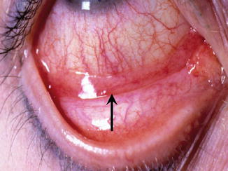
Fig. 3.1
Allergic conjunctivitis. Arrow indicates area of chemosis in the conjunctiva
3.2.4 Patient Evaluation, Diagnosis, and Differential Diagnosis
An individual suspected of having allergic conjunctivitis should have a thorough ocular, medical, and medication history. This will help greatly in differentiating AC from other ocular processes (Table 3.1). This history should establish whether the process is acute, subacute, chronic, or recurrent. It should further delineate whether the symptoms/signs are unilateral or bilateral and whether they are associated with any specific environmental or work-related exposure. Ocular symptoms such as tearing, irritation, stinging, and burning are nonspecific. A history of significant ocular itching and a personal or family history of “hay fever,” allergic rhinitis, asthma, or atopic dermatitis are suggestive of ocular allergy. Because AC is secondary to environmental allergens as opposed to transmission by eye-hand contact (infectious etiology), unless occurring in the context of petting an animal then rubbing one’s eye, SAC and PAC usually present with bilateral symptoms. This is in contrast to transmissible infections caused by viruses and bacteria that in general initially present in one eye, with the second eye becoming involved a few days later. Itch is an uncommon complaint during infectious conjunctivitis episodes. Furthermore, viral conjunctivitis may cause subepithelial corneal infiltrates not seen in AC. Palpable preauricular nodes would also signify infectious etiology for the ocular symptoms.
The type of ocular discharge (watery, mucoid, or grossly purulent) can also be helpful in determining the underlying cause of conjunctival inflammation. A watery discharge is most commonly associated with viral or allergic ocular conditions. A mucoid or purulent discharge, with morning crusting and difficulty opening the eyelids, would strongly suggest a bacterial infection.
In allergic inflammation, the eye appears red. Vision, pupil shape, ocular movement, light reactivity, and the red retinal reflex remain normal in allergic conjunctivitis. Dry eye (secondary to a decrease of the aqueous portion of the tear film) gives symptoms suggestive of foreign body in the eye and may result in conjunctiva redness. Similar symptoms are possible from anticholinergic side effects of systemic medications. Typically, itch is not reported with dry eye.
Medication history should include questions concerning the patient’s use of over-the-counter topical ocular medications, cosmetics, contact lenses, and systemic medications. Any of these can produce acute or chronic conjunctivitis. This inquiry should include direct questions and should not rely on the patient to volunteer information. Many individuals do not appreciate the potential for nonprescription topical ocular medications to cause eye symptoms or partially treat AC. Differentiation of AC from the more chronic and sight-threatening forms of allergic eye disease is discussed below in the context of the specific conditions.
3.2.5 Treatment
Medications approved for use in allergic eye disease are found in Table 3.3 . Allergic conjunctivitis can be debilitating and may cause the individuals affected to seek any type of help for relief of symptoms. Itching and tearing may be unbearable and sleepless nights frequent. Allergic conjunctivitis symptoms may be worse than the nasal symptoms in those suffering from rhinoconjunctivitis. Furthermore, treatment of the nasal symptoms with topical nasal steroids may help the rhinitis, but may not be effective for relieving ocular symptoms.
Table 3.3
Topical treatments and activity of compounds
Drug and classification | Inhibition of mediator release from human conjunctival mast cells | Inhibitory effects on other cells | References |
|---|---|---|---|
Antazoline | No effect | Inhibits IL-6, IL-8 release from conjunctival epithelial cells in vitro | |
H1 receptor antagonist | |||
Pheniramine | No effect | Inhibits IL-6, IL-8 release from conjunctival epithelial cells in vitro | |
H1 receptor antagonist | |||
Emedastine | No effect | Inhibits IL-6, IL-8 release from conjunctival epithelial cells in vitro | |
H1 receptor antagonist | |||
Levocabastine | No effect | Inhibits IL-6, IL-8 release, ICAM-1 expression on conjunctival epithelial cells in vitro | |
H1 receptor antagonist | |||
Olopatadine | Histamine, tryptase, PGD2, TNFα in vitro | Conjunctival mast cell TNFα-mediated upregulation of ICAM-1 on conjunctival epithelial cells in vitro | |
H1 receptor antagonist | |||
Mast cell stabilizer | |||
Ketotifen | Histamine in vitro | Chemotaxis and activation of eosinophils in vitro | |
H1 receptor antagonist | |||
Mast cell stabilizer | |||
Azelastine | In vitro data not available | Activation of eosinophils in vitro | |
H1 receptor antagonist | Eosinophils and neutrophils in tears | ||
Mast cell stabilizer | ICAM-1 expression in vivo | ||
Cromolyn | Not inhibitory for histamine release in vitro | Chemotaxis and activation of eosinophils, neutrophils, monocytes in vitro | |
Mast cell stabilizer | Tryptase in tears | ||
Lodoxamide | In vitro data not available | Chemotaxis and activation of eosinophils in vitro | |
Mast cell stabilizer | histamine and tryptase in tears | Eosinophils, neutrophils, T cells in tears | |
ICAM-1 expression on conjunctival epithelial cells in vitro | |||
Nedocromil | Not inhibitory for histamine release in vitro | IgE synthesis from B cells | |
Mast cell stabilizer | ICAM-1 and HLA-DR expression on conjunctival epithelial cells in vitro | ||
Activation and survival of eosinophils in vitro | |||
Pemirolast | Not inhibitory for histamine release in vitro | Activation of eosinophils and neutrophils in vitro | |
Mast cell stabilizer | |||
Alcaftadine | Histamine in vitro | Chemotaxis and activation of eosinophils in vitro | No reference |
H1 receptor antagonist | |||
Mast cell stabilizer | |||
Bepotastine | Histamine in vitro | Chemotaxis of eosinophils in vitro | No reference |
Mast cell stabilizer |
Management of allergic conjunctivitis is, therefore, primarily aimed at alleviating symptoms. The best treatment is avoidance of the specific allergen, which, unfortunately, is usually not possible. Avoidance of scratching or rubbing, application of cool compresses and artificial tears, and refrigeration of topical ocular medications are practical interventions to alleviate discomfort. While oral antihistamines may help to relieve eye itch, first-generation drugs may also decrease tear production, causing more ocular symptoms. Topical medications are generally considered more effective to relieve ocular itching than oral medications and may be additive to relief gained from oral antihistamines.
The treatment of choice for mild to moderate AC is a dual-acting topical ocular medication. The mast cell-stabilizing component of these drugs benefits patients most if treatment is started before the height of symptom onset. Patients usually note rapid onset of relief of itch upon drop instillation, as most dual-action medications have high H1 receptor affinity. Drug dosing varies from one to four times per day, and efficacy is judged best by symptom relief.
In severe disease, combination therapy is recommended. This therapy may include topical medications (antihistamines, mast cell stabilizers, NSAIDs, or combinations) and oral antihistamines. Nonsteroidal drugs inhibit cyclooxygenase resulting in decreased formation of prostaglandins and thromboxanes, but not leukotrienes. Therefore, these compounds are useful in controlling itching and some inflammation, but not the infiltration of inflammatory cells. In extreme cases, the use of a topical steroid four times a day should be considered. All patients receiving topical steroids should have their intraocular pressure measured every 3 months if on topical steroids and be evaluated for cataract annually. Specific injection immunotherapy performed by an allergist may be beneficial in decreasing the severity of future ocular allergy symptoms. Sublingual immunotherapy specifically for perennial allergic conjunctivitis has been demonstrated to be effective in relieving symptoms (Potter 2006; Calderon et al. 2011). There is no retrograde passage of nasal steroids up through the lacrimal sac so any ocular effect is considered to be secondary to systemic absorption. Intranasal stimulation is a common method to stimulate reflex tearing, and this is considered a nonspecific reaction. The stimulation may be mechanical, by chemical irritant, or by allergen.
3.3 Atopic Keratoconjunctivitis
3.3.1 Historical Perspective
Atopic keratoconjunctivitis (AKC) is a bilateral, chronic inflammation of the conjunctiva and lids associated with atopic dermatitis. Hogan, in 1953, was the first to describe the findings of chronic conjunctivitis and keratitis in patients with atopic dermatitis (Hogan 1953). Three to 17 % of the population has atopic dermatitis (Garrity and Liesegang 1984; Spergel 2010). From 15 to 76 % of patients with atopic dermatitis have ocular involvement, usually AKC (Garrity and Liesegang 1984; Dogru et al. 1999; Moscovici et al. 2009).
3.3.2 Epidemiology
The onset of disease is usually in the second through fifth decade although the majority of patients with atopic dermatitis are diagnosed by age 5 years. Series report the onset of symptoms between the ages of 7 and 76 (Foster and Calonge 1990; Power et al. 1998; Tuft et al. 1991). The highest male to female ratio is reported as 2.4:1 (Foster and Calonge 1990; Tuft et al. 1991). No racial or geographic predilection is reported.
3.3.3 Clinical Features
Itching is the major symptom of AKC. This may be more pronounced in certain seasons or it may be perennial. Other symptoms, in decreasing order of frequency, include watering, mucous discharge, redness, blurring of vision, photophobia, and pain (Tuft et al. 1991). Exacerbation of symptoms most frequently occurs in the presence of fur-bearing animals and pets (Tuft et al. 1991).
Signs of AKC include skin, lid margin, conjunctival, corneal, and lens changes (Fig. 3.2). The periocular skin often shows a scaling, flaking dermatitis with a reddened base. The skin of the lids may become leatherlike, developing cicatricial ectropion (turning outward of the lid from skin scarring) and lagophthalmos (incomplete closure of eyelids). Lateral canthal ulceration and cracking as well as lash loss (madarosis) may also be present. This may be the principal manifestation in a minority of cases. The lid margins may show meibomitis, keratinization, and punctal ectropion. The conjunctiva of the tarsal surfaces can manifest a papillary reaction and possibly pale white edema. In contrast to VKC, the papillary hypertrophy of AKC is more prominent in the inferior conjunctival fornix. Subepithelial fibrosis is present in many, fornix foreshortening in some, and symblepharon (scar of conjunctival surface of lid to conjunctiva of the globe) in a few. The bulbar conjunctiva may have few findings besides erythema and chemosis. A perilimbal, gelatinous hyperplasia may occur. Horner-Trantas dots (whitish dots that are aggregates of degenerated epithelial and eosinophils that accumulate at the surface of the hyperplastic, gelatinous limbus) have been reported to occur in AKC (Friedlaender 1979).
Fig. 3.2
Atopic keratoconjunctivitis. Periocular skin lesions of atopic dermatitis
Significant vision loss in this disease usually results from pathologic conditions of the cornea. Punctate epithelial keratopathy is the most common corneal finding. Persistent epithelial defects, scarring, microbial ulceration, and neovascularization are the main corneal causes for decreased vision (Table 3.1). Penetrating keratoplasty or cornea transplant is at risk of the same surface problems but has been shown to improve vision in some (Ghoraishi et al. 1995). Herpetic keratitis is reported to occur in 14–17.8 % of patients (Foster and Calonge 1990; Tuft et al. 1991). Keratoconus, a noninflammatory progressive thinning of the cornea occurs in 6.7–16.2 % of patients (Foster and Calonge 1990; Tuft et al. 1991).
Anterior uveitis and iris abnormalities are not reported. The prevalence of cataract associated with AKC is probably increased, since steroids are so frequently used in the treatment of the disease. The lens opacity typically associated with AKC, however, is an anterior or subcapsular cataract. This cataract often has the configuration of a multilobed opacity resembling a “milk splash.” Retinal detachment with or without previous cataract surgery is the principal posterior manifestation of AKC reported (Hurlbut 1961; Yoneda et al. 1995; Klemens 1966).
3.3.4 Patient Evaluation, Diagnosis, and Differential Diagnosis
Paramount to both diagnosis and treatment in AKC is a careful history. The patient typically describes severe, persistent, periocular itching associated with dermatitis. There is usually a family history of atopic disease in one or both parents and commonly other atopic manifestations in the patient, such as asthma (65 %) or allergic rhinitis (65 %) (Power et al. 1998). A history of seasonal or exposure-related exacerbations is usually present. History and examination reveal features to help differentiate AKC from other atopic ocular conditions. The lack of contact lens wear aids in differentiating AKC from GPC. AKC patients are usually older and have major lid skin involvement compared to patients with VKC. SAC patients have no or markedly diminished symptoms out of their season and show no evidence of chronic inflammation in the conjunctiva. The significant past history or concurrent presence of eczema cannot be emphasized enough as a finding in patients with AKC. The serum level of IgE is often elevated in patients with AKC. A Giemsa stain of a scraping of the upper tarsal conjunctiva may reveal eosinophils. Meibomian gland obstruction is noted to be significant and may be found to exceed that seen in obstructive Meibomian gland disease patients (Ibrahim et al. 2012).
3.3.5 Treatment
The approach to treatment is multifaceted and includes environmental controls as well as topical and systemic medications (Table 3.3 ). It is unlikely that the AKC patient will see the ophthalmologist without also being under the care of a primary care physician and allergist. However, the patient must remove environmental irritants in both the home and the employment or school setting. The nature of the irritants may be better defined through allergy testing.
The topical application of a vasoconstrictor-antihistamine combination may bring transient relief of symptoms but is unlikely to alter the immunopathologic process or its sequelae. There is potential for overuse due to the chronic nature of the disease. The potent topical antihistamines offer much greater H1 receptor antagonism than over-the-counter antihistamines. The topical administration of steroids such as prednisolone acetate eight times per day for 7–10 days is clearly beneficial in controlling symptoms and signs. These agents, of course, must be used judiciously, since the chronic nature of the disease may encourage overuse. The patient must be instructed that steroid use must be transient only and must be carefully monitored for efficacy; he or she must also be warned of the potential for causing cataract and glaucoma. Nonsteroid medications have been shown to be effective in reducing itching, tearing, and photophobia. Topical mast cell stabilizers one to four times daily are recommended year-round in patients with perennial symptoms. If an exacerbation occurs and the patient is not taking a mast cell-stabilizing agent topically, its use should be initiated one to four times daily concurrent with a short burst of topical steroids (for 7–10 days). Mast cell stabilizers alone such as cromolyn, nedocromil, lodoxamide, or mast cell stabilizer antihistamine combinations such as olopatadine, azelastine, epinastine, and ketotifen may be helpful. Cyclosporine-A and tacrolimus, both orally and topically have been shown effective at treating AKC as well as reducing the amount of topical steroid use (Mohammed Al-Amri 2013; Miyazaki et al. 2009; Rikkers et al. 2003; Hoang-Xuan et al. 1997; Hingorani et al. 1998; Stumpf et al. 2006; Anzaar et al. 2008). Tear levels of eosinophilic cationic protein are reduced following treatment with topical tacrolimus (Wakamatsu et al. 2011). Foster and Calonge recommend maximizing the use of systemic antihistamines (Foster and Calonge 1990). Itch is a major complaint, and newer H1 receptor antagonists are fairly specific. Only in rare cases of uncontrolled dermatitis with vision-threatening complications are oral steroids indicated. The role of allergen desensitization is similar to that in AC and VKC. Plasmapheresis has been effective in the treatment of AKC (Aswad et al. 1988).
Stay updated, free articles. Join our Telegram channel

Full access? Get Clinical Tree


