When one third of the diabetic population does not appreciate that they have diabetes, it is common for retinal specialists to see patients who present with a vitreous hemorrhage.1 The diagnosis of proliferative diabetic retinopathy (PDR) in such a clinical setting is made by examining the fellow eye. If the fellow eye has retina findings characterized by small hemorrhages, cotton-wool spots, macular edema and hard exudates rings, or other characteristics of diabetic retinopathy, then the patient is at risk for similar level of diabetic retinopathy in the eye with a vitreous hemorrhage. For a new patient who does not know his or her diabetes status, laboratory testing for HbA1c, fasting blood sugars (FBSs), lipid profiles, and creatinine must be done. Further testing with 3-hour glucose tolerance tests can be performed at the endocrinologist’s office. The ophthalmologist may be the first physician to diagnose diabetes in this type of new patient with vision loss.
The risk of PDR is the greatest in those patients who have severe nonproliferative diabetic retinopathy (NPDR). Severe NPDR is described as having soft exudates (cotton-wool spots), intraretinal microvascular abnormalities (IRMAs), venous beading, extensive retinal hemorrhages, or microaneurysms (see Chapter 7). IRMAs can be new intraretinal vessels or dilated preexisting vessels. IRMAs are very difficult to see clinically and photographically. They are perhaps best seen with red-free photographs in the seven standard fields. Severe NPDR is defined as having three of the aforementioned four characteristics, each involving at least two quadrants of the fundus.2 Once this occurs, the progression to PDR is within 15 months2 (Figs. 9-1 to 9-4).
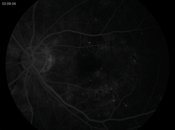
FIGURE 9-1. Soft exudate or cotton-wool spot.
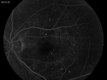
FIGURE 9-2. Intraretinal microvascular abnormality.
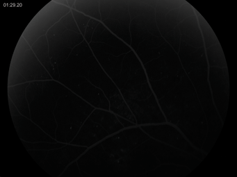
FIGURE 9-3. Venous beading.

FIGURE 9-4. Extensive retinal hemorrhages or microaneurysms.
One of the reasons that IRMAs may be difficult to identify is that they can be transient. Cotton-wool spots usually disappear in 6 to 12 months. Blot hemorrhages and IRMAs disappear after extensive capillary closure. Capillary closure can be identified when the small vascular branches decrease in number and small arterioles become white threads, producing a picture described as “featureless retina.” On fluorescein angiography, these areas are known to “capillary nonperfusion” (Figs. 9-5 and 9-6).
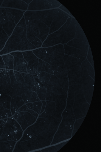
FIGURE 9-5. “Featureless retina”
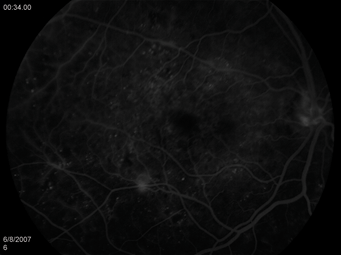
FIGURE 9-6. “Featureless retina” and the fluorescein angiographic photo denoting capillary nonperfusion.
New vessels can arise anywhere but if they occur at the optic nerve (NVD), neovascular vessels are seen within 45 degrees from the optic nerve. They are commonly seen on the optic disc. Studies have documented 69% to 73% of neovascularization in PDR eyes is found on the optic disc.3,4
Neovascularization disc (NVD) can be seen as fine threadlike loops of vessels lying on the surface of the disc or bridging across the physiologic cup. They are usually easily identified but in their earliest stages, they may be overlooked with the low magnification of indirect ophthalmoscopy. They may be seen with a slit lamp examination using a 90 D or 60 D lens. They may be most easily identified with fundus photographs or fluorescein angiography. Fluorescein angiography can delineate new vessels at the optic disc because of their intense hyperfluorescence, which fills an increasingly larger area with each successive frame of the fluorescein angiogram (Figs. 9-7 and 9-8).
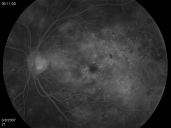
FIGURE 9-7. Fluorescein angiogram of disc neovascularization.
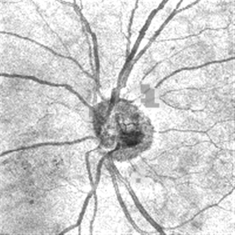
FIGURE 9-8. OCT, disc neovascularization, seen as a frond at the optic disc.
Techniques for finding the neovascularization elsewhere (NVE) and NVD and other characteristics in severe NPDR and PDR include the use of binocular indirect ophthalmoscopy, direct ophthalmoscopy, slit lamp biomicroscopy with a 90 or 60 D lens, and Goldmann contact lens. The goal is to find neovascular fronds 45 degrees from the optic nerve and examine the area 5 to 6 disc diameter (DD) from the optic nerve. It is essentially the area of the posterior pole to the equator, the area subtended by the seven standard photographic fields.
A fundus drawing with detailed notations in color is useful: red for new vessels elsewhere, noting if it is equatorial or anterior to the equator and the clock hour. The floating sheets of red blood cells imbedded in the vitreous gel can be color coded with shades of green, marking its location in space, with a diagram of the eye in cross section. NVE can be described in terms of the clock hour and the extent and the presence in the equator, in the posterior pole. Using light red to code the light vitreous hemorrhage enables the clinician to follow the progression of the hemorrhage at later follow-up appointments. In later examinations, the site of the original vitreous hemorrhage may reveal fronds of NVE, explaining the origin of the vitreous hemorrhage. In addition, this area may herald the site of future fibrovascular proliferation, also colored in green. Dense vitreous hemorrhage can be colored in dark green.
The Goldmann contact lens is excellent for examining the periphery if the clinician suspects NVE in the presence of a faint, diaphanous vitreous hemorrhage. The three mirror lenses of the Goldmann contact lens allow navigation to the periphery and lucid areas where there is no vitreous blood.
In diabetic patients who are myopic, or who have had trauma to the orbit, the vitreous hemorrhage may be due to a retinal hole. Also in the setting of mild NPDR, short duration of diabetes, and good glycemic control, a vitreous hemorrhage may mean a retinal hole instead of PDR. Thus, examination of the periphery is important when the diabetic retinopathy does not equal the level of PDR and yet there is a vitreous hemorrhage. Always be suspicious that there is a retinal tear if the PDR or severe NPDR markers are not found on examination. Scleral depression and Goldmann contact lens examination of the retinal periphery in the eye with a vitreous hemorrhage lead to the discovery of a retinal hole.
When an eye has a vitreous hemorrhage, aim for a large pupillary dilation of 6 mm or 7 mm or more. Maximal pupillary dilation allows a view of the periphery which can give clues to the level of severe diabetic retinopathy with or without the presence of a retinal hole.
Stereoscopic fundus photographs using the 30 or 35 degree camera setting show early neovascularization of the optic disc and elsewhere. The seven standard fields are useful as well. Maximal pupillary dilation is important for a good fundus photograph. For follow-up visits, these photographs aid the clinician in pinpointing the progression of diabetic retinopathy.
Finding NVE and IRMAs may be difficult. In addition, differentiating between NVE and IRMA is a challenge. IRMAs may be extensive, and the NVE may not show classic frondlike patterns or wheel-spoke presentations. Frank NVE presents as extensions across arterial and venous systems in the equator and anterior to the equator. Older NVE is associated white fibrotic condensations adherent to the vitreous which are known as fibrovascular proliferations. These fibrovascular changes can occur as the NVE regresses or the fibrovascular changes occur after the resolution of a previous localized vitreous hemorrhage.
Using a Goldmann contact lens and the slit lamp biomicroscope, look at the equatorial mirror and then at the peripheral mirror for these areas of NVE. Start with low illumination but with a broad slit beam. This allows time for the examiner’s eyes to be dark adapted. Then, use a very thin, bright slit beam to see more clearly if the area of neovascularization has an abnormal adherent relationship to the vitreous. Here, the three-dimensional quality of the NVE can be seen by using the slit beam at an angle to the viewing axis and in line with the viewing axis to see the characteristics about the NVE or IRMA and retinal surface.
Fluorescein angiography and fluorescein angioscopy can identify neovascularization versus IRMA. Neovascularization manifests as profound hyperfluorescence on fluorescein angiography. Fluorescein angioscopy is difficult to see in NVE in areas of equator and anterior to the equator because of the structural limitations of the fundus camera.
Lastly, if the clinician is suspicious of NVE and IRMAs, ask the patient to return in 1 month or sooner if his vision deteriorates. In careful follow-up in subsequent visits, the “suspicious” lesions become more pronounced and the level of diabetic retinopathy manifests.
New vessels may be barely visible at their inception. The caliber of the new vessels is 1/8 to 1/4 of that of a major vein at the optic disc margin. New vessels may be amorphous, without a pattern. Commonly, NVD are radiating spokes from the carriage wheel’s center, with an associated circumferential vessel surrounding its periphery. NVD lie over vessels and seem to drain into veins. The superotemporal vein, seen in field 4, is the most common site of NVE, 3,4 occurring in about 27% of the 1,158 eyes under the DRS.5 The inferotemporal field, field 5, has 17% of NVE seen out of 1,158 study eyes.5 The inferonasal view of field 7 has 21% of the NVE seen out of 1,158 study eyes.5
The rate of growth of the NVE is variable. The change can vary between 1 week and several months. Early in the life of the patch of NVE, the vessels are bare. However, later, white fibrous tissue is adjacent to them. The fibrous proliferation contains fibrocytes and glial cells.6,7 Their life cycle is that of proliferation followed by partial or complete regression.3,8,9 Regression occurs when the wheel-shaped NVE has a decreasing number of vessels and caliber of the vessels in the center of the wheel. Next, there is a partial replacement of the NVE with fibrous tissue. Or paradoxically, the NVE patch enlarges but the peripheral vessels narrow. The regressing NVE can show sheathing of the vessels as well. The width of the sheath in the vessels of the NVE increases such that the NVE patch becomes a network of white lines without any red blood cells seen in the lumen. At this stage, there are new vessels forming at the edge of the patch of NVE. New vessels are seen at different stages of growth in different areas of the retina in the same eye.
The fibrovascular proliferation maybe translucent and are misinterpreted as inactive. Watch for any changes in the surrounding area. With more NVE, contraction of the fibrovascular proliferation can lead to separation from the retina, causing anterior-posterior traction. This type of traction retinal detachment is glistening white and concave. At this point, surgery is an option.
If contraction of the fibrovascular stalk does not occur but rather the NVE passes through all of its stages without affecting the patient’s vision, the patient will be undergoing a quiescent stage of his or her PDR. The patient will have spontaneous regression of NVE leaving no adverse sequela for the patient.10
The posterior vitreous face touches the NVE patch and the NVE patch appears to be elevated. Adjacent to the patch of NVE adherent to the vitreous is a localized posterior vitreous detachment. The small localized vitreous detachment and the rest of the vitreous gel is close to the retina. As the posterior vitreous detachment becomes more extensive, the curved contour of the vitreous face is parallel to the surface of the retina and the NVE. The vitreous face is 0.5 to 2 DD from the surface of the retinal. Meanwhile, anatomically, the surface of the new vessels are adherent to the vitreous. As NVE networks get larger, the new vessels can be pulled away and can create a contraction and a traction retinal detachment.
The thickness of the posterior vitreous surface changes. At the site of NVE, the clinician can see the vitreous as a thick band using slit lamp biomicroscopy. This is probably due to fibrovascular proliferation along the posterior vitreous surface. There are alternating thicker and thinner adhesions to the vitreous, giving an undulating, textured, and irregular appearance to the vitreous and internal limiting membrane (ILM) interface. Lastly, the posterior vitreous surface is subtle and can be seen by using the binocular slit lamp’s narrow slit beam to glide smoothly over the retina. The slit beam moves over the surface of the retina, while you are looking at the different types of light reflexes. Blood in the fluid vitreous, posterior to the detached vitreous gel, can languish on thew retina for weeks and months. However, blood inside the formed vitreous tends to lose color and becomes yellow or white before absorption is complete. This type of bleeding into the formed vitreous may require months to resorb. Unresolved vitreous hemorrhage ultimately needs surgery.
The contraction of the abnormal vitreous interface and fibrovascular proliferation may cause the macula to be dragged and distorted. Most commonly, the macula can be dragged nasally and vertically with overlying glistening white fibrous membranes.
Contraction of the extensive sheet of fibrovascular proliferation may lead to retinal detachment. The subsequent avulsion of a retinal vessel, usually a vein, can be accompanied by a vitreous hemorrhage. The retinal detachment may involve the entire macula causing a concave traction retinal detachment. Or in severe cases, sheets of fibrovascular proliferation are cross layered over the macula producing a traction retinal detachment. Associated holes can occur in the periphery as well, but this is rare.
Extensive NVE with large caliber new vessels accompany heavy fibroproliferation with tight vitreoretinal adhesions. Usually, these occur after a vitreous hemorrhage in the setting of an intact posterior hyaloid, adherent to the ILM. Contraction is followed by extensive retinal detachment. In those vitreous hemorrhages where there is little fibrovascular proliferation, NVE, and a posterior vitreous detachment, there is less risk for a traction retinal detachment. These vitreous hemorrhages when resolved, have a better visual prognosis than those vitreous hemorrhages with an intact hyaloid and severe fibrovascular proliferation.
Chief Complaint
The patient may present with abrupt loss of vision or gradual loss of vision. The vision could be distorted reflecting the abnormality of the retinal surface and traction retinal detachment.
Past Medical History
In those instances of known diabetic patients who present with decreased vision, it is important to ask about their past history of fasting blood glucose, HbA1c, cholesterol levels, triglyceride levels, and creatinine levels.11 In addition, ask about vision loss, past history of laser, macular laser, panretinal laser photocoagulation, and past vitrectomy surgery. In patients with moderate to severe diabetic retinopathy, it is important for the clinician to assess the risk of accelerating diabetic retinopathy. Does the patient have PDR, yet to be detected?
Physical Examination
PDR is the most severe type of retinal complication in a diabetic patient. As we are all aware, diabetic retinopathy is progressive. Thus, this patient presenting to the ophthalmologist’s office has the most severe form of disease. He or she must have either had eye care in the distant past. Alternatively, he or she may never have received any eye care at all in his or her life.
If the patient has never received eye care from any eye care practitioner, the patient may be in denial. It is the responsibility of the ophthalmologist’s office to provide patient education materials, all of which are available on the American Diabetes Association Web site.1
Patient History and Review of Systems
In taking the patient history, one can assess if the patient is aware of his or her status of diabetes. By the time the PDR appears, the patient has impaired vision and there are other risk factors to ascertain.11 Thus, the ophthalmologist and retinal specialist play an important role as an educator about diabetic retinopathy and diabetic eye disease. The ophthalmologist’s office serves as a resource for community social services for diabetic patients with PDR. He or she may need to find a nutritionist, podiatrist, and psychologist. There is a high rate of depression among the chronically ill diabetic patients who have severe degrees of diabetic retinopathy.12
In one study, the combination of cognitive behavioral therapy and supportive diabetes education is an effective nonpharmacologic treatment for depression in Type 2 diabetic patients (Table 9-1). The behavioral therapy and education are associated with improved glycemic control.13
TABLE 9-1 Education of Proliferative Diabetic Patient
1. Potential for severe vision loss if noncompliant with internal medicine physician.
2. Obesity and metabolic syndrome play a role in diabetic retinopathy
3. Lipid profiles
4. HbA1c and FBS
5. Kidney function
If the patient cannot remember the name of his or her medication or how long he or she has had the disease, he or she must have been newly diagnosed or unfamiliar with his or her diagnosis. In addition, the diagnosis of PDR could be new for him or her. Prior to this visit, the patient may not have had any deterioration of vision affecting his or her activities of daily living. Or he or she could be in denial. In any case, we need to emphasize that the patient and the medical doctors are a “team.” If the patient follows the instructions of his or her team of doctors, he or she will have a good treatment outcome. However, if the patient does not follow the treatment regimen for his or her diabetes, the treatment outcome may be disappointing.11 Consequently, the patient may suffer accelerated, severe vision loss along with systemic complications involving the heart and kidneys.11,14
Similarly, in asking about the review of systems, the clinician gains an understanding of the patient’s level of cardiovascular health, hypertension, and lipid metabolism.5 All these factors play a role in taking care of the patient (Table 9-2). Weight issues and obesity play a role in diabetes and cardiovascular disease. According to the CDC, it is estimated that 33.3% of the men and 35.3% of the women in the United States are obese.5 Smoking plays an adverse role in the development of diabetes.14 Smoking has been associated with the development of NPDR.14,15
TABLE 9-2 Basic Health Literacy for the Diabetic Patient
1. Definition of Type 1 and Type 2 diabetes
2. The symptoms of hyperglycemia and hypoglycemia
3. Correlation of glycemic control and diabetic retinopathy
4. Correlation of glycemic control and systemic side effects: silent ischemia, diabetic retinopathy, renal insufficiency, hyperlipidemia, diabetic neuropathy, foot ulcers, and amputation
5. Diabetic diet: calorie counting, decreased portion size at meals, and ADA guidelines for diet
6. Oral medications and need for constant monitoring of diet
7. Insulin
8. See an ophthalmologist or retina specialist regularly if the patient has PDR.
Stay updated, free articles. Join our Telegram channel

Full access? Get Clinical Tree


