QUESTIONS
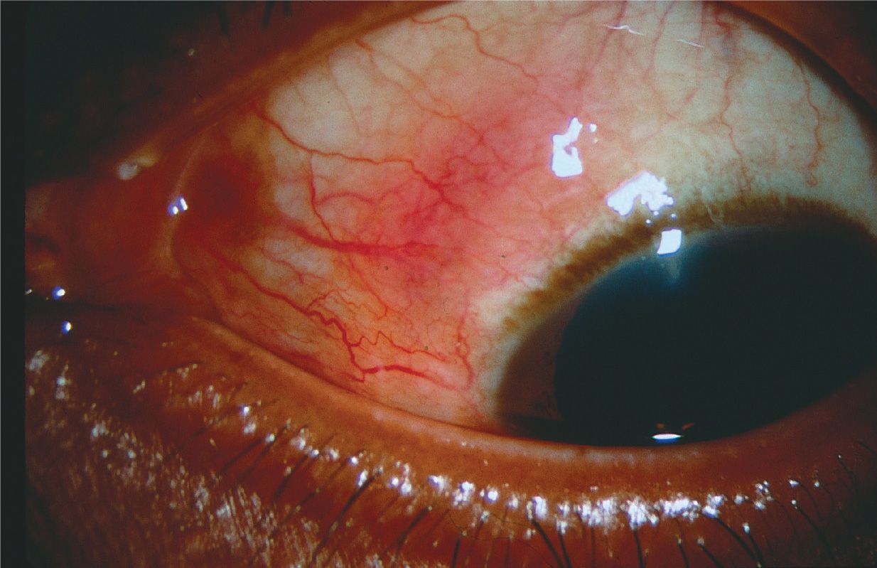
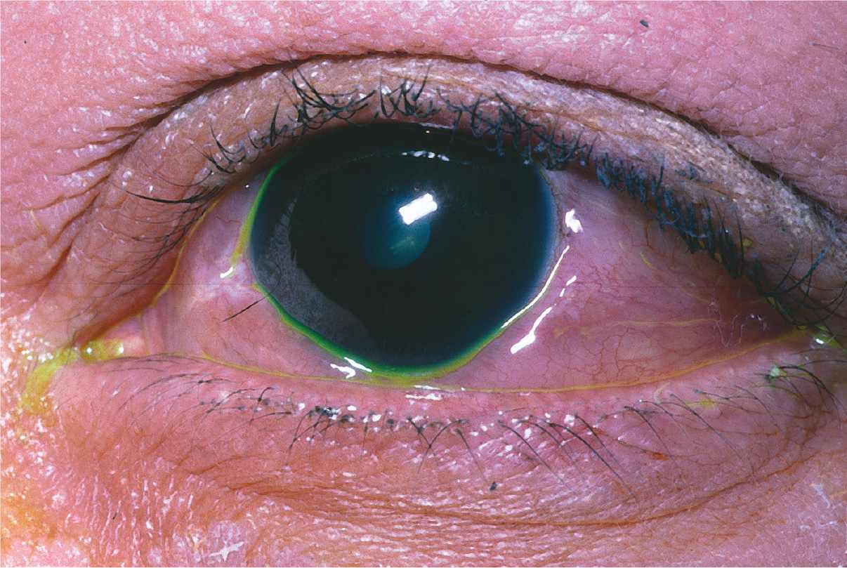
1 A 64-year-old white woman presents with complaints of pain, decreased vision, and a red eye for the past 2 days (Fig. 8-1). Her medical history reveals that she had a trabeculectomy for primary open-angle glaucoma in this same eye 6 months before. What would a Gram stain of the vitreous fluid of this patient most likely show?
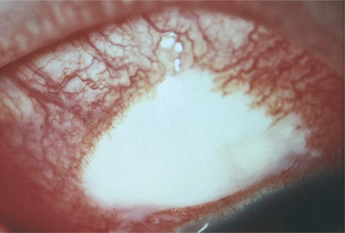
A) Gram-negative coccobacilli
B) Gram-positive rods
C) Branching pseudohyphal forms
D) Gram-positive cocci in clusters
QUESTIONS 2–4 A 69-year-old white man presents with mild discomfort and reports a gradual decrease in the vision of his right eye over the course of 1 week. His ocular history is remarkable for cataract extraction and posterior chamber intraocular lens (IOL) insertion in this eye 4 months previously. Because of the low-grade nature of the inflammation, topical steroids are begun, and the inflammation responded favorably. After discontinuing the steroids, the symptoms return and the patient again presents with a granulomatous anterior chamber inflammation, including a small hypopyon and a mild anterior vitritis.
2 What would examination of the white plaque present at the equator of the posterior lens capsule most likely reveal?
A) Candida albicans
B) Propionibacterium acnes
C) Staphylococcus epidermidis
D) Mycobacterium tuberculosis
3 Which one of the following is the most appropriate therapy for this condition?
A) Vitrectomy and intravitreal injection of amphotericin B
B) Observation
C) Vitrectomy, removal of the IOL, and intravitreal injection of gentamicin
D) Vitrectomy, posterior capsulectomy, and intravitreal injection of vancomycin
4 What is the least likely microbial cause for this syndrome?
A) P. granulosum
B) Achromobacter
C) Klebsiella
D) Corynebacterium
QUESTIONS 5–7 A 32-year-old African American woman presents with floaters and blurred vision in her right eye. Slit lamp examination shows nodules on the iris (Fig. 8-2). A recent chest radiograph revealed hilar adenopathy.
5 Biopsy of her lacrimal gland would likely reveal:
A) epithelioid cells within necrotic tissue
B) multinucleated giant cells of the Touton type
C) multinucleated giant cells of the Langhans type
D) diffuse lymphocytic infiltration
6 Other ocular manifestations include all of the following except:
A) papillitis
B) scleritis
C) follicular conjunctivitis
D) granulomatous keratic precipitates
7 Ocular complications in this condition as a result of steroid treatment include all of the following except:
A) cataract
B) retinal neovascularization
C) glaucoma
D) scleromalacia
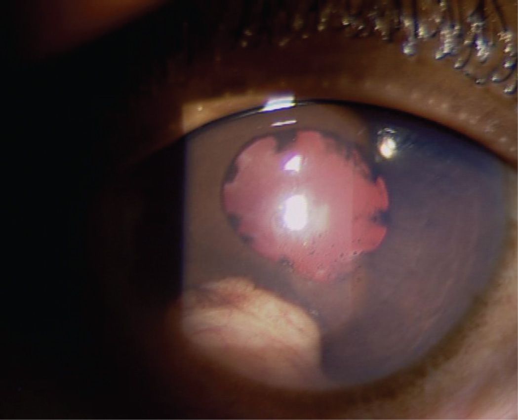
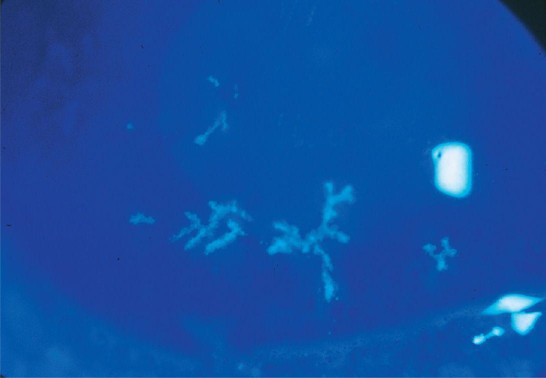
QUESTIONS 8 and 9 An 11-year-old white girl presents with reports of floaters in her left eye and a mild decrease in vision. On examination of her anterior chamber, you notice a mild anterior chamber inflammatory reaction and the corneal changes shown in Figure 8-3. Dilated fundus examination reveals whitish fibrous material in the inferior periphery.
8 Which one of the following is associated with this condition?
A) Anterior subcapsular cataracts
B) HLA-B27
C) Unilaterality in the majority of cases
D) Multiple sclerosis
9 Pathologic examination of the “snowbank” would reveal:
A) lipid exudates
B) fibroglial and vascular components
C) an aggregation of epithelioid and multinucleated giant cells
D) a conglomeration of lipofuscin and chronic inflammatory cells
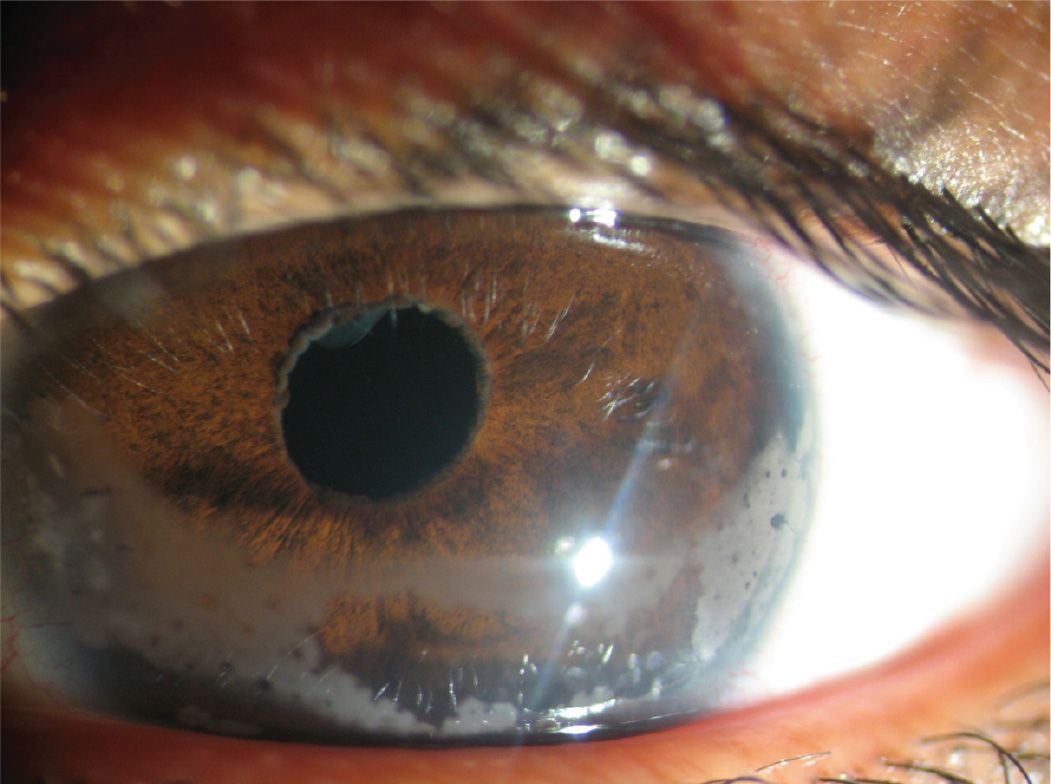
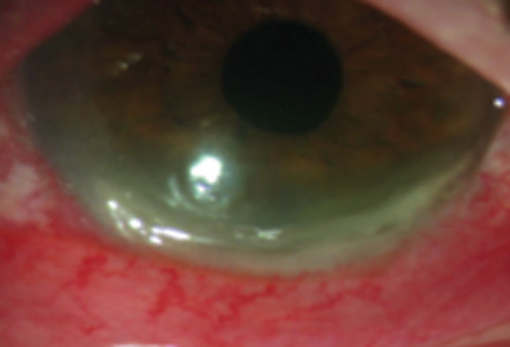
10 A patient presents with the eye illustrated in Figure 8-4. All of the following conditions can cause this except:
A) uveitis–glaucoma–hyphema (UGH) syndrome
B) herpetic iridocyclitis
C) HLA-B27 anterior uveitis
D) traumatic iridocyclitis
11 Which one of the following conditions is not typically associated with diffusely distributed keratic precipitates?
A) Fuchs heterochromic iridocyclitis
B) Sarcoidosis
C) Vogt–Koyanagi–Harada syndrome
D) Syphilis
12 Which one of the following conditions is not associated with HLA-B27–linked acute iritis?
A) Behçet disease
B) Psoriatic arthritis
C) Crohn disease
D) Reactive arthritis
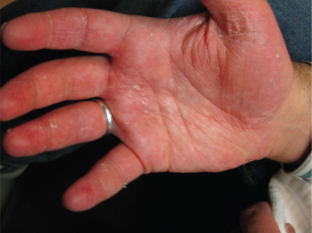
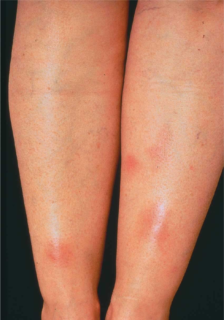
QUESTIONS 13–15 A 35-year-old man presents with ocular pain, pain in his wrists and feet, pain on urination, and aphthous ulcers. Ocular examination is significant because it reveals a mucoid conjunctival discharge and a mild cellular reaction in the anterior chamber. A peculiar skin lesion is also noted (Fig. 8-5).
13 Which one of the following findings would not be expected in this patient?
A) Balanitis
B) Prostatitis
C) A recent history of diarrhea
D) Positive rheumatoid factor
14 Which organism has not been implicated in triggering this condition?
A) Chlamydia
B) Ureaplasma urealyticum
C) Yersinia
D) Rochalimaea
15 The classic skin lesion associated with this condition, pictured in Figure 8-5, is:
A) erythema chronicum migrans
B) pustular psoriasis
C) keratoderma blennorrhagicum
D) eczema
16 A 10-year-old girl with a history of arthralgia and a chronic anterior chamber inflammation of both eyes presents to your office. She has no cells, but mild flare is present in the anterior chamber. Given the natural history of this disease, which one of the following statements is most likely true?
A) This patient is antinuclear antibody (ANA)–negative.
B) This patient has pauciarticular disease involving the hands and wrists.
C) This patient has pauciarticular disease involving the lower extremities without involvement of the wrist joints.
D) This patient is rheumatoid factor–positive.
QUESTIONS 17–19 A 40-year-old white man is referred for ophthalmologic examination because the vision in one eye could not be refracted better than 20/40. On examination, you note a mild unilateral anterior chamber inflammation and stellate keratic precipitates diffusely dispersed over the posterior surface of the cornea. The patient has hazel irides with a mild difference of iris pigmentation between the eyes.
17 Which one of the following statements is most likely true?
A) The involved eye is darker brown than the uninvolved eye.
B) Less than 2% of patients with this condition will have bilateral involvement with no obvious heterochromia.
C) Pathologic specimens of this condition have revealed the presence of plasma cells within the ciliary body.
D) Posterior synechiae may be prominent.
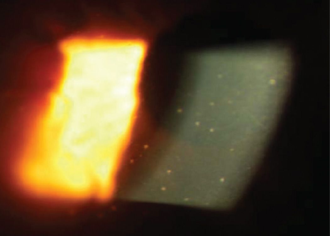
FIGURE 8-6
18 Which one of the following infectious agents has been suggested as having an association with this condition?
A) Histoplasma
B) Toxoplasma
C) Toxocara
D) Epstein–Barr virus
19 Which of the following topical medications used unilaterally can cause a similar appearance?
A) Dipivefrin
B) Cyclosporine A
C) Latanoprost
D) Brimonidine
20 All of the following major immunoglobulin classes are found in human tears except:
A) IgD
B) IgE
C) IgG
D) IgM
21 Which one of the following statements concerning the classic immune hypersensitivity reactions in diseases that affect the eye is true?
A) The granulomatous response seen in sarcoid uveitis is primarily a type I hypersensitivity reaction.
B) Phacoanaphylaxis is a type III hypersensitivity reaction.
C) Hay fever is an example of a type II hypersensitivity reaction.
D) Type IV hypersensitivity reactions are mediated by cytotoxic antibodies.
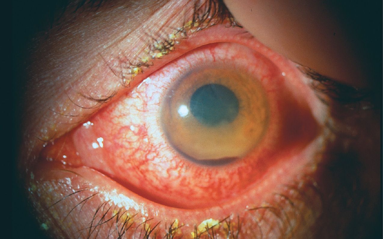
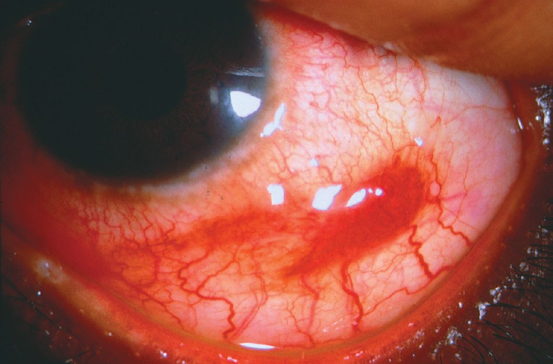
QUESTIONS 22 and 23 An elderly African American patient comes with the complaint of a whitish area on her eye for the last several weeks (Fig. 8-7). Her eye has been comfortable. There is no discharge or crusting of the lashes.
22 All of the following blood tests would be useful in making the diagnosis except:
A) HIV antibodies
B) rheumatoid factor
C) anti-neutrophil cytoplasmic antibody
D) PPD skin test
23 There is a small trickle of fluid from the peripheral corneal defect. What treatment might be most helpful in the acute setting?
A) Aqueous suppressant
B) Application of cyanoacrylate glue
C) Corneal patch graft
D) Topical corticosteroids
24 Which one of the following characteristics regarding serpiginous choroidopathy is true?
A) Recurrent, indolent course
B) Primarily affects children
C) Multifocal lesions
D) Responds promptly to corticosteroids
25 A 25-year-old man was hammering a nail into a piece of wood in his garage when he noticed a sudden sharp pain in his right eye with only a mild decrease in his vision. He presents to your office 2 days later with reports of gradually increasing pain and a severe loss of vision in that eye. On examination, you notice a small peripheral corneal laceration and a hypopyon. Which one of the following statements concerning endophthalmitis in this setting is true?
A) The most common infecting organism in these cases is Staphylococcus aureus.
B) A visual acuity of 20/400 or better is likely to be retained in the majority of patients after appropriate treatment and rehabilitation.
C) Endophthalmitis would be expected to occur in less than 10% of similar trauma cases.
D) Endophthalmitis with Bacillus species has a good visual prognosis.
26 You examine a 78-year-old white man with complaints of decreased vision in his right eye. Three weeks earlier, he had undergone coronary artery bypass surgery with a complicated postoperative course requiring prolonged ventilatory support. He has been receiving hyperalimentation and IV antibiotics since the time of his surgery. On dilated fundus examination of his right eye, you note a fluffy white choroidal lesion under the macula. There is minimal overlying vitritis present. Which one of the following statements concerning the organism most likely responsible for this lesion is true?
A) The responsible organism grows on blood agar and Sabouraud glucose within 24 to 48 hours.
B) This organism exists exclusively as an oval budding cell known as a blastoconidia.
C) Ocular infection caused by this organism is generally accompanied by positive blood cultures.
D) This organism often colonizes the respiratory tract and is a frequent cause of pneumonia.
27 A 25-year-old homeless man presents to your office with reports of decreased vision and pain in his right eye for the past 2 days. Examination reveals marked conjunctival injection, a 2-mm hypopyon, and dense vitreous opacities on B-scan ultrasound. You suspect endogenous endophthalmitis, and on further questioning, the patient admits to IV drug abuse. Given the patient’s history, which one of the following organisms is the least likely to be involved in this patient’s endophthalmitis?
A) C. albicans
B) Staphylococcus species
C) Bacillus cereus
D) Haemophilus influenzae
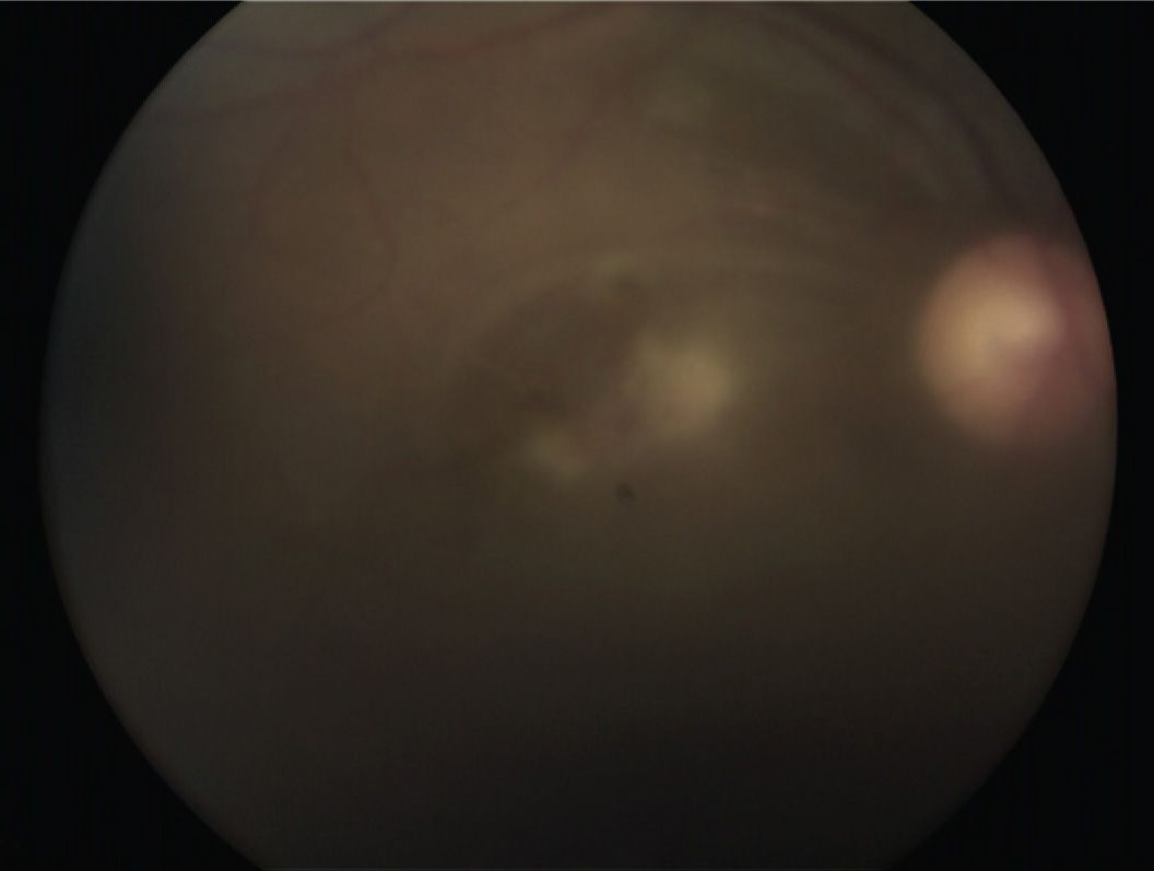
QUESTIONS 28–30 A 27-year-old Japanese exchange student presents to your office with reports of decreased vision in both his eyes. On examination, a small hypopyon OD and a moderate cellular reaction OS is found. Fundus examination of his left eye is shown in Figure 8-8. On further questioning, you elicit a history of arthritis of his knees and wrists, and painful lesions in his mouth and around his genitals. On examination of his lower extremities, you note the lesion seen in Figure 8-9.
28 Skin lesions common to this condition include all of the following except:
A) acne-like lesions over the back and face
B) erythema nodosum
C) psoriasis
D) thrombophlebitis
29 Which one of the following statements concerning the therapeutic treatment of this condition is true?
A) Oral corticosteroids alone are usually effective in preventing the relapse of ocular inflammation.
B) Periocular steroids alone are usually effective in preventing the relapse of ocular inflammation.
C) Colchicine may be helpful in preventing recurrences.
D) Cyclosporine may be useful in this condition, but the incidence of liver toxicity may limit its use.
30 Which one of the following statements concerning the ocular inflammation associated with this condition is true?
A) Patients generally develop chronic, unremitting ocular inflammation if this condition is not aggressively treated.
B) A granulomatous inflammation is typically present.
C) The retinitis associated with this condition may be confused with a viral retinitis.
D) Inflammation in this condition predominantly affects the choroid.
31 What is the most common cause of acute, noninfectious, hypopyon iritis?
A) Behçet disease
B) Idiopathic anterior uveitis
C) HLA-B27–associated iritis
D) Sarcoid iridocyclitis
32 In which one of the following is the HLA-B27 antigen least likely to be present?
A) Men with ankylosing spondylitis
B) Young women with pauciarticular juvenile rheumatoid arthritis (JRA)
C) Men with reactive arthritis
D) Men with psoriatic arthritis
33 Which form of uveitis is most common in ocular sarcoidosis?
A) Panuveitis
B) Intermediate uveitis
C) Anterior uveitis
D) Choroiditis
34 Which of the following statements is most accurate regarding the lesions pictured in Figure 8-10?
A) Live virus can be recovered from epithelial lesions.
B) This disease is usually seen in children or young adults.
C) Topical corticosteroid drops are effective in eliminating this disease.
D) These lesions are frequently bilateral at presentation.
QUESTIONS 35–37 Select the answer that corresponds to the conditions listed.
35 Iris nodules
A) Granulomatous uveitis
B) Nongranulomatous uveitis
C) Both
D) Neither
36 Epithelioid cells
A) Granulomatous uveitis
B) Nongranulomatous uveitis
C) Both
D) Neither
37 Fuchs heterochromic iridocyclitis
A) Granulomatous uveitis
B) Nongranulomatous uveitis
C) Both
D) Neither
QUESTIONS 38–40 (Figs. 8-11 to 8-14)

38 Biopsy of which lesion would show a monotonous proliferation of B cells?
A) Figure 8-11
B) Figure 8-12
C) Figure 8-13
D) Figure 8-14
39 Which would present with pain and localized tenderness?
A) Figure 8-11
B) Figure 8-12
C) Figure 8-13
D) Figure 8-14
40 Human herpesvirus 8 (HHV-8) has been implicated in causing which condition?
A) Figure 8-11
B) Figure 8-12
C) Figure 8-13
D) Figure 8-14
41 What is the most characteristic side effect of oral cyclophosphamide?
A) Secondary infections
B) Secondary malignancies
C) Hemolytic anemia
D) Hemorrhagic cystitis
42 Side effects of systemic corticosteroid therapy include all of the following except:
A) aseptic necrosis of the hip
B) hypoglycemia
C) exacerbation of hypertension
D) gastric ulceration
QUESTIONS 43–45 Match the following immunosuppressives to their class.
43 Cyclosporine
A) Cytotoxic antimetabolite
B) Cytostatic anti-inflammatory
C) Immune modulator of interleukin 2
D) Cytotoxic alkylating agent
44 Prednisone
A) Cytotoxic antimetabolite
B) Cytostatic anti-inflammatory
C) Immune modulator of interleukin 2
D) Cytotoxic alkylating agent
45 Methotrexate
A) Cytotoxic antimetabolite
B) Cytostatic anti-inflammatory
C) Immune modulator of interleukin 2
D) Cytotoxic alkylating agent
46 Which one of the following is the most common retinal finding in AIDS?
A) Cotton wool spots
B) Cytomegalovirus (CMV) retinitis
C) Pneumocystis choroiditis
D) Acute retinal necrosis
47 The life form of the Toxoplasma gondii organism that is responsible for stimulating inflammation is/are:
A) cyst
B) bradyzoite
C) tachyzoite
D) all of the above
48 Which one of the following statements about the drugs used to treat ocular toxoplasmosis is true?
A) Pyrimethamine blocks the production of dihydrofolate from para-aminobenzoic acid.
B) Sulfadiazine inhibits the dihydrofolate reductase enzyme.
C) Clindamycin can effectively kill Toxoplasma organisms.
D) Systemic and periocular corticosteroids are contraindicated because they may cause proliferation of Toxoplasma organisms.
49 JRA-associated iridocyclitis is most common in:
A) early-onset pauciarticular disease
B) late-onset pauciarticular disease
C) still disease
D) late-onset polyarticular disease
50 Schwartz syndrome is caused by:
A) retinal pigment epithelial cells blocking the trabecular meshwork
B) forward rotation of the lens–iris diaphragm
C) ciliary body and choroidal edema
D) photoreceptor outer segments blocking the trabecular meshwork
51 Posner–Schlossman syndrome:
A)
Stay updated, free articles. Join our Telegram channel

Full access? Get Clinical Tree


