TRIAMCINOLONE
Intravitreal Triamcinolone and Vitrectomy
Intravitreal triamcinolone is used for visual demarcation of the vitreous at the time of vitrectomy. Kumagai and Burk1,2 performed this technique using 40 mg of triamcinolone in a 3-mL syringe, connected to a three-way joint (Millipore filter), which in turn is connected to a 10-mL syringe containing balanced salt solution (BSS) (Alcon laboratories, Fort Worth, TX). The aqueous phase of the triamcinolone solution is discarded through the filter, leaving the particulate matter of the triamcinolone in the 3-mL syringe. Triamcinolone particles are mixed and washed in the 3-mL syringe. This is done three times, discarding, mixing, and washing. The final volume of 0.1 mL of desired 3-mL triamcinolone solution is suspended in the BSS and then injected into the vitreous cavity. The triamcinolone particles are trapped in the vitreous gel and enable visualization of the vitreous.1,2
Kumagai and Burk1,2 used 0.2-mL triamcinolone acetonide (40 mg/mL), which was filtered through a 5-μm filter and rinsed with 2 mL of BSS and resuspended and recaptured; this was repeated for a total of three times before it was injected into the eye.1,2 Furino et al.3 used triamcinolone acetonide (40 mg/mL), which was injected in midvitreous to visualize the posterior hyaloid once core vitrectomy is performed. They used 0.1 to 0.2 mL of triamcinolone acetonide to visualize the retinal surface’s epiretinal membranes. No adverse reactions were observed with the use of triamcinolone acetonide without the use of the BSS.
Triamcinolone acetonide has been considered for use in the treatment of diabetic macular edema.4 Triamcinolone acetonide has been used without surgery and found to improve the macular edema in 28 eyes of 20 patients. Jonas et al.5 found no adverse glaucomatous side effects and reported improved visual acuity after 7 months of follow-up.
The use of triamcinolone acetonide in vitrectomy surgery serves as an aid for visualization during complete vitrectomy in cases of macular edema. In addition, triamcinolone acetonide reduces postsurgical inflammation which may exacerbate macular edema.6 Matsumoto et al.7 have found that intravitreal triamcinolone improves the removal of posterior vitreous hyaloid by using electron microscopy to examine the surgical specimens. Intravitreal triamcinolone acetonide decreased blood ocular barrier breakdown and improved visual acuity postoperatively in eight diabetic macular edema eyes and ten proliferative diabetic retinopathy eyes of patients who underwent pars plana vitrectomy.8 They used an anterior chamber laser flare detector to study the side effects.
Intravitreal Triamcinolone Acetonide and Diabetic Macular Edema
Intravitreal triamcinolone is used for the treatment of refractory diabetic macular edema. The treatment has its origins in 2002 when clinically significant diabetic macular edema eyes failed to respond to previous laser photocoagulation sessions. After 6 months of initial laser therapy, the response is measured by OCT and clinical examination. If central macular thickness of 300 μm is attained (normals 200 μm), 4 mg of intravitreal triamcinolone acetonide is injected. In one small study, 16 patients were evaluated, and the average increase in Snellen visual acuity was two lines of visual improvement and a mean of 50% decrease of thickness on OCT scans.4 Vitreous vascular endothelial growth factor (VEGF) and stromal-derived factor-1 levels were measured by enzyme linked immunosorbent assay in samples obtained immediately before and 1 month after the injection of intravitreal triamcinolone.9,10
In a Phase III, multicenter, randomized study, the Diabetic Retinopathy Clinical Research Network (DRCR.net) investigated the safety and efficacy of preservative-free intravitreal triamcinolone acetonide (1 and 4 mg) and compared these treatments with standard ETDRS focal/grid photocoagulation for the treatment of diabetic macular edema. The study enrolled 840 eyes divided into three treatment arms. After a 2-year follow-up, the authors concluded that the focal/grid photocoagulation treatment was more effective and displayed fewer side effects than either the 1- or 4-mg doses of preservative-free triamcinolone acetonide for diabetic macular edema patients. Thus, focal/grid photocoagulation is still the benchmark against which other treatments are compared in diabetic macular edema studies.11
VEGF INHIBITORS
VEGF is a potent enhancer of vascular permeability and a key inducer of angiogenesis. VEGF is identified in tumor models as a vasopermeability factor and an initiator of angiogenesis upregulated by hypoxia. VEGF is the strongest endothelial mitogen and can also induce the expression of plasminogen activator in vascular endothelial cells, which, in turn, are important in the extracellular proteolysis necessary for capillary formation and breakdown of blood retinal barrier.12 VEGF, also known as VEGF-A or VEGF-1, is expressed as five mRNA splice variants: isoforms 121, 145, 165, 189, and 206.13 VEGF isoform 165 is a critical isoform for both developmental and pathologic retinal angiogenesis. VEGF is expressed by retinal endothelial cells and retinal pigment epithelial cells in vitro and is upregulated by hypoxia.14,15 Other modulators of VEGF expression are hypoglycemia, beta-estradiol, and rod outer segments.16–19
Clinical studies have demonstrated that VEGF plays a role in ischemic retinal diseases. Vitreous from surgical specimens show increased VEGF levels in patients with proliferative disease from diabetes or retinal vein occlusion.20,21 VEGF protein levels decrease after successful laser retinal ablation.22
PEGAPTANIB (MACUGEN)
Pegaptanib, a novel RNA aptamer, originally used in the treatment of AMD, binds and inactivates VEGF isoform 165. In animal models, the VEGF isoform 165 can induce diabetes-like ocular pathology. Pegaptanib has been shown in animal models to reverse the blood retinal barrier breakdown associated with diabetes. Aptamers are short nucleic acid sequences of a specific strand shape and length, which can bind to their target polypeptides and change their final form.23 Target polypeptides can be RNA or DNA which are chemically synthesized through a process, named systemic evolution of ligands by exponential enrichment.24,25 In pegaptanib, a large library of RNA sequences ranging from 1014 to 1015 are incubated with the target molecule, VEGF isoform 165. The unbound sequence is washed away, and the target molecule, VEGF isoform 165, is uncoupled from the candidate strands; this process is repeated 8 to 15 times.24,25 The end product has increased specificity of the aptamer for the target, VEGF isoform 165. The molecule is selectively binding only to VEGF isoform 165. The binding of the pegaptanib aptamer to VEGF isoform 165 lasts 6 weeks.
In the course of evaluating the properties of VEGF, two are particularly relevant in the pathogenesis of diabetic retinopathy. VEGF is upregulated by hypoxia and thus creates neovascularization as a response to hypoxia. Secondly, VEGF is a potent inducer of vascular permeability, 50,000 times more potent than histamine. VEGF causes fenestrations in the endothelium, dissolution of tight junctions, induction of leukostasis, and subsequent injury to the endothelium.26 These characteristics of VEGF support a direct involvement of VEGF in the diabetic macular edema, a form of diabetic retinopathy.
These observations were the basis of the Phase II clinical trial involving pegaptanib and diabetic macular edema.27 Participants had the best corrected visual acuity of 20/50 to 20/320 in the study eye with diabetic macular edema involving the center of the macula, for whom the investigator judged laser photocoagulation could be safely withheld for 16 weeks. Intravitreal pegaptanib was injected at the onset of study, week 6, and week 12 with additional injections and/or focal photocoagulation as needed for another 18 weeks. Final assessments were done at week 36. Outcome measures were visual acuity and central subfield thickness measurements using OCT. A total of 172 patients were enrolled in the study. Compared to sham injections, the patients given 0.3 mg had a better visual acuity (20/50) versus sham treated patients (20/63). Mean central thickness of the retina as measured by OCT showed mean reduction of 68 μm in retinal thickness in the 0.3 mg-treated group versus an increase of 4 μm in the sham-treated group at week 36. Thus, early data showed that pegaptanib may have a beneficial role in the treatment of diabetic macular edema.27
VEGF also plays a role in neovascularization leading to evaluation of pegaptanib for the treatment of proliferative diabetic retinopathy. Using the patient information from the Phase II clinical trial of pegaptanib and diabetic macular edema,27 data were retrospectively analyzed to discern the effects of pegaptanib on retinal neovascularization.28 Patients in the analysis had the best corrected vision of 20/50 to 20/320 and had received a sham injection or intravitreal pegaptanib (0.3, 1, or 3 mg) at onset, week 6, and week 12. Changes in neovascularization were assessed by fundus photography and fluorescein angiography. Of the 172 original study patients, 19 had retinal neovascularization in the study eye at baseline. One of these 19 patients was excluded because he had scatter laser photocoagulation 13 days before randomization; two others were excluded because fundus photographs were not available. The median age of the remaining 16 patients was 55.5 years. Eight of the thirteen patients in the pegaptanib-treatment group showed regression documented by fundus photographs or fluorescein angiography at 36 weeks versus none in the sham group. No fellow (nonstudy) eyes showed regression of neovascularization at 36 weeks on fundus photographs or fluorescein angiography. This small study showed that pegaptanib treatment can lead to regression of diabetic neovascularization.27,28
BEVACIZUMAB (AVASTIN)
Bevacizumab is a mouse-derived monoclonal antibody to all isoforms of VEGF. In animal studies using tumor cell lines and models with ocular neovascularization, bevacizumab was effective against primary permeability and proliferative effects of VEGF. It has a molecular weight of 148 kDa, and it is a larger molecule than ranibizumab with 48 kDa.
There have been small studies evaluating bevacizumab and proliferative diabetic retinopathy. Forty-five eyes of 32 patients with retinal and/or iris neovascularization secondary to diabetes were given intravitreal 6.2 μg to 1.25 mg. All of the patients showed regression of neovascularization as seen with fluorescein angiography within 1 week. About 73% had complete resolution of disc neovascularization; 82% of the eyes had resolution of iris neovascularization. Follow-up was 11 weeks (Figs. 6-1 and 6-2).29–31

FIGURE 6-1. Monoclonal antibody blocking VEGF.
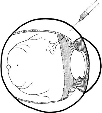
FIGURE 6-2. Intravitreal injection, cross- sectional view.
Two patients with proliferative diabetic retinopathy and vitreous hemorrhage were given intravitreal avastin.30 Both patients manifested regression of retinal neovascularization after 1 month of follow-up. Vitreous hemorrhage in each patient showed partial resolution at 1 week and nearly complete regression in 1 month.30
These were the earliest studies of bevacizmab—in the treatment of proliferative diabetic retinopathy in 2006 when the drug was just being released for the treatment of age-related macular degeneration.
Since that time, bevacizumab (Avastin) has been used by retinal surgeons in the United States to great success, despite being a non–FDA-approved drug for diabetic retinopathy. In another study, bevacizumab treatment of patients with diabetic macular edema yielded better visual outcome than laser photocoagulation.31 Twelve weeks of follow-up was done in 37 eyes treated with bevacizumab versus 33 eyes treated with laser. Combination therapy of bevacizumab and triamcinolone eyes had a beneficial effect at 6 weeks but not at 12 weeks. Thus, bevacizumab alone was found to be more efficacious in this small study.31
The Pan American Collaborative Retina Study Group evaluated 78 eyes of 64 patients with a follow-up of 6 months, from six centers from six countries.32 The patients were treated with at least one intravitreal injection of 1.25 or 2.5 mg of bevacizumab and were tested using ETDRS vision testing, OCT, and fluorescein angiography at baseline and follow-up visits. Sixteen eyes needed a second injection at a mean of 13.8 weeks (range was 4 to 28 weeks); six eyes needed a third injection at a mean of 11.5 weeks (range was 5 to 20 weeks). Final best corrected visual acuity analysis by subgroups showed that 41.1% of the eyes remained stable and 55.1% improved or had greater than or equal to two ETDRS lines of visual acuity improvement. Mean central macular thickness at baseline OCT was 387.0 ± 182.8 μm and decreased to a mean 275.7 ± 108.3 μm at the end of follow-up. The authors concluded that primary intravitreal bevacizumab at doses of 1.25 and 3.5 mg provided stable or improved vision at 6 months of follow-up, using outcome measures of visual acuity, OCT, and fluorescein angiography.32
RANIBIZUMAB (LUCENTIS)
Ten patients were studied; five patients received 0.3 mg and five patients received 0.5 mg of ranibizumab intravitreally. At month 3, four of ten patients gained ≥10 letters of visual acuity, and eight of the ten patients gained ≥1 letter. At month 3, the mean decrease in OCT retinal thickness measurements was 45.3 ± 196.3 μm for the low-dose group and 197.8 ± 85.9 μm for the high-dose group.33
Ranibizumab is the antigen binding portion of the monoclonal antibody directed against VEGF. This monoclonal antibody fragment weighs 48 kDa and effectively blocks increased fluorescein leakage after laser in animal eyes. Ranibizumab is not FDA approved for diabetic macular edema, and ongoing studies at the time of this writing are being performed for the treatment of diabetic retinopathy and diabetic macular edema.33
A multicenter clinical trial sponsored by Juvenile Diabetes Research Foundation, named the READ Trial, is currently ongoing. READ-2 Study (Ranibizumab for Edema of the Macula in Diabetes) had its beginnings in 2006. Its purpose is to evaluate ranibizumab in diabetic patients with DME. Phase I of the study involved 20 patients with DME treated with repeated intravitreal injections of 0.5 mg of ranibizumab at baseline, months 1, 2, 4, and 6. Results showed that at month 7, the median reduction of excess of foveal thickness was 97% and a median improvement of ten letters (Fig. 6-3).34

FIGURE 6-3. Intravitreal injection, external view.
Phase II of the READ Study enrolled 126 diabetic patients, with an average age of 62 years, with documented macular edema at enrollment; 115 patients had completed the trial at 6 months. Patients were included in the study if they had at least 250 μm on OCT scan of central macular thickness. Previous treatment was allowed if it had been given at least 3 months prior to the onset of the study. The majority had 20/80 average vision at enrollment. The subjects received ranibizumab 0.5 mg alone or with focal laser treatment or laser alone. At 6 months, improvement in visual acuity was noted to be the mean of 8 letters in the ranibizumab group, 3.8 letters improvement in the combination group of ranibizumab and laser, and 1 letter loss in the laser-only group. The OCT showed 56% decrease in excess retinal thickness in the ranibizumab-treated group versus 11% reduction in those who received laser alone or laser and ranibizumab.34–36
Acetazolamide
Carbonic anhydrase inhibitors (CAI) have been known to work on retinal fluid absorption for the past two decades. Initial observations were based on experimental data which suggested that acetazolamide can increase fluid absorption across the retinal pigment epithelium (RPE). CAI have direct effects on retinal and retinal pigment epithelial cell function by inducing an acidification of the subretinal space, a decrease in the standing potential as well as an increase in retinal adhesion. It is thought that acidification of the subretinal space is responsible for the increase in fluid resorption from the retina through the RPE into the choroid.37
A normal clinical dose of CAI of 500 mg per day should be continued for 1 month to see an effect. The dose may be reduced by the patient over the course of the therapy. Metaphylaxis to the drug may occur with a rebound of edema despite continuation of treatment.37–41
NONSTEROIDAL ANTI-INFLAMMATORY DRUGS (NSAIDS)
Nonsteroidal Anti-inflammatory Eye Medications
Bromfenac (Xibrom ISTA) and nepafenac (Nevanac) are now available for diabetic macular edema, age-related macular degeneration, and cystoid macular edema. The prodrug formulation of the new NSAID such as nepafenac is thought to be more effective than the older generation of flurbiprofen 0.03% (Ocufen), ketorolac 0.5% (Acular), and diclofenac 0.1% (Voltaren).42–44
The older generation drugs, Ketorolac and Diclofenac, are FDA approved for the treatment of macular edema after cataract surgery. They have been used for the treatment of diabetic macular edema as well. Since diabetic macular edema is often refractory in diabetic patients with poor glycemic control, NSAIDs have been used along with topical prednisolone acetate and steroid injections in the vitreous such as triamcinolone.4
PHARMACOTECHNOLOGY
Intravitreal Implants and Drug Delivery Systems
Intravitreal implant, Iluvien, previously known as Medidur FA (Alimera Sciences, Alpharetta, GA) is a nonerodable insert. Iluvien is in Phase III clinical trials for the delivery of fluocinolone acetonide in patients with diabetic macular edema. Iluvien is 3.5 mm long and 0.37 mm in diameter. A 25-gauge insertion system facilitates intravitreal delivery in the office setting. The insert, once inside the globe, delivers an initial daily release rate of either 0.23 or 0.45 μg of fluocinolone acetonide, lasting 36 months. By October 2007, 956 patients were enrolled for a follow-up of 24 months, with a final analysis after 36 months. This is the FAME study, Fluocinolone Acetonide in Diabetic Macular Edema.45–47
CORTICOSTEROIDS
Fluocinolone Acetonide (Retisert)
Fluocinolone acetonide is a nonbiodegradable intravitreal corticosteroid implant. It is designed to release slowly over a 3-year time period. Retisert was originally designed for noninfectious uveitis affecting the posterior segment, affecting 175,000 people in the United States. On April 11, 2005, the FDA approved the use of Retisert for the treatment of chronic noninfectious uveitis affecting the posterior segment of the eye. The most common side effects are eye pain, cataract progression, and intraocular pressure elevation.48
Currently, Retisert has enrolled 80 patients who are randomized to receive either an implant 0.5 or 2.0 mg, or standard of care macular laser, or observation. These patients had refractory macular edema. At 12 months of follow-up, 49% of the eyes that are treated with 0.5-mg implant had resolution of the macular edema compared to 25% of the standard of care group (p < 0.5). More patients in the standard of care group had worsening diabetic retinopathy at 12 months than the 0.5-mg-implant group (30% vs. 5%). Serious ocular adverse events include increased intraocular pressure and vitreous hemorrhage, and cataract formation is significantly higher in the implant group (59%) than in the standard of care group (11%). This is an ongoing trial.49
Another steroid drug delivery system now being investigated is the biodegradeable dexamethasone in a posterior segment delivery system, Posurdex (Allergan, Irvine, CA). Posurdex is being evaluated for patients with diabetic macular edema, vein occlusions, pseudophakia, and uveitis. The implant is designed to last 37 days.50
In the Phase II trial, 306 patients were randomized to receive a 350-μg or 700-μm implant or observation. The eyes were divided as follows: 165 diabetic macular edema, 102 retinal vein occlusion, 25 Irvine Gass syndrome, and 14 uveitis. The average pretreatment vision was 20/80 to 20/85. At the 6-month endpoint, 36% of the 700-μg group and 27% in the 350-μg group had at least a two-line improvement of vision, as compared to the observation group which had 19% with two or more lines of improved vision. In the 700-μg group, 19% of patients had three or more lines of improvement, which was statistically significant compared to the 8% of the observation group who had the same improvement. The average change in retinal thickness was 142 μm for the 700-μg group and a decrease in retinal thickness of 61 μg in the 35-μg group. There was an increase of 11 μm in the central retinal thickness in the observation group.50–52
Another fluocinolone acetonide implant is Medidur. Medidur is a narrow 3 mm long micro-cylinder containing fluocinolone acetonide to be injected intravitreally in the office setting, using a 25-gauge injector. Its indications are being codeveloped by pSivida (Watertown, MA) and Alimera Sciences (Alpharetta, GA). Medidur has completed its initial 20-patient safety trial, and it is currently enrolling patients. The 20 patients were on a low dose of 0.23 μg a day and 17 patients on a high dose, approximately 0.45 μg a day. The 3-month results showed that 20% of the low-dose patients and 18% of the high-dose patients improved compared to controls.53
Another drug delivery system, I-Vation (Surmodics, Eden Prairie, MN), has been tested in a 30-patient Phase I study, with the delivery of triamcinolone acetonide in patients with diabetic macular edema. I-Vation requires surgical implantation at the pars plana region, under the conjunctiva, for rapid retrieval if needed. The Phase I study found that 100% of the patients either maintained or improved vision at 6 months. Eighty-seven percent or 26/30 patients who were treated with I-Vation triamcinolone acetonide had either the same or an improved vision. Forty-three percent or 13/30 patients achieved visual acuity of 20/40 or better compared to 20% or 6/30 patients at time of entry into the study. Mean retinal thickness decreased 164 µm or 37% and excess retinal thickness decreased 63% from baseline retinal thickness.54
Topical Eyedrops
CoMentis, Inc. (South San Francisco, CA) is interested in ATG-3 (mecamylamine), for the treatment of diabetic macular edema. ATG-3 works by inhibiting angiogenesis mediated by VEGF. This drug would be considered an adjunct to intravitreal ranibizumab.45 The drug, ATG-3, is in the Phase II clinical trial. It is an antagonist of the nicotinic acetylcholine receptor pathway that may counter the vascular leakage and angiogenesis mediated by VEGF.45
TG100801 (tetrahydrozoline HCL) is another drug for diabetic macular edema. Its manufacturer is TargeGen, currently sponsoring Phase II clinical trials at the time of this writing. Tetrahydrozoline HCL is designed to suppress VEGF-mediated leakage associated with inflammation, edema, and angiogenesis of diabetic macular edema.55
Sirolimus is rapamycin, a macrolide immunosuppressive agent. It has been used to prevent coronary restenosis in the coating of coronary stents for angioplasty. Sirolimus has a target, mTOR, a protein kinase that regulates cell growth and metabolism in response to the presence of growth factors or other changes in the environment. Cell processes such as protein synthesis and nutrient transport are upregulated by mTOR.56
In addition, sirolimus has immunosuppressive, antiangiogenic, and antiproliferative properties. It inhibits VEGF by decreasing the endothelial cell response to VEGF activation through down-regulation of intracellular signaling. The inhibition of mTOR inhibits the activity of HIF-1 alpha which decreases VEGF production and inhibits VEGF-induced endothelial cell proliferation. For all of these properties, sirolimus is thought to have the potential in treating neovascularization in diabetic retinopathy.56
Sirolimus (Rapamune, Wyeth) is in a Phase I study for diabetic macular edema.56,57 In its Phase 1 trial of 50 patients, the results showed improvements in visual acuity. Patients on the two lowest doses of sirolimus showed greater than or equal to 7.4 letters from baseline. Foveal thickness improved for 180 days after a single dose of sirolimus. One focus of the study is on the intravitreal or subconjunctival route as an effective method of drug delivery.
CLINICAL CORRELATIONS
Patient complaints: Loss of vision in one eye or sudden loss of vision, despite having had macular laser photocoagulation.
Caveats: Loss to follow-up with ophthalmologist, relocation to another city, and interruption of medical care
Case Study (Figs. 6-4–6-11)
Stay updated, free articles. Join our Telegram channel

Full access? Get Clinical Tree


