HISTORY
When taking the history of the diabetic patient, be aware of other entities that can contribute to the severity of diabetic retinopathy, such as hypertension, kidney disease, hyperlipidemia, ischemic heart disease, or peripheral vascular disease.1–3 Look for metabolic syndrome in patients who are overweight, obese, diabetic and have a history of dyslipidemia. Lastly, always inquire about their cholesterol and triglyceride levels in these diabetic patients. Smoking history is important as nicotine can cause acceleration of the diabetic retinopathy. Ask about the fasting blood sugar and glycosylated hemoglobin (HbA1c) as an assessment of the overall status of diabetes in the patient about to be evaluated for diabetic retinopathy. If the patient cannot remember his glucose level or HbA1c or any of his past blood sugar levels, the patient is probably not fully informed about the importance of such laboratory indices with respect to his diabetic retinopathy.
Duration of Diabetes
Duration of diabetes is an important question for the patient. The longer the duration of diabetes, the greater the risk of diabetic retinopathy. In Type 1 diabetes, the longer the duration of diabetes, the greater the risk for increasing severity of diabetic retinopathy. In Type 2 diabetes, after 15 years of diabetes, there will be increased risk of diabetic retinopathy. After diabetes has been present for 20 years, almost all persons in whom the onset of diabetes occurred before age 30 years have some evidence of diabetic retinopathy, and about 50% have proliferative diabetic retinopathy. In persons who are 30 years or older when diabetes is diagnosed, while they are at lower risk for retinopathy, retinopathy may be their first symptom of diabetes.4
Past Ocular History
Ask the patient about previous laser or eye surgery history. When did the diabetic patient first note eye problems or vision problems?
Review of Systems: These are some of the pertinent and related issues that have some influence on vision and diabetic retinopathy.
Glaucoma can be present in patients with longstanding diabetic retinopathy or in patients with rapid deterioration of vision due to proliferative diabetic retinopathy. There is neovascular glaucoma to consider in those diabetic patients whose vision is decreasing rapidly over a short period of time. Open-angle glaucoma can occur in Type 2 diabetic patients.
Thyroid disease does not affect the retina unless it is severe and causes optic disc edema. Thyroid disease can occur in diabetic patients and present as severe dry eye. If there is proptosis in the setting of thyroid disease and diabetes, send the patient for an endocrinology evaluation.
Other endocrine abnormalities can occur with diabetes. This one caveat should be considered when the diabetic patient sees an endocrinologist at the time of diagnosis.
Obesity has been linked to the epidemic of diabetes in teens in America for the past decade. Adipocytes are now thought to play a role in insulin resistance in Type 2 diabetic patients. Thus, obesity is a risk factor in the development of diabetes.
Poor lipid metabolism with its associated hypercholesterolemia and hypertriglyceridemia plays a role in diabetic complications, such as heart disease, heart attacks and transient ischemic attacks. If there is loss of vision, the dilated funduscopic examination may show areas of ocular ischemia.
Family History
Do members of the family have diabetes, Type 1 or Type 2 diabetes? Do family members who are diabetic have eye disease that lead to blindness or amputation or kidney dialysis? Is there a history of stroke and/or cardiovascular disease in the family? Ask about glaucoma in family members. Open-angle glaucoma is common among Type 2 diabetic patients. Rubeotic glaucoma is common in Type 1 diabetics who have been in poor glycemic control (Table 5-1).
TABLE 5.1 Examination of the Diabetic Eye Patient
Correlate vision loss to the retina findings
Eyelids and pupils:
III nerve palsy and eyelid abnormalities
Papillary abnormality
Slit lamp examination:
Rubeosis iridis, iris atrophy
Cataract
Fundus examination
Review of system checklist
Hypertension (silver wiring or copper wiring, poor flow seen on angiogram)
Hyperlipidemia (hard exudate rings)
Cardiovascular disease (transient ischemic attack by history with loss of vision suddenly, emboli)
Smoking (attenuated arterioles or capillary nonperfusion or ischemic retina)
Obesity and Metabolic Syndrome (expect poor glycemic control and retinopathy)
Diabetes history
Duration of Diabetes (the greater the duration, the worse the retinopathy)
Type 1 diabetes versus Type 2 diabetes
Gestational diabetes in the past (acceleration of diabetes over pregnancy)
Diabetes and pregnancy (acceleration of diabetes over pregnancy)
Poor glycemic control (expect diabetic retinopathy at first visit to ophthalmologist’s office)
EXAMINATION
Visual Acuity
Visual acuity can tell you if the patient is seeing 20/40 or better and therefore able to perform the activities of daily living or continue to drive legally. Visual acuity at 20/40 to 20/60 range may signify macular edema. Visual acuity at the level of 20/200 or worse signifies legal blindness. Vision worse than 20/200 suggests that the patient might have proliferative diabetic retinopathy and requires close monitoring with an ophthalmologist.
Pupils
The pupillary examination will show brisk response to light and accommodation. Diabetic patients who have never had previous eye surgery but exhibit a sluggish but symmetrical papillary response may have iris atrophy or an abnormality of the iris at the sphincter muscle. Thus, there is an inability of the iris to contract properly. In these patients with sluggish but equal papillary responses, it is important to look for rubeosis iridis on the slit lamp examination (Fig. 5-1). It is these same eyes that will have a poor ability to respond to dilating and diagnostic drops.
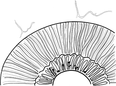
FIGURE 5-1. Iris neovascularization, diagram.
Extraocular Movements
The extraocular movements may reveal a III nerve palsy or VI nerve palsy from diabetes. Look for superior oblique palsy for IV nerve palsy. Is there a new onset of diplopia or head tilt in a diabetic patient?
Eyelids
Look for symmetric lid position of the eyelids. If there is ptosis of long-standing duration, the ophthalmologist will see creases of the ipsilateral frontalis muscle.
SLIT LAMP EXAMINATION
Cornea
Is the cornea clear? Are there signs of previous scars from old recurrent corneal abrasion? Is there corneal edema? Is there abnormal punctuate staining of the cornea from dry eye? Look at Descemet membrane for folds suggesting corneal dysfunction in diabetic patients with long-standing diabetic retinopathy. Is the patient at risk for corneal epithelial defect in the future? Are there areas of corneal thinning?
Anterior Chamber
Is there evidence of cell and flare which one would find in rubeosis iridis? Is there chronic flare which would mean long-standing inflammation in the diabetic eye? The long-standing inflammation could be due to rubeosis iridis or recent panretinal laser photocoagulation. Is the chamber deep or is there a risk of angle-closure glaucoma?
Iris
Look for rubeosis iridis under low-power and then under high-power magnification. Look in the crypts of the iris to find rubeosis iridis. Look at the iris margin to find early pinpoint rubeosis iridis. In brown-eyed patients, it is especially difficult to identify small tufts of neovascularization. Looking for iris neovascularization would entail a darkened room with an intense but narrowed slit beam at the pupillary margin.
Lens
Look for a diabetic cataract in noncompliant diabetics or diabetic patients who have been suddenly out of control of long-standing Type 1 diabetes. Type 1 diabetics usually exhibit early cataract formation. Look at the Y suture in the center of the lens. There may be isolated abnormalities near the Y suture suggesting a severe illness in childhood, especially common in Type 1 diabetic patients. The lens opacity may correlate to the first time the patient was diagnosed with diabetes in childhood.
Gonioscopy
Look at the angle by Goldmann three-mirror or the Zeiss four-mirror lens. Angle neovascularization is very common in diabetic patients with proliferative diabetic retinopathy. Press on the angle to view small fronds of neovascularization which might be hidden from the view.
EXAMINATION OF THE RETINA (SEE CHAPTER 3)
In examining the retina, the use of the direct ophthalmoscope, indirect ophthalmoscope, Goldmann contact lens, hand held 60-D lens, and 90-D lens are all useful. The 90-D lens is small, fits into a pocket, and can be used at all times. Direct ophthalmoscope has been less utilized in a retina practice since retinal specialists have myriad equipment and cameras to examine the retina and the optic nerve.
However, the direct ophthalmoscope is useful as a screening tool for evaluating the optic nerve before we examine the optic nerve using the 90-D lens. The direct ophthalmoscope is a useful teaching tool for medical students and general internal medicine colleagues who are not trained to use the indirect ophthalmoscope.
The retina is extremely important part of the diabetic eye examination since so much pathology can occur while the patient may be relatively asymptomatic. For example, optic nerve cupping, optic disc neovascularization, trace macular edema, cotton-wool spots in the periphery, and microaneurysms all can be existent while the patient sees 20/20.
Optic Nerve
Starting out with the optic nerve head, the examination can begin at the slit lamp. The 90-D lens or 60-D lens can do the job. The optic nerve can be examined for fine fronds of neovascularization (Fig. 5-2), flame-shaped hemorrhages as in glaucoma. Glaucoma can occur with diabetic patients. While the etiology is unclear about the association of glaucoma and diabetes, optic disc pathology and abnormalities can be seen in the examination of a diabetic patient.5 A recent study involving 76,000 women in the Nurses Health Study shows there is a 70% increased risk of developing open-angle glaucoma in women with diabetes as compared to women without diabetes.6–8 At the optic nerve, look for abnormalities of the caliber of the vessels or unusual circumferential vessels which would also indicate increased cupping and therefore increased glaucomatous risk.
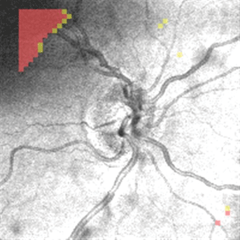
FIGURE 5-2. Optic nerve neovascularization, diagram.
Then, look at the nerve fiber layer emanating from the optic nerve. Is it abnormal? This is the baseline information. If this area changes sequentially over time, this would make the clinician think about glaucoma in the setting of diabetes.
The optic nerve can demonstrate atrophy from long-standing quiescent proliferative diabetic retinopathy. Optic nerve atrophy can be seen in patients with a history of heavy panretinal photocoagulation, overall retinal atrophy, and attenuation of the retinal arterioles in all four quadrants. These patients may have had 25 years or more of diabetes and may have 20/20 to 20/40 vision in the center, but there is a pale optic nerve. It is unclear whether the atrophy is from open-angle glaucoma or from low-tension glaucoma due to chronic poor blood flow to the optic nerve.
Optic disc edema can occur in diabetic patients. This is usually nonarteritic anterior ischemic optic neuropathy (NAION).9 The systemic diseases associated with this entity include hypertension, diabetes, and prior history of cardiovascular disease such as a past history of a myocardial infarction. Arteritic AION is due to giant cell arteritis which has severe risk of visual loss. In NAION, the visual loss is less severe, but NAION is more common than the arteritic AION. The vision loss in NAION is painless. The patient may complain of loss of vision upon awakening.
In arteritic AION, the disc is chalky white, pale, and swollen. Ischemia in multiple vascular regions can occur as central retinal artery occlusion, choroidal infarction, anterior segment ischemia, and extraocular muscle ischemia causing diplopia. Arteritic AION is associated with pain, scalp tenderness, pain when brushing hair, jaw claudication, and pain while chewing. Temporal arteries may be prominent and tender. Sedimentation rate can be obtained to verify the diagnosis.
In NAION, atherosclerosis, arteriolar sclerosis is thought to affect the circulation of the optic nerve head. The posterior ciliary arteries feed the optic nerve head, and these are affected in hypertension and diabetes.
In arteritic AION, similar anatomy is affected. Giant cells affect the posterior ciliary arteries, the orbital vessels, and the central retinal artery (Table 5-2).
TABLE 5.2 Checklist for the Retinal Examination: Optic Disc
Field 1 optic disc
Disc edema (think nonarteritic AION)
Disc pallor (think arteritic AION, low-tension glaucoma)
Optic nerve neovascularization
Cup to disc ratio
Flame-shaped hemorrhage at the optic disc (think glaucoma)
Optociliary shunt vessels at the disc (think old branch vein occlusion)
Look for emboli in the lumen of vessels in TIA complaints
Stay updated, free articles. Join our Telegram channel

Full access? Get Clinical Tree


