Summary
Connective tissue and its collagen support are critical to the functioning of the extraocular muscles, intrinsically within the muscles themselves, as well as to the function of the surrounding orbital pulleys and muscle pulley sheaths. Those with weak, lax native (unoperated) connective tissue are subject to pulley displacement, which may cause profound disruption of eye muscle function. Those with stiff, tight connective tissue may be more prone to restrictive strabismus due to inflammation (thyroid ophthalmopathy and sinus-related strabismus) and trauma.
Wound healing and final scar strength may also vary between individuals, ranging from weak wound healing with the risk of scar stretch and scar migration, to aggressive hypertrophic scar formation with the potential for fibrosis and restrictive strabismus.
Preoperative knowledge of an individual’s connective tissue type can help the strabismologist to plan for these contingencies and tailor surgical techniques to the patient’s collagen and wound healing status. Active strabismus procedures are less likely to succeed in the weak collagen group of patients, so surgical planning should steer to passive procedures whenever possible. The use of nonabsorbable sutures, more thorough dissection of perimuscular tissues, and avoidance of steroids would be indicated in the weak healing group, but careful preservation of layers and judicious use of steroids would be considered in the aggressive healing group.
Surgical principles designed to optimize wound healing have been well studied in other surgical specialties but have been largely overlooked in ophthalmology. The aim of most ophthalmic surgery is to inhibit healing as much as possible in order to prevent vision-reducing scars in the cornea, or bleb-closing scars in glaucoma surgery. The focus of these subspecialties is to minimize scar formation as much as possible. Strabismologists primarily deal with tendon transfers; therefore, precisely directed scar formation, with enough strength to withstand years of muscle pulling, is required. The aim of strabismus surgery should be to develop strong scar in the location(s) needed to maintain desired muscle position(s), but to prevent scar formation in other directions, which would detract from the surgical goals.
5 Collagen and Healing
5.1 The Biology of Collagen
The word collagen finds it origin in ancient Greece. The tendons, muscles, and other tissues rich in collagen of horses and other animals were boiled to make glue, hence the name “κόλλα” (kolla), meaning “glue,” and “γέν” (gen), meaning “forming.” 1 Collagen’s role in making glue used to hold together wood furniture, fix murals, and bind books is quite appropriate considering the role it plays in the human body. Found in the extracellular matrix in and around every tissue in the body, it is central to our structural integrity. Collagen is the molecule that holds our whole body together. Thus, while being a microscopic molecule, collagen has major effects on the macroscopic integrity of muscle fibers, tendons, and connective tissue. To better understand this relationship, knowledge of collagen on a biochemical, cellular, and tissue level is needed. From this, the variations seen on a macroscopic scale can be explained on a microscopic scale as well.
While having a wide variety of function-specific variations, the basic biochemical structure of all collagen proteins is a triple helix of polypeptides forming a tropocollagen molecule. Tropocollagens are then arranged into fibrils, which further aggregate into fibrillar bundles, which form functional tissues. 2 The nature of the interactions between the amino acids, collagen monoproteins, tropocollagens, fibrils, bundles, and then finally tissues is a wide and deep field of study, with more than 100,000 published papers on the subject over the last 40 years. 3 Variations on these interactions are responsible for the many types of collagen, forming many more different functional tissues. As this book is written primarily for ophthalmologists, not collagen researchers, most of this is out of our scope. However, as the oculomotor system relies heavily on muscles and tendons, collagen type I is particularly important.
Collagen I is the essential molecule for the strength, stability, and flexibility of muscle fibers, tendons, and connective tissue. This stems from its integral role in the extracellular matrix (ECM) of all these tissues. In particular, muscles are particularly reliant on collagen I—up to 10% of dry muscle mass is collagen. 4 Structurally, the ECM surrounding muscle fibers has been found to be much stiffer than the muscle fibers themselves, meaning on a macroscale, flexibility is largely determined by the collagen in muscle rather than the muscle tissue itself. 5 Additional evidence for this comes from increased stiffening of muscles in the setting of muscular dystrophies, aging, diabetes, or immobilization. Histologic examination of muscles reveals that increased proliferation of ECM collagen fibers highly correlates with fibrosis and increased stiffness. 6 , 7 These findings, however, are not very specific, as not only the amount of collagen, but also the composition of collagen, is important in determining flexibility. Several studies have found biochemical changes in collagen such as increased cross-linking to be associated with increased tissue stiffness in a pathologic setting. 8 , 9 , 10 It can thus be concluded that variability of muscle and tendon stiffness comes from both the quantity and the quality of collagen, particularly type I.
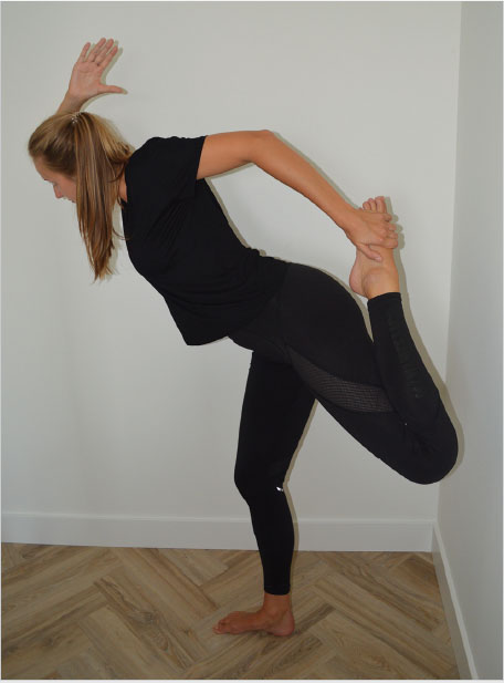
This variance in stiffness, while studied mostly in a pathologic setting, appears in the normal population as well. This can be seen in the wide variety of flexibilities among normal, healthy individuals (Fig. 5‑1, Fig. 5‑2). The basis for these variances in amount and character of collagen comes largely from the enzymes and cells supporting collagen development, proliferation, maturation, modification, and maintenance. 11 , 12 While collagen type I’s basic molecular structure is determined by just two genes (one for each of the heterotrimers, consisting of two molecules), the possibility for genetic variation grows exponentially when considering all the genes determining the function and expression of all of the modifying and supporting role players. To illustrate this, an example from pathology can be used. Ehlers-Danlos syndrome (EDS) is a spectrum of disorders in which dysfunctional collagen results in a host of problems due to tissue instability. Muscle and joint flexibility is often increased in EDS, although to a variable degree. The genetics of collagen disorders are a focus of much current research. While EDS is a collagen disorder, only half of the mutations known to cause EDS affect the actual collagen molecule gene. The others affect enzymes critical to normal collagen formation. 13 This large genetic variation, which results in a syndrome that also has great clinical heterogeneity of muscle and tendon flexibility, speaks to the complexity of collagen disorders. There is probably also just as much genetic variance, and therefore variability of flexibility, within the normal population.

The classification of collagen status could be very useful for strabismus surgeons, as different types of collagen call for different surgical approaches. While guidelines exist to assess for pathologies like EDS, there are few data about the normal variability in collagen, and few guidelines for assessing these differences in a nonpathologic setting. This doesn’t mean, however, that strabismologists shouldn’t search for these differences. Borrowing from the EDS exam, physicians can use a joint mobility test to assess collagen integrity. Not only does hypermobility of joints indicate a deficient or looser collagen structure, 14 but hypomobility is associated with increased amounts of collagen and increased cross-linking when compared to normal. 15 Goniometry, commonly used in orthopaedics, could be used to assess the mobility of joints, from which an approximation of collagen characteristics could be made. A protractorlike device is used to assess the range of motion of various joints around the body, and these values are compared to reference ranges. While joint mobility testing is correlated with collagen stiffness, there are other factors at play, such as elastin and muscle strength and integrity. To be more specific in testing collagen integrity, perhaps some novel methods could be designed.
From dermatology, we know that confocal fluorescence microscopy can be used to assess the three-dimensional arrangement of collagen fibers. 16 This is a microscopic technique commonly used in cytologic assessments of skin cancers, but it can also be used to assess collagen in the reticular dermis. The loss of normal collagen structure in the dermis is associated with aging, scarring, diabetes, and notably, collagen disorders such as EDS. 17 As such, it could potentially be inferred that a loss of collagen structure at the dermis might suggest a similar loss of collagen structure and integrity in the extraocular muscle (EOM) system. Would it be possible to predict collagen status of the eye muscles by testing collagen three-dimensional structure and integrity in the periocular skin with confocal microscopy? This is merely speculation and would require further research to create guidelines. Staining with Alcian blue for glycosaminoglycan can be used to assess how cross-linked a collagen sample is, 18 and could potentially have use as a test of collagen status. If a biopsy is done, single collagen fibers can be tested for their mechanical properties using newly developed methods. 19 As the field of collagen genetics evolves, it may one day also be possible to screen for collagen differences genetically.
5.2 Collagen, Connective Tissue, and Healing in Strabismus Surgery, Including Stretched Scar
5.2.1 Introduction
Connective tissue forms the structural “scaffolding” that supports the EOMs in their paths to allow their proper function. The anatomy and function of these structures, termed the “pulleys,” have been shown to be as important as the muscles themselves in maintaining normal alignment and motility. (This work is summarized in Chapters 4 and 19.)
Our new appreciation of the importance of the connective tissue framework to EOM function must then lead us to conclude that collagen and connective tissue disorders may contribute primarily to the development of strabismus. (This tendency has been observed in this author’s practice, but has yet to be subjected to scientific study.) Collagen disorders may also cause abnormal responses to strabismus surgery if healing is either too aggressive or too weak. 20 , 21 , 22
5.2.2 Collagen and Connective Tissue in Strabismus
5.2.2.1 Weak and Lax Collagen
There are multiple types of collagen, the discussion of which is beyond the scope of this book. The collagen dysfunction in genetic disorders such as EDS, Marfan’s syndrome, and osteogenesis imperfecta is well known, but there are also less-understood intrinsic variations in the normal population. Some individuals are highly flexible (e.g., hypermobile joint syndrome) 23 (Fig. 5‑1), whereas others are stiff (Fig. 5‑2). Stretchy, weak connective tissue could lead to pulley displacement and mechanical strabismus, and this association between hypermobile joint syndrome and strabismus has been observed in cases of acquired esotropia in children with displaced lateral rectus (LR) pulleys. If connective tissue pulley support of the EOMs is lax due to collagen weakness, then gravity could possibly induce pulley shifts and change of alignment. This has not been studied, but Marfan’s syndrome patients were shown to have pulley shifts during gaze position shift. 24
This author has begun to retest alignment in the supine position in a few patients with suspected pulley disorders, with surprisingly large differences between upright and supine measurements. This is a new observation and has not been studied systematically.
The association of Arnold-Chiari malformation with esotropia is known, and although some patients have resolution of strabismus with neurosurgical correction, others do not, and require strabismus surgery or prisms. 25 , 26 , 27 , 28 Arnold-Chiari malformation has been identified as a possible hereditary connective tissue disorder, and is found to be associated with occipitoatlantoaxial hypermobility. 29 Of interest is that comitant divergence insufficiency esotropia is seen in over half of these cases, 26 , 27 which is the same pattern manifest with LR pulley displacement (Chapter 19). 30 , 31 Could some cases of esotropia with Arnold-Chiari malformation actually represent a comorbidity due to collagen weakness with pulley displacement rather than a primary neurologic disorder?
Collagen production and content diminishes with age throughout our tissues. 9 , 30 , 31 These changes are both genetically and environmentally influenced. Long-term smoking, chronic disease, steroid usage, and nutritional abnormalities are just some of many factors shown to weaken connective tissue. 20 , 32 Age-induced pulley shifts have been demonstrated to cause divergence insufficiency esotropia as well as cyclovertical deviations. 30 , 31
5.2.2.2 Stiff and Tight Collagen
Excessive collagen formation and fibrosis can shorten and stiffen the muscles themselves as well as the perimuscular tissues and pulleys, restricting motility. This is seen in inflammatory disorders such as thyroid ophthalmopathy (Chapter 12), severe pterygium (Chapter 14), and chronic sinusitis (Chapter 13).
5.2.2.3 Active and Passive Strabismus Procedures: Planning Strabismus Surgery with Collagen and Healing Considerations
In addition to considering the patient’s collagen status when diagnosing strabismus, it needs to be considered in planning strabismus surgery, particularly with regard to the potential for strong stable healing, and the ability of the tissues to support sutures. Grouping procedures into active and passive strabismus surgeries is a helpful way to think about these concepts. If native collagen is so thin and stretchy that sutures simply pull out, then procedures designed to tighten or strengthen muscles and/or tissues (active surgery) are destined to fail. If any slight injury is met with an aggressive healing response and marked fibrosis, then the opposite problem will occur, and strengthening procedures will work too well and overcorrect the problem. The converse holds true for procedures designed to weaken or relax muscles and tissues (passive surgery). Healing abnormalities and native collagen abnormalities do not always coexist. Some people with weak collagen and sagging pulleys are able to mount a normal, strong wound healing response, and some with strong native collagen have poor healing. Of course, dense collagen and aggressive healing may be combined, as may weak collagen and weak healing. The processes and disorders are complex and multifactorial.
5.2.2.3.1 Active Strabismus Surgery
Active procedures are those in which the muscle is asked to actively increase its action to produce the desired effect, and a strong surgical bond is critical to the outcome. These include muscle resections and advancements, myopexies, inferior oblique (IO) anterior transposition, rectus muscle transpositions, repair of displaced muscles, pulley posterior fixation, faden operation, pulley sleeve fusion, tether flap creation, plications, and tucks. Nonabsorbable sutures should be considered for these procedures, especially when weak connective tissue is suspected. (Superior oblique [SO] tendon tenotomies with extender band or “chicken suture” do tend to stretch apart unless nonabsorbable sutures are used, so these should be considered in the active procedures group despite their muscle weakening effects.)
5.2.2.3.2 Passive Strabismus Surgery
Passive strabismus procedures are those in which the aim is to reduce active pulling by the muscle or relieve retrictions. These include recessions, myotomies and tenotomies, flap tear repair, and conjunctivoplasty with conjunctival autograft or amniotic membrame graft. Suture choice in these cases is based upon other considerations, such as suture reactivity and ease of use, but suture strength is not as critical, and there is less tension on the surgical site. (The SO insertion is a special case, as it tends to be a low-tension location, and not prone to scar stretch or migration. Even SO advancements hold well with absorbable suture despite a tendency toward weak healing in other body locations, or stretched scar of other muscles. One may therefore consider SO insertional surgery in the passive group. Additionally, although flap tear repair relieves tension and flap reattachment is done under little tension, flaps have been observed to redetach when absorbable sutures were used, and nonabsorbable sutures are therefore recommended.)
5.2.3 Healing after Strabismus Surgery
Surgery is an iatrogenic act of targeted tissue trauma to repair or remove an abnormality. It necessarily creates injury to tissue, which then undergoes the complex staged process of healing. 20 , 33 , 34 The ultimate integrity of the tissue requires formation of a scar strong enough to resist normal stresses. 35 , 36 The initial injury leads to coagulation, with vasoconstriction and platelet-induced clot formation. The inflammatory phase begins at about 24 hours with the arrival of polymorphonuclear leukocytes into the wound, followed by macrophages, which perform wound débridement and fibroplasia. The fibroplastic (or proliferative) phase is next, during which collagen is formed and cross-linking begins. Angiogenesis, formation of granulation tissue, and wound contracture occur during this phase. The longest phase is remodeling, during which collagen bundles increase in size and inflammation decreases. Cross-linking of collagen bundles increases, and collagen fibers align along tension planes. The phases of healing overlap with each other and are complex multifactorial processes involving numerous cells, growth factors, and proteins. Disruption of any of these stages can interfere with the ultimate scar (either causing weak scarring or leading to excessive scar formation) and therefore alter the surgical result. 32 , 33 , 34
5.2.4 Principles of General Surgery
The goal of most ophthalmologic research into wound healing has been to minimize scar formation in order to preserve corneal clarity or to prevent blockage of filtering blebs and glaucoma drainage valves. In most other surgical specialties the goal is to promote formation of a strong, healthy, properly directed scar. Strabismus surgery is more analogous to the tendon transfer procedures of orthopaedics than to corneal or intraocular surgery. The surgical principles that guide orthopaedists and general surgeons are therefore worth consideration by the strabismus surgeon.
A surgical wound is created by apposition of iatrogenically injured tissue. Meticulous technique is directed toward minimizing tissue trauma and inflammation. Excessive cautery causes tissue necrosis, but hematoma formation also interferes with healing, so cautery should be used judiciously and precisely. Sutures tied too tightly will damage the incorporated tissue, and crushing tissue with forceps or clamps is also destructive. All these tissue insults worsen the chances of a clean narrow scar and decrease ultimate tissue functionality. As longer or wider scars are weaker, it is also important to prevent gap formation within the wound. Layered closure of tissues and prevention of tissue damage will minimize gap development. During tendon anastomoses, the strongest results were obtained with sutures placed around the circumference of the tendon, avoiding the central cut tissue altogether. 37
The principle of a two-sided wound is universal in general surgical procedures. 18 The situation present in strabismus surgery, in which an injured tissue (the disinserted eye muscle tendon) is attached to an uninjured tissue (the sclera), does not have a parallel in general surgery. 16 Direct suturing of the tendon to sclera may create some tissue injury at the needle entry and exit sites, but there is no scleral wound in hang-back or adjustable suture surgery. (This may explain the higher incidence of stretched scar after adjustable suture strabismus surgery). 20 , 21
General surgeons are taught to recognize and dissect within tissue planes. This skill is fundamental to good surgical skill, and has been shown to reduce hemorrhage (as tissue planes are avascular) and improve surgical outcome. 38 Successful repairs of flap tear, lost muscles, and slipped muscles require recognition of tissue planes within the orbit. 22 , 39 , 40 , 41
Tension on a wound can cause suture failure and wound dehiscence in the short term, and scar stretching and widening in the long term. 19 , 42 Surgeons employ many techniques to reduce tension across wounds throughout the body, such as splints, stay sutures, deep imbricated sutures, tissue undermining, and bandaging and taping techniques. 43 , 44 , 45 , 46
Scar tissue never achieves the strength of native collagen. Healed tendons achieve about 10% of normal tensile strength, 37 knee ligaments 40%, 47 skin 25 to 80%, 48 , 49 and fascia 50 to 80% in different animal studies. 20 , 50
Cells subjected to tension while in tissue culture have been shown to increase DNA production and cell division, collagen production, and synthesis of protein, chondroitin sulfate, hyaluronate, and prostaglandin. 51 , 52 , 53 , 54 , 55 Collagen is subject to creep under tension, and this tendency is increased in scar tissue over normal tendon. 56 , 57 Normal tissue has been shown to stretch and grow when subject to tension, with fibroplasia, increased collagen production, rearrangement of collagen fibers, and vascularization. 58 , 59 , 60 Plastic surgeons take advantage of this property with their use of tissue expansion techniques in reconstructive procedures. 61
5.2.5 Collagen and Strabismus Surgery
The strabismus surgical plan should take into account the strength of the patient’s collagen and any history of wound healing abnormalities. If collagen is weak, there may be insufficient support to hold the sutures in the tissue, and the strabismus will recur. Other surgical procedures that attempt to deal with the complications of weak collagen include those for incisional abdominal hernia, 62 joint hypermobility, 23 and uterine and bladder prolapse. 63 Surgical correction of these conditions struggles with similar concerns.
At the other end of the spectrum are patients who mount an unusually aggressive healing response. Although fibrosis is often related to traumatic surgical technique, it can also be integral to that patient’s healing process. Knowledge of hyperkeratotic scar formation from trauma or prior surgery could warn the strabismus surgeon to design and perform surgery as atraumatically as possible.
5.2.5.1 Weak Collagen
5.2.5.1.1 Stretched Scar in Strabismus
When scar tissue is subjected to tension, it may gradually increase in width or length due to stretch, growth, or a combination of the two. Stretching is a thinning of tissue due to mechanical stretching (collagen creep), and growth involves formation of new tissue. Both processes have been shown to occur under tension in all scar tissues of the body, 20 , 44 , 64 with the best studied being the incisional abdominal hernia. 62 The newly formed eye muscle insertion after strabismus surgery is subject to the same problem, with an incidence (2–8%) 20 , 21 , 22 similar to incisional abdominal hernia (3–20%). 62 This phenomenon has been termed “stretched scar” for convenience, but a more correct term might be “stretched and lengthened scar.” If the original procedure were a muscle recession, the resulting strabismus pattern would be a consecutive overcorrection, and stretched scar after a resection would cause recurrence of the original deviation. Balanced stretching of the medial rectus (MR) and LR after a recess/resect procedure usually leads to exotropia due to the MR’s greater force 63 , 64 , 65 , 66 , 67 (and therefore longer stretched scar segment), but has also caused proptosis and globe subluxation. 68 Just as repair of an abdominal hernia requires nonabsorbable support to prevent inevitable recurrence, stretched scar of an EOM insertion requires nonabsorbable suture repair.
Pathologic evaluation of numerous stretched scar segments showed dense connective tissue (Fig. 5‑3), which lacks the regular orientation of normal EOM tendon (Fig. 5‑4). There are no findings to distinguish the different stretched scar mechanisms. Excised stretched scar segments lose their linear appearance as tension is eliminated (Fig. 5‑5), unlike excised normal rectus muscle tendon (Fig. 5‑6). EOM stretched scar segments were also shown to orient themselves more randomly in cell culture than cells from normal tendon. They also produced more extracellular matrix than tendon cells. 20


Scar stretch has been shown to increase in frequency when the tension on the insertion is high. The inferior rectus (IR) insertion with its powerful force 65 , 66 , 67 is the most prone to develop postoperative scar stretch, followed by the medial and superior rectus muscles. 20 , 21 , 22 LR and IO stretched scars are less common, and SO stretches have not been observed. Large resections and transpositions can put high stress on the new insertion, predisposing to scar stretch and migration. The use of nonabsorbable suture for the primary procedure is advised in these cases to prevent stretched scar.
The use of nonabsorbable sutures in repair was shown to significantly decrease the rate of recurrence after stretched scar repair. 20 , 21 , 22 This improvement over absorbable sutures was also demonstrated in an animal model. 20
Many conditions predispose to scar stretch, including primary collagen disorders (see above), pregnancy, use of corticosteroids, smoking, alcoholism, chronic illness, malnutrition (especially vitamin deficiencies of C, D, E, and A), connective tissue disease, hypothyroidism, hypoxia, chemotherapy, and uremia. 20 , 35 The breakdown of previously well-healed scars in sailors suffering from scurvy (vitamin C deficiency) has been known for centuries. 69
5.2.5.1.2 Cooper’s Dictum
Historically, the most common clinical presentation of stretched scar was consecutive exotropia after previous MR recession. The logical solution to this complication would seem to be to advance the previously recessed muscle(s), but this approach was consistently met with failure in the 1940s and 1950s (probably due to unrecognized scar stretch). This led Cooper to recommend operating on fresh antagonist muscles, as in recession of the LR muscles. He advocated planning reoperation with no consideration of the prior surgery. 70 This advice became known as “Cooper’s dictum.” Since exotropia was the typical pattern seen with stretched scar after bilateral MR recession, and bilateral LR recession was the typical approach to surgical correction of exotropia, this was, and has remained, a common choice of procedure for many strabismologists. It is technically easier than stretched scar repair and produces adequate initial success in these patients, but they may then develop limitation of motility in adduction and abduction, and in rare cases, proptosis and globe subluxation. If stretching continues to progress (as is usually the case), the MR scars usually stretch faster than the LR scars, causing recurrent exotropia. Now the patient is faced with difficult four-muscle repair, or the risk of regular recurrence of strabismus. It is time to replace Cooper’s dictum with proper repair of stretched scar with nonabsorbable suture at the outset.
Diagnosis of stretched scar involves a combination of the patient’s history, office examination, and direct inspection of the muscle insertion intraoperatively (Fig. 5‑7, Fig. 5‑8, Fig. 5‑9, Fig. 5‑10, Fig. 5‑11, Video 5.1, Chapter 27). The history should include questioning about underlying collagen abnormalities or predisposing illness. Ask whether scar widening was seen after other surgical procedures (Fig. 5‑12), and whether there have been any orthopaedic problems, uterine or bladder prolapse, frequent sprains, or other indications of weak collagen or healing. The time course of scar stretch or lengthening follows several different patterns: early/subacute stretch, early/gradual stretch, late/subacute stretch, and slow stretch. The patients with early/subacute stretch begin to show gradual change of the initial postoperative alignment after the absorbable suture loses strength, at 3 to 4 weeks. Alignment changes over weeks to months and then stabilizes in the long term. These patients probably have delayed scar maturation, which is not enough to maintain alignment in the face of the constant pulling of the muscle during the early stages of healing. Once their collagen remodeling strengthens, the stretching stops and the alignment stabilizes (perhaps slower-dissolving absorbable sutures would perform as well in this group as nonabsorbable sutures, but these have not been tried). In the early/gradual stretch group, the gradual alignment change also begins when the suture strength declines, but the alignment continues to worsen over time without stabilizing. These patients never achieve sufficient scar tissue strength to resist the pulling from the muscle. The genetic collagen abnormalities fall into this group. Those in the late/subacute stretch group have good stable alignment for a long period following the original surgery, followed by decompensation and stretched scar development over a relatively short period of weeks to months. This can usually be attributed to a process or illness known to weaken collagen, as listed above. Once the cause of collagen weakening is improved, the stretching, and therefore the worsening of strabismus, will slow or stop. Those with slow stretch have a very gradually progressing deviation over time, which they find impossible to date. They probably have an underlying collagen/connective tissue abnormality, which is less severe or has a different mechanism than that of the early/gradual group, but which creates a similar picture over a longer period of time. (Similar temporal variability in the development of abdominal hernia has also been reported. 71 , 72 , 73 )Pulley shifts with age could mimic late onset stretched scar in some cases, as could flap tear. A careful history as well as imaging can help sort this out preoperatively, but sometimes the diagnosis cannot be definitively made until surgical exploration is performed.


Stretched scar is not generally a complication of strabismus surgery in the aged, although this observation has not been subjected to analysis. The elderly may have old stretched scars from prior surgery, but do not usually develop new stretches late in life. Whether their old stretched scars require nonabsorbable suture repair is not known. Collagen loses elasticity in the elderly, and this may prove protective against scar stretch after muscle surgery.
5.2.5.1.2.1 Steroids
Corticosteroids are potent inhibitors of wound healing and collagen formation. They should be avoided in stretched scar cases, and this author avoids them in most strabismus surgery, unless severe fibrosis is the problem.
Stay updated, free articles. Join our Telegram channel

Full access? Get Clinical Tree


