FIGURE 25.1. Algorithm regarding the management of neovascular glaucoma.
CAUSES OF ANTERIOR CHAMBER NEOVASCULARIZATION
While many pathologic ocular processes can initiate fibrovascular proliferation, ischemic retinal disease is the most common cause of anterior chamber neovascularization (Table 25.1).1 Proliferative diabetic retinopathy and central retinal vein occlusion are each responsible for about one third of cases of neovascularization of the iris (NVI).2 Retinal ischemia from various disorders such as rhegmatogenous retinal detachment or vascular pathology account for the remaining one third of cases. Only about 3% of NVG cases are due to nonischemic causes, such as tumors or ocular inflammation.2 The crystalline lens, lens capsule, and vitreous appear to act as natural barriers to the diffusion of angiogenic mediators into the anterior chamber; consequently, anterior segment neovascularization is often seen after vitrectomy or disruption of the posterior capsule in eyes with predisposing ocular pathology.3,4,5,6 The ocular pathology causing NVG is typically obvious, identifiable by careful slit-lamp biomicroscopy, gonioscopy, and fundus examination. Occasionally, the etiology of NVG is unclear, and ancillary testing is necessary to detect occult pathology. Recognition of the underlying proangiogenic pathology is critical for successful treatment of NVG in order to effectively reduce the angiogenic mediators that promote anterior chamber neovascularization.
Table 25.1.
Diseases associated with neovascular glaucoma
Retinal ischemic diseases
Leber congenital amaurosis Coats disease Eales disease Sickle cell retinopathy Retinal hemangioma Persistent hyperplastic primary vitreous Norrie disease Wyburn-Mason Carotid-cavernous fistula Dural shunt Stickler syndrome X-linked retinoschisis Takayasu aortitis Juxtafoveal telangiectasia Surgically induced Carotid endarterectomy Cataract extraction Pars plana vitrectomy/lensectomy Silicone oil Scleral buckle Neodymium: yttrium-aluminum-garnet capsulotomy Laser coreoplasty Tumors Iris: melanoma, hemangioma, metastatic lesion Ciliary body: ring melanoma Retina: retinoblastoma, large cell lymphoma Choroid: melanoma Conjunctiva: squamous cell carcinoma Radiation External beam Charged particle: proton, helium Plaques Photoradiation Inflammatory diseases Uveitis: chronic iridocyclitis, Behcet disease Vogt-Koyanagi-Harada syndrome Syphilitic retinitis Sympathetic ophthalmia Endophthalmitis Miscellaneous Vitreous wick syndrome Interferon-α |
Data from Sivak-Callcott JA, O’Day DM, Gass JDM, et al. Evidence-based recommendations for the diagnosis and treatment of neovascular glaucoma. Ophthalmology. 2001;108: 1767–1778.
Microscopic analysis has revealed that the new, abnormal blood vessels arise from iris stromal vessels and spread onto the anterior iris surface.7 Anterior segment neovascularization is characterized microscopically by thin, fenestrated blood vessels that grow in irregular patterns.8,9,10 Clinical and microscopic analysis has identified fibrovascular tissue developing over anterior segment structures, including the trabecular meshwork and in intertrabecular spaces.7,8,9,10,11
CLINICOPATHOLOGIC STAGES
A spectrum of clinicopathologic stages may be identified in the development of NVG and can be useful for diagnostic classification, for developing treatment plans, and for prognostic direction (Fig. 25.2).3 Anterior chamber neovascularization in the setting of normal IOP characterizes the preglaucoma stage. In the open-angle stage, IOP elevates when a fine fibrovascular membrane obstructs a gonioscopically open angle. The closed-angle stage occurs when the fibrovascular membrane contracts, causing varying degrees of peripheral anterior synechiae (PAS). Ultimately, obstruction of aqueous outflow causes elevated IOP, acutely or chronically, and is the primary mechanism by which glaucomatous optic neuropathy and ultimate visual field and vision loss may occur.
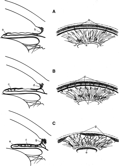
FIGURE 25.2. Clinicopathologic stages of neovascular glaucoma include (A)the preglaucoma stage with normal IOPs in the setting of iris (a) and angle neovascularization (b). B: The open-angle stage characterized by IOP elevation in the setting of iris neovascularization (c) and a fine fibrovascular membrane obstructing a gonioscopically open angle (d). C: The closed-angle stage occurs when the iris (f) and angle fibrovascular membrane contract, causing varying degrees of PAS (g) and ectropion uvea (e). (Reproduced from Allingham RR, Damji KF, Freedman S, et al. Shields’ Textbook of Glaucoma. 5th ed. Philadelphia, PA: Lippincott Williams & Wilkins; 2004;330–335, with permission.)
A high index of suspicion in the setting of common predisposing conditions, and a careful and comprehensive ocular exam, including gonioscopy, are necessary to detect NVI and/or neovascularization of the angle (NVA). Iris or gonioscopic fluorescein angiography may be useful to detect NVI or NVA when rubeosis is not visualized by slit-lamp biomicroscopy.12 Although NVI is typically appreciated prior to NVA, NVA may precede NVI in about 10% of eyes with retinal ischemia, highlighting the importance of gonioscopy in high-risk eyes.13–15
TREATMENT MODALITIES
Panretinal Photocoagulation
Panretinal photocoagulation (PRP) is the standard antiangiogenic treatment for NVG caused by ischemic retinal disease.16–19 Extensive PRP, usually 1,500 to 2,000 or more spots over two sessions, is necessary to decrease vasoproliferative factors.20 PRP is believed to work by reducing the production of angiogenic mediators by the ablated and scarred retina.21 In addition, PRP may work by creating a shunt through the scarred retinal tissue that improves oxygen delivery to the retina from the choroid.22,23 While PRP is necessary for the long-term control of angiogenesis, it may take multiple sessions and 2 to 4 weeks after the laser therapy for regression of anterior chamber neovascularization, during which time aqueous outflow obstruction and synechial angle closure may persist or progress.24 Intraocular anti-VEGF injections can offer rapid and effective adjunctive therapy to reduce anterior chamber neovascularization.
Anti-VEGF Drugs
Early anti-VEGF pharmacotherapy, either bevacizumab (Avastin [Genentech, South San Francisco, CA]) or ranibizumab (Lucentis [Genentech]), has been shown to promptly eliminate clinical evidence of anterior chamber neovascularization (Figs. 25.3–25.5).25–29 Pathophysiologic analysis of trabecular meshwork sections, obtained at the time of trabeculectomy surgery from patients with NVG who received bevacizumab, demonstrated that despite clinical regression of NVA, neovascular tissue in angle structures persisted, but the remaining vascular tissue lacked normal fenestrations.11 Early regression of angle neovascularization in the preglaucoma and open-angle stages of NVG can prevent the formation of PAS and preserve normal aqueous outflow routes. Maintenance of normal angle anatomy may eliminate the need for long-term IOP lowering therapy or may decrease the need for medical therapy or incisional surgery to treat NVG.30,31
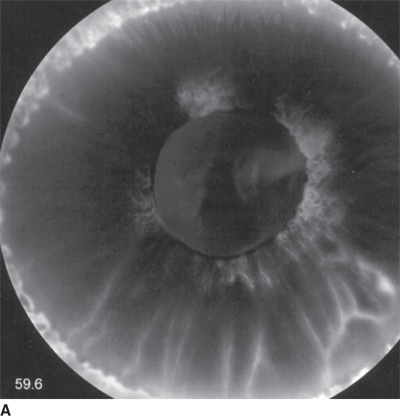
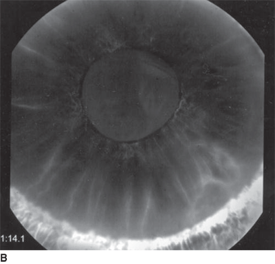
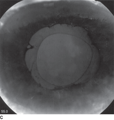
FIGURE 25.3. A: Iris fluorescein angiogram revealing florid rubeosis iridis before bevacizumab injection. B: Iris fluorescein angiogram showing marked improvement of rubeosis iridis 5 days after bevacizumab injection. C: Iris fluorescein angiogram demonstrating nearly resolved rubeosis 8 weeks after bevacizumab injection. (Reproduced from Davidorf, et al. Retina. 2006;26:354–356, with permission.)
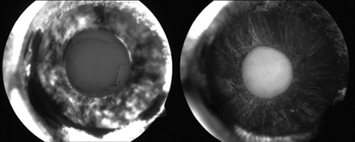
Figure 25.4 Iris rubeosis due to occlusive ischemic retinopathy. Iris fluorescein angiography of the right eye taken 5 days before (Left) and 2 days after (Right) intracameral injection of bevacizumab. Pictures were taken in the early phase (10 seconds) of angiography. (Reproduced from Grisanti, et al. Am J Ophthalmol. 2006;142:158–160, with permission.)
Stay updated, free articles. Join our Telegram channel

Full access? Get Clinical Tree


