25-Gauge Vitrectomy for Macular Surgery
Steve Charles
Sutureless, 25-gauge (25G) vitrectomy was initially utilized by most surgeons for epimacular membranes, macular holes, and vitreomacular traction syndromes, as well as for procedures now shown to be ineffective: branch vein decompression and radial optic neurotomy. Virtually all techniques utilized for 20-gauge (20G) vitrectomy can be utilized with 25G vitrectomy (1, 2, 3, 4, 5, 6, 7, 8, 9, 10, 11, 12, 13, 14, 15, 16, 17 and 18) using new tools and technique modifications discussed in this chapter. Some techniques to be discussed are applicable to 25G surgery and selected 23-gauge (23G) and 20G procedures as well.
ADVANTAGES OF 25G VITRECTOMY
There are many advantages to eliminating both scleral and conjunctival sutures, including reduced discomfort, photophobia, tearing, squeezing, conjunctival hyperemia, and bleeding. Faster visual recovery occurs because astigmatism from the sclerotomy sutures is eliminated and the ocular surface suffers less trauma. Although faster visual improvement does not translate into better long-term visual outcomes, it is important if there is impaired vision in the other eye. Elimination of sutures and conjunctival incisions greatly reduces damage to the conjunctiva, Tenon’s capsule, and episclera, which is crucial for eyes with a glaucoma filtering procedure and eyes likely to have glaucoma surgery in the future. Reduced conjunctival damage is also advantageous for patients with ocular surface disorders.
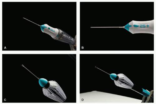 Figure 17-1. A and B: Shows new vitrctomy probe 25-G Plus. C and D: Shows Grieshaber 25-G Plus disposable forceps. |
It was initially thought that 25G fluidics would be limiting but this has not proven to be the case. As will be discussed later in this chapter, 25G fluidics are safer than 20G or 23G fluidics.
The new series of 25+™ (25 Plus) instruments from Alcon allows complex surgical maneuvers such as membrane peeling using cutter rather than a forceps, avoiding the use of scissors. The high cut rate (up to 5000 cpm), strong aspiration and escellent IOP control of the Constelation® system allows shaving the vitreous base with negligible traction to the surrounding tissues and retina. In the 25+™ vitrectomy probe design, the tip has been redesigned to place the port closer to the distal end of the probe, allowing vitreous shaving closer to the retina.
The port size has also been enlarged to increase aspiration rate, And the shaft has been stiffened to reduce flexure and improve control (Figs. 17-1 and 17-2).
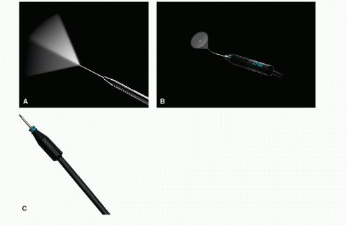 Figure 17-2. A: Illuminated pick 25-G Plus B: Illuminated fotocoagulation probe 25-G Plus C: Chandelier -w-sleeve -close 25-G Plus. |
WOUND CONSTRUCTION
Displacement of the conjunctival wound relative to the sclerotomy site before insertion of the trocar cannula ensures that conjunctiva will cover the self-sealed sclerotomy (Fig. 17-3


Stay updated, free articles. Join our Telegram channel
Full access? Get Clinical Tree
