FIGURE 24.1. Avascular cornea contains sflt-1 bound to VEGF-A. A: Photo of human eye demonstrates abrupt termination of blood vessels in the conjunctiva (CJ) at its border with the cornea (C), the limbus (asterisks). B: Representative nonreducing Western blot of mouse cornea reveals immunoreactive bands of VEGF-A at 100 to 130 kDa corresponding to bound forms, and negligible immunoreactivity at 45 to 50 kDa corresponding to the free form. n = 5. C: sflt-1 (lane 1) and VEGF-A (lane 3) transcripts in mouse cornea identified by representative polymerase chain reaction with reverse transcription (RT-PCR). Lane 2 is water (template-negative) control. n = 5. bp, base pairs; aa, amino acids. D, E: sflt-1 mRNA detected by in situ hybridization in mouse corneal epithelium (epi) and stroma (str). Antisense RNA probes show purple-brown reactivity. Sense RNA probes show negligible reactivity. F: Immunolocalization (brown) of sflt-1 protein in mouse cornea. G: Representative reducing Western blots (WB) with an antibody against the amino (N) terminus of flt-1 that recognizes both mbflt-1 and sflt-1 and an antibody against the unique C terminus of sflt-1 show that mouse cornea (a) contains primarily sflt-1 (60 kDa), whereas the conjunctiva (b) contains mainly mbflt-1 (190 kDa). H: Representative Western blot of two independent mouse cornea samples immunoprecipitated (IP) with anti-VEGF-A antibody and immunoblotted (WB) with a biotinylated antibody against the N terminus of flt-1 that recognizes both mbflt-1 and sflt-1 shows that VEGF-A interacts with sflt-1 (60 kDa). Subsequent immunoblot with a biotinylated anti–VEGF-A antibody confirms the pull-down of VEGF-A by the immunoprecipitating antibody. n = 6. (Reproduced from Ambati., et al. Nature. 2006;443:993–997, with permission).
In a recent report, mouse corneas were treated with a novel tetrameric tripeptide to interrupt sFlt-1 binding to VEGF-A. As in prior experiments, where different strategies were used to disrupt sFlt-1 function, these treatments resulted in significant KNV.45 Examination of several human subjects using immunohistochemistry and immunoprecipitation techniques revealed that normal avascular human corneal tissue contains high levels of sFlt-1, which normally binds and sequesters VEGF. In contrast, corneal tissue from subjects with KNV resulting from diverse etiologies including alkali burns, aniridia, ocular cicatricial pemphigoid, and interstitial keratitis contained low levels of sFlt-1 with correspondingly higher levels of free VEGF.46 These finding are an important extension to the initial discovery outlined above and demonstrate that the model of sFlt-1–dependent VEGF sequestration is likely to be important in a wide variety of corneal disease states involving neovascularization.
For many years, it was thought that a collection of redundant mechanisms, rather than a single molecular factor, was responsible for the preservation of corneal avascularity. While the data summarized above indicate that several biologic pathways are involved in corneal protection, it is now clear that sFlt-1–dependent binding and sequestration of VEGF-A is the dominant mechanism. Thus, although it is critically important for the field to consider multiple targets in the development of future strategies to combat KNV, the sFlt-1 pathway is certainly the single most attractive candidate at this time. Of even greater significance is the prospect of leveraging this regulatory system to treat other common disease states where stimulation or inhibition of angiogenesis may be beneficial.
CAUSES OF CORNEAL NEOVASCULARIZATION
It is estimated that 1.4 million people in the United States develop KNV in a given year and that 4% of the US population has KNV.47 In simple terms, the overall cause of KNV is a loss of balance between angiogenic and antiangiogenic growth factors.48 When extensive KNV develops, corneal and visual clarity declines. Any surface ocular disease involving inflammation predisposes a patient to developing KNV. Chronic infections are more likely to cause KNV because of the associated macrophage infiltration. Many causes associated with KNV will be described in four categories: inflammatory, infectious, traumatic injury, and models used for research (Fig. 24.2).
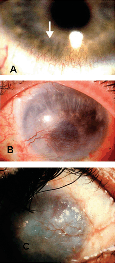
FIGURE 24.2. Common causes of corneal neovascularization. A: contact lens overwear, B: HSV keratitis and C: trachoma.
Inflammatory
Inflammatory causes of KNV vary from iatrogenic in surgery to graft rejection to aberrant immune diseases. Associated conditions discussed here include graft rejection, optimizing surgical technique, ocular cicatricial pemphigoid, Stevens-Johnson, and conjunctivitis.
KNV in graft rejection occurs in many cases of keratoplasty. One study showed the incidence of KNV in corneal buttons analyzed after keratoplasty at almost 20%.49 Many different risk factors have been identified to help minimize the probability of graft rejection. Graft rejection has been associated with host corneas with more than two quadrants of prior neovascularization, herpes simplex keratitis, uveitis, silicone oil keratopathy, previous rejected grafts, young host age, and having multiple surgical procedures during the graft placement.50 Embedding suture knots within the host stroma instead of in the graft, eyes with larger graft beds in the host, and active blepharitis are significantly associated with KNV.51 Other risks identified include atopic dermatitis, tear insufficiency, and short duration of graft storage.52 The risk factors do not significantly change if the transplant is a first graft or a regraft.53
Optimizing surgical technique can minimize the incidence of KNV in transplant cases. Risk factors associated with KNV include shorter distance from the limbus of both the graft tissue and the sutures.54 Use of nonmechanical trephination using an excimer laser may decrease the incidence of KNV within the 1st year.55
Donor tissue rejection occurs following the loss of immune privilege of the cornea. In animal studies, unless topical steroids were instituted sufficiently early, the course of neovascular response progressed to graft failure regardless of treatment.56 The most profound outgrowth of KNV seems to occur in the 1st week following transplant and requires vigilant follow-up and aggressive treatment to avoid progression.54 In mouse studies, short capillary growth began 1 day following cauterization and KNV peaked on days 4 and 5 and then regressed. Host immune cells access the corneal tissue and macrophages secrete VEGF,57 which acts to swing the balance of growth factors in favor of angiogenesis. If treatment attempts fails, the rejection progresses to graft failure and necessitates subsequent transplants.
Ocular cicatricial pemphigoid is an autoimmune subepithelial blistering disorder.58 The subepithelial bulla that forms can rupture and healing leads to scar formation. The immune reaction is a type II reaction with linear deposition of immunological factors in the epithelial basement membrane of the cornea. Most patients present later in the course of the disease. The prognosis is generally good when treatment is aimed at limiting inflammation and promoting healing. Systemic immunosuppressive therapy is the most important aspect of limiting disease progression. There are many extraocular manifestations of the disease that must be addressed as well.
Stevens-Johnson’s (erythema multiforme major) is a severe immune reaction to an unknown pathogen involving mucous membranes. Many different causes have been implicated including infections with viruses, bacteria, and fungi or the use of certain medications. When severe, the skin lesions can last up to 6 weeks.59 The eye is generally very dry and needs supportive lubrication. Systemic corticosteroids are used to treat and control acute disease.60 Healing may lead to scar formation and lifelong sequelae.
Causes of acute conjunctivitis are either allergic or infectious. Allergic conjunctivitis is divided into both acute and chronic diseases. Acute allergic conjunctivitis includes both seasonal allergic conjunctivitis (SAC) and perennial allergic conjunctivitis (PAC). SAC is only seasonal while PAC generally presents all year with seasonal exacerbations of symptoms. Chronic atopic keratoconjunctivitis is a chronic inflammatory disease of the cornea and conjunctiva. A majority of patients have comorbid asthma or eczema.61 Patients present with symptoms of intense itching, tearing, and blurred vision. Vernal keratoconjunctivitis is a recurrent inflammation that is associated with seasonal allergy, asthma, or eczema. Generally, the inflammation is worse in warmer months and remits in cooler months. For all of these conditions avoidance of the antigen stimulating the immune system is first-line therapy along with treatment of symptoms using artificial tears or cold compresses. Various medications can be used and selection depends on the patient’s tolerance and severity of the symptoms.
Infectious
Many infectious organisms are directly associated with KNV.62 Sixty percent of cases of keratoconjunctivitis have associated KNV.63 These infectious diseases can be difficult to eradicate. Among the viral illnesses, the two main infections associated with KNV are in the herpes family of viruses: herpes zoster and herpes simplex. Bacterial causes include pseudomonas, Chlamydia trachomatis, and syphilis. Common fungal infections include Candida, Fusarium, and Aspergillus. Onchocerca volvulus infection is also known to induce KNV. All of these infections are associated with KNV in the literature. Now we will look at some common surface diseases that involve infect.
Infections including viral, bacterial, fungal, and parasitic can all lead to ocular surface injury. The fear of any of these infections is the progression to ulcerative keratitis. The most common pathogens of bacterial conjunctivitis include Staphylococcus aureus, Streptococcus pneumoniae, and Haemophilus influenzae. Treatment consists of topical antibiotic drops or ointments. Infection is generally self-limiting, but treatment can enhance recovery. Viral conjunctivitis is very common with adenovirus identified as the leading pathogen. In immunocompetent hosts the infection is self-limiting within 5 to 15 days.64 Fungal and parasitic conjunctivitis are rare but require aggressive treatment to avoid corneal damage such as ulceration.
Neurotrophic keratopathy involves the loss of innervation to the cornea from the ophthalmic division of the trigeminal nerve. The absence of innervation to the cornea leads to decreased sensation and neurotrophic factors that normally support cell health.65 The damage occurs commonly with varicella zoster and herpes simplex keratitis (HSV-1 more common than HSV-2). The nerve damage can lead to ulceration, melting, and perforation. The goal of treatment is to avoid the development of persistent epithelial defects, which can progress to ulceration, melting, and perforation necessitating keratoplasty. Treatment of epithelial defects includes lubrication and elimination of all topical toxicity. Ulcer treatment involves application of neuronal mediators including substance P and IGF-1.66 Placing an amniotic membrane patch is used to decrease exposure and dryness, promoting wound healing.67
Traumatic Injury
In developed countries hypoxia-induced KNV is reported as a major cause of corneal loss leading to corneal transplant. As early as 1980, doctors recognized that wearing contact lenses for extended periods of time leads to corneal damage and KNV.68 In 1983, a warning to patients fit with extended wear contact lenses was made.69 These patients require frequent and diligent follow-up and should not be fitted with extended wear lenses if they are not able to manage their care properly. Many contact lenses causing a problem are too tight on patients. Any patient wearing contact lenses, regardless of the type needs follow-up eye exams to ensure proper use.70 More commonly, KNV associated with contact lens use is more superficial. The hypoxia caused by wearing contact lenses for extended periods of time leads to chronic inflammation and the stimulation of growth factors that initiate new deep vessel growth.71–73 Much of the vessel growth is superficial and generally responds well to medical treatment. Deeper vessel growth is more serious and can lead to hemorrhage, scarring, and loss of corneal transparency. Macrophages involved in the chronic inflammation play a role in the release of VEGF which stimulates the growth of new vessels. The presence of Macrophage Migration Inhibitory Factor (MIF), abundantly expressed in corneas with neovascularization, causes macrophages to infiltrate the cornea and secrete the angiogenic factors.74
KNV plays a role in wound repair in cases of trauma only when the angiogenic factors dominate the corneal environment.13 Alkali burns occurring in patients involved in automobile accidents with deployment of airbags can lead to KNV.75,76 This serious ocular injury is well treated if caught early following induction of the wound.
Alkali or acid injuries vary in the damage to the surface of the eye. The damage can vary from minor irritation of the eye to permanent visual loss. Alkali injuries are more common than acid injuries.77 The common chemicals causing alkali injuries include ammonia, lye, potassium hydroxide, and magnesium hydroxide. Causes of acid injuries include sulfuric, sulfurous, hydrofluoric, acetic, and hydrochloric acid. Irrigating the surface of the eye following the insult as soon as possible will limit the damage. Damage is directly related to the exposure time of the chemical on the cornea. Removal of particles causing continued irritation and debridement of necrotic area promotes wound healing.
RESEARCH MODELS OF CORNEAL NEOVASCULARIZATION
Research of KNV necessitates creating various animal models that are reproducible and economic ways to investigate angiogensis and corneal neovasculariazation and to create theraputic targets:
Growth Factor–Induced KNV
Implantation of Hydron pellets induces development of angiogenesis in the cornea upon exposure to doses of bFGF or VEGF and sucralfate.44
With the number of people in the United States alone affected by KNV, the importance of recognizing and treating this condition in the early stages cannot be overemphasized. Studies are increasing throughout academia to find ways of preventing and treating KNV. While much is now known about the causes and associations of diseases with KNV, much more research is needed to increase our ability to slow its incidence.
Suture-induced Corneal Neovascularization
This model was produced by placing a silk suture centrally in pigmented mice corneas causing limbal vasculature to develop. This inflamatory response was found to correlate with the increased expression of matrix metalloproteinase-2 (MMP-2), a proteolytic enzyme that regulates angiogenesis. MMP-2–deficient mice exhibited significant delay of neovascularization in comparison with wild-type mice.78
Alkali Burns
In this model animal corneas were subjected to alkali-burning with NaOH and the effects of altering the eicosanoid precursor pool via dietary manipulation was monitored. The results of this study suggested that neovascular responses and chronic inflammation occurring in the clinical setting of severe structural damage may be modulated by nutritional alteration of the eicosanoid precursor pool. A similar in vivo model of alkali burn was used to monitor the process of corneal neovascularization from the early to the regressive stages.79,80
Spontaneous Vascularization
In this model, corneas of corn1 and Pax6+/- mice, which are found to be deficient in sflt-1, exhibited spontaneously vascularized corneas. Adminstration of recombinant sflt-1 restored corneal avascularity in these mice.1 This model is similar to aniridia and spontaneous corneal neovascularization exhibited in humans with mutations in Pax6.
NEOVASCULARIZATION TREATMENT
Maintaining the avascular homeostatic state in the cornea has become a major focus of neovascularization research. Understanding the relationship between proangiogenic and antiangiogenic regulators has led to a number of antineovascularization pharmaceutical strategies. These therapeutic designs have targeted both the general inflammatory cascade and the inhibition of selective proangiogenic factors.
Steroids
In the clinic, steroids have remained the mainstay therapeutic strategy in the prevention of corneal neovascularization. Topical and subconjunctival administration of triamcinolone is known to inhibit corneal neovascularization.81,82 However, steroids carry with them serious side effects including glaucoma, cataract formation, and an inherent risk of infection.83 Likewise, corticosteroids sometimes are ineffective in inhibiting neovascularization, making alternative therapeutic strategies necessary.
VEGF Inhibitors
VEGF has been known to play a key role in angiogenesis in the human cornea. Comprised of five isoforms, VEGF promotes several steps within normal vascular growth including the induction of angiogenesis, endothelial cell proliferation, enhanced inflammatory response, proteolytic activities, and increased vascular permeability.36,84,85
Several cellular components within the human cornea have been found to produce VEGF when under inflammation or duress including corneal endothelial and epithelial cells, fibroblasts, macrophages, and limbal vessel vascular endothelial cells.42,86,87 VEGF antagonists disrupt these pathways, thus decreasing or even preventing corneal neovascularization.
Bevacizumab and Ranibizumab
Bevacizumab is a full-length, recombinant monoclonal antibody that binds all VEGF isoforms (Fig. 24.3). First approved in 2004 as an antineovascularization strategy in the treatment of colon cancer, bevacizumab effectively inhibits the VEGF-receptor interaction, and thus inhibiting vascular permeability, and capillary formation.88 A related compound, ranibizumab, is a high-affinity recombinant monoclonal antibody derived from the same parent murine antibody as bevacizumab. It is much smaller than the parent molecule and neutralizes all isoforms of VEGF-A.89
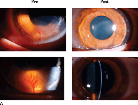
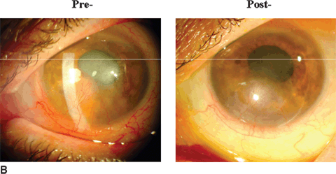
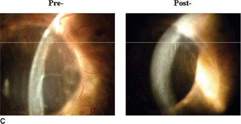
FIGURE 24.3. Bevacizumab therapy in corneal neovascularization showing pretreatment and posttreatment status (From author’s own practice).
Stay updated, free articles. Join our Telegram channel

Full access? Get Clinical Tree


