SYSTEMIC DISEASE AND DIABETES
Type 1 diabetes, also known as juvenile onset diabetes or insulin-dependent diabetes mellitus (IDDM), is a chronic condition of the pancreas in which there is little or no insulin, a hormone needed to convert sugar into energy. Although Type 1 diabetes can occur at any age, it appears during childhood or adolescence.
Currently, the American Diabetes Association recommends using the nomenclature that Type 1A diabetes is used for immune-mediated diabetes with its destruction of the islet beta cells of the pancreas,1,2 whereas non–immune-mediated diabetes with severe insulin deficiency is termed Type 1B diabetes.3
Recent epidemiological studies show that the incidence of diabetes in children is now comparable in adults.4 The international variation in incidence is now recognized. A child in Finland is almost 40 times more likely to develop Type 1 diabetes than a child in Japan, and the same child in Finland is 100 times more likely to get the disease than a child in the Zunyi region of China.5 The EuroDiab collaborative study, registry involving 44 countries in Europe, indicates an annual rate of increase in the incidence of Type 1 diabetes of 3.4%, with a larger increase in some central and eastern European countries.6 The largest rate of increase is seen in children aged 0 to 4. Type 1 diabetes is associated with other autoimmune conditions, the most common association is with thyroid disease.7 The Belgian Diabetes Registry indicates that the prevalence of thyroid peroxidase autoantibodies is 22% in patients with Type 1 diabetes. Approximately 1 in 10 patients with Type 1 diabetes express transglutaminase IgA autoantibodies and more than half of these patients have celiac disease on intestinal biopsy. Approximately 1 in 50 people with Type 1 diabetes have 21-hydroxylase autoantibodies and approximately 25% of these patients progress to Addison disease.8
GENETICS
Genetic susceptibility and environmental factors are thought to provide the fundamental element for disease and potential targets for both prediction and prevention of disease.9 More than 90% of patients who develop Type 1 diabetes have DR3, DQ2 or DR4, or DQ8 haplotypes, whereas fewer than 40% of normal controls have these haplotypes.9 The DAISY (Diabetes Autoimmunity Study in the Young) in Denver, Colorado, found that bovine milk ingestion, enteroviral infection, or vaccination does not contribute to risk of diabetes. It is unclear that early ingestion of cereal or gluten increases Type 1 risk and needs further studies. This may involve aberrant immune response to cereal antigens in an immature gut system in susceptible individuals.
Alpha interferon has been implicated as an important cytokine linking viruses to the initiation of Type 1 diabetes. There are case reports of patients developing anti-islet autoantibodies, who then later manifest Type 1 diabetes, autoimmune endocrine disorders. Thus, neutralizing this cytokine, alpha interferon may potentially prevent the disease.
Compounds such as poly IC (a viral RNA mimicker) can induce alpha interferon which then can generate insulitis (selective beta cell destruction) and diabetes in animal models. Thus, there is a link between induction of diabetes and alpha interferon.10
PATHOGENESIS OF TYPE 1 DIABETES
Role of autoantibodies in the actual pathogenesis of Type 1 diabetes has not been established in humans. In general, Type 1 diabetes is considered a T-cell mediated disease. Examination of islet tissue obtained from pancreatic biopsy from patients with recent onset of Type 1 diabetes confirms insulitis, with the presence of an infiltrate composed of CD4 and CD8 T lymphocytes, B lymphocytes, and macrophages, suggesting that these cells have a role in the destruction of the beta cells.11 Therapies directed against T cells have been shown to halt the disease process and prevent recurrent beta cell destruction after islet cell transplantation. Less is known about the nature and function of these T cells. The cause of the loss of tolerance to islet autoantigens is unknown. We understand neither why the immune system fails to suppress autoreactivity nor which autoantigens are critically involved in the initiation or progression of the disease. Some T cells are pathogenic, but others can regulate the disease process and thus form new targets for immunointervention.12
Examination of islet tissue obtained from pancreatic biopsy from patients with recent onset of Type 1 diabetes shows insulitis, with the presence of infiltrate composed of CD4 and CD8 T lymphocytes, B lymphocytes, and macrophages, suggesting that these cells have a role in the destruction of beta cells.11
Studies in mice show that anti-CD3 treatment prevented diabetes, thus subsequently spawning a new trial using humanized anti-CD3 antibody in patients with new onset Type 1 diabetes.13
To date, no treatment has been shown to prevent Type 1 diabetes in humans. More than 100 different treatments prevent Type 1 diabetes in the Nonobese diabetic (NOD) mouse model.14 In the United States, the Diabetes Prevention Trial began in 1994 with the goal of determining whether antigen-based treatment with insulin (oral and parenteral insulin in relatives of high to moderate risk) would prevent or delay diabetes. The European Nicotinamide Diabetes Intervention Trial found no difference in the protection from diabetes when participants were assigned to either oral nicotinamide or placebo treatment.1
Thus, insulin is the main treatment of Type 1 Diabetes. The Diabetes Control and Complications Trial (DCCT) demonstrates the importance of strict metabolic control in delaying and preventing complications.15
The introduction of rapidly absorbed insulin analogs has reduced variability of insulin absorption and allows insulin administration in young children after meals. The insulin, glargine, functions as a very long acting insulin (peakless basal insulin).16 Combinations of engineered very long acting insulin and rapid acting insulin can provide control and convenience similar to that obtained with insulin pumps.
The use of metformin alongside insulin has increased in patients with Type 1 diabetes. Recent studies have suggested that metformin might benefit Type 1 diabetes patients who are overweight, are receiving large doses of insulin, or have an HbA1c >8%.17 The coexistence of insulin resistance in patients with Type 1 diabetes is a new area of interest. Islet cell transplantation is a consideration for the limited but important subset of patients with recurrent severe hypoglycemia not responsive to medical management.18 However, inability to control autoimmunity and a lack of donor organs limit the application of islet transplantation.19
PATHOGENESIS OF TYPE 2 DIABETES MELLITUS
Type 2 diabetes is a result of insulin resistance, impaired beta cell insulin secretion. Excessive visceral adipose tissue in obesity contributes to these metabolic abnormalities. Coronary artery disease (CAD), a life threatening consequence of atherosclerosis, is the most common vascular consequence of insulin resistance.19 The prevalence of CAD in Americans with Type 2 diabetes is two to four times greater than that in a nondiabetic patient population. The manifestations of CAD such as angina pectoris, myocardial infarction, and sudden death are twice as common in patients with Type 2 diabetes as in nondiabetic patients. The incidence of these manifestations is six times more common in diabetics versus nondiabetics.20
Type 2 Diabetes, Insulin Resistance and Obesity
Normal glucose metabolism requires the coordination of glucose production and glucose uptake. Hepatic glycogenolysis and gluconeogenesis become balanced with glucose uptake into skeletal muscle and adipose tissue in the fasting and postprandial states under the direction of neural influences and circulating hormones, insulin, glucagons, and incretins. There is a close association between obesity and insulin resistance. Intra-abdominal fat or adipose tissue is an active secretory organ. They secrete adipokines such as leptin and adiponectin. Leptin causes decrease insulin-mediated glucose uptake. Adiponectin increases insulin release and plays a role in anti-inflammatory effects in the vasculature. In obesity, leptin is increased and adiponectin is decreased.21
Adipose tissue releases free fatty acids (FFAs), cytokines, such as tumor necrosis factor–alpha (TNF-alpha), signaling proteins (angiotensin II), hormones, complement factors, and regulators of atherothrombosis (e.g., plasminogen activator inhibitor-1). Changes in the circulating levels of these substances play a role in the insulin resistant state of Type 2 diabetic patients who are overweight. In addition, these metabolites exert adverse effects in vascular endothelial and smooth muscle cells. TNF-alpha is elevated in adipose tissue in experimental models of obesity, suggesting that TNF-alpha plays a role in the systemic insulin resistance of Type 2 diabetes.22 Both obesity and endothelial dysfunction are linked to Type 2 diabetes in the early inflammatory stages of atherogenesis. It is the endothelial cell dysfunction that mediates the two major long-term complications, cardiovascular disease and diabetic retinopathy.22
The increase in FFA in obesity leads to higher levels of fatty acid molecules within skeletal muscle and liver cells, which in turn cause insulin resistance. This is mediated by the activation of the signaling kinase, protein kinase C. Some of protein kinase C isoforms oppose the insulin action cascade.23
In response to insulin resistance associated with visceral obesity, pancreatic beta cell insulin secretion is augmented to maintain normal glucose level. Consequently, most obese patients are insulin resistant and hyperinsulinemic at the same time. These overweight Type 2 diabetics have impaired fasting glucose and impaired glucose tolerance.
LIFESTYLE MODIFICATIONS OF TYPE 1 DIABETES
Type 1 diabetic patients can be educated on estimating the carbohydrate content in their diets so that they can accurately match insulin administration during meals to carbohydrate intake by using insulin to carbohydrate ratios. Methods available for estimating carbohydrate intake are experience-based estimation, the exchange system, and carbohydrate counting. For those patients who use fixed daily doses of insulin, carbohydrate intake should be consistent in terms of the time of day and amount of food consumed each day.24,25
For planned exercise, patients should reduce their premeal bolus based on their level of anticipated activity to prevent hypoglycemia. Moderately intense exercise has been shown to increase glucose use by 2 to 3 mg/kg/min above usual requirements. Thus, a 70-kg person would need an additional 10 to 15 g of carbohydrate during moderate physical activity. More intense exercise would require the intake of more carbohydrates. As a result, unplanned physical activity usually requires supplemental carbohydrate intake to prevent hypoglycemia.24
LIFESTYLE MODIFICATION FOR TYPE 2 DIABETES
For patients with Type 2 diabetes, lifestyle modifications would be aimed at improving glycemia and dyslipidemia and reducing blood pressure and overall cardiovascular risk. The goal would be reduction of the intake of saturated and trans-fatty acids, cholesterol, and sodium and increase in physical activity.
A diet rich in fruits, vegetables, whole grains, nuts, and fiber would improve dyslipidemia and lower blood pressure and decrease cardiovascular risk. Patients should be encouraged to limit carbohydrate consumption by calculating the carbohydrate content of each meal based on grams of carbohydrate consumed or switching servings of food based on carbohydrate equivalents from exchange lists.26
For Type 2 diabetic patients using fixed doses of insulin, their carbohydrate intake should be consistent with the time of day and amount consumed on a daily basis.
Gestational Diabetes
Medical nutrition therapy, at the time of diagnosis, is aimed at food choices that allow for adequate energy intake to provide appropriate weight gain, normoglycemia, and absence of ketones. Weight loss during pregnancy is not recommended. However, obese or overweight women may benefit from modest exercise and carbohydrate restriction.
There are stringent glycemic targets during pregnancy; thus, frequent glucose monitoring and the maintenance of a dietary log will provide valuable information for possible meal plan adjustments.
Benefits of Exercise
The ADA recommends at least 150 min/week of moderate aerobic exercise (50% to 70% of maximal heart rate) and resistance exercise that targets all major muscle groups three times per week.27 One exercise session can cause an acute decrease of plasma glucose levels in Type 2 diabetes patients. Postexercise enhancement of glucose metabolism, possibility for increased insulin sensitivity in muscle tissue may last for hours, even days. It is thought that more effective glycemic control may result from the additive effect of the exercise sessions and not from increased fitness.28
RISK FACTORS FOR DEVELOPMENT OF DIABETIC RETINOPATHY
Duration of Diabetes Mellitus
The Wisconsin Epidemiology Study of Diabetic Retinopathy (WESDR) found that 3.6% of patients with Type 1 diabetes whose disease occurred prior to age 30 are legally blind compared with 1.6% of patients with Type 2 diabetes whose disease occurred after age 30.29
Glycemic Control
Two studies, DCCT15 for Type 1 diabetes and the United Kingdom Prospective Diabetes Study (UKPDS)30 for Type 2 diabetes, demonstrate the efficacy of good glycemic control in preventing diabetic retinopathy. In the DCCT, the Type 1 diabetic patients, after a mean follow-up of 6.5 years, the groups of patients treated with intensive insulin therapy (multiple insulin injections during each day) have a mean glycosylated hemoglobin of 7.2% and have a reduction of progression of diabetic retinopathy or delayed development of diabetic retinopathy by 27% versus patients treated in a conventional manner (nonintensive insulin therapy). In the Type 2 diabetic patients in the UKPDS, intensive glycemic control reduces microvascular complications including diabetic retinopathy by 25% as compared to conventional therapy. Every percentage point of decrease of the HbA1c is associated with 35% risk reduction. Thus, the effort to achieve good glycemic control at the time of diagnosis is important.
Elevated Blood Pressure
In the WESDR, higher diastolic blood pressure is associated with increased progression of diabetic retinopathy and macular edema.29 In the UKPDS, Type 2 diabetics with blood pressure of <150/85 have a 34% reduction in progression of diabetic retinopathy and a 47% decreased risk of deterioration in visual acuity compared with patients with blood pressure >185/105.30
Hypertension impairs autoregulation of retinal blood flow in diabetics compared with nondiabetics. It enhances endothelial damage and expression of vascular endothelial growth factor (VEGF) and its receptors in poorly controlled diabetes.
Hyperlipidemia
The WESDR shows that retinal hard exudates development is 50% more likely in patients with elevated total cholesterol or triglycerides and a significant risk factor for moderate visual loss.31 The Early Treatment Diabetic Retinopathy Study shows an association between elevated baseline LDL and retinal hard exudates.32
Diabetic Nephropathy and Diabetic Retinopathy
Microalbuminuria is associated with diabetic retinopathy. In patients with known renal disease, their risk of diabetic retinopathy is increased and their diabetic retinopathy is accelerated as compared to those diabetic patients without renal disease.33
TREATMENT OF TYPE 2 DIABETES
The therapeutic goal of diabetes management is to achieve a HbA1c close to the normal range (4% to 6%), at least <6.5%. This requires the fasting blood glucose level of <110 mg/ dL and 2 hour postprandial glucose of <140 mg/dL. These biochemical targets confer benefits in the reduction of diabetic complications, for example, nephropathy, neuropathy, retinopathy, and cardiovascular morbidity and mortality. Unfortunately, Type 2 diabetes is a chronic and progressive disease which requires lifelong pharmacotherapy in addition to diet and exercise.
Oral Antiglycemics
Biguanides, Sulfonylureas, Thiazolidinediones (TZDs)34
Biguanides
Biguanides (Metformin, Glucophage, Glucophage XR, Glumetza) work by increasing insulin sensitivity in the liver by inhibiting hepatic gluconeogenesis, thereby reducing hepatic glucose production.34
Sulfonylureas
Sulfonylureas are the most widely prescribed drugs for Type 2 diabetes.34 They are chlorpropamide (Diabinase), glipizide (Glucotrol, Glucotrol XL), glyburide (DiaBeta, Micronase). The mechanism of action of sulfonylureas involves the augmentation of insulin release from pancreatic beta cells and the potentiation of action on its target cells.
Nonsulfonylurea Secretagogues
Repaglinide (Pradin), a benzoic acid derivative, and nateglinide (Starlix) are distinguished from the sulfonylureas because the former has short half-lives and does not contain the sulfonic acid moiety.36 Thus, these are useful in patients with sulfa allergies.
Thiazolidinediones
TZDs are selective agonists for the nuclear receptor peroxisome proliferator-activated receptor–gamma (PPAR).34 The role of PPARs in humans involves the regulation of gene transcription by two mechanisms: (a) transactivation and (b) transrepression. Transactivation is where activation occurs in the cytoplasm: PPARs bind to a retinoid X receptor/retinoic acid receptor. The formation of this complex allows PPARs to be transported into the nucleus of an atom and bind to the promoter region of the regulated gene. The second mechanism is transrepression which is a DNA-independent pathway. This pathway interferes with other transcription pathways and may be responsible for the anti-inflammatory actions of TZDs.35
These mechanisms of action of TZDs create an increased insulin-stimulated glucose uptake by skeletal muscle cells, decreased hepatic glucose production, decreased lipolysis, and enhanced adipocyte differentiation.
Alpha-glucosidase Inhibitors
Alpha-glucosidase inhibitors delay the absorption of complex carbohydrates and inhibit postprandial glucose peaks and consequently lower postprandial insulin levels. In the United States, there are two alpha-glucosidase inhibitors: acarbose (Precose) and miglitol (Glyset).34
Dipeptidyl Peptidase IV Inhibitors
Dipeptidyl peptidase IV (DPP-IV) inhibitors are exenatide (Byetta) and sitagliptin (Januvia). As a background, glucagon-like peptide-1 (GLP-1) is an insulinotropic hormone secreted by L cells of the small intestine.34 GLP-1 has several important biologic actions including the stimulation of insulin secretion in a glucose-dependent manner, inhibition of gastric emptying, and suppression of glucagon secretion and central anorexic activity. Patients with Type 2 diabetes exhibit reduced levels of active GLP-1 along with an impaired GLP-1 response to a glucose load. Endogenous GLP-1 is inactivated by the enzyme DPP-IV. DPP-IV inhibitors block this enzyme and thereby increase GLP-1.
ANATOMY OF THE RETINA
The retina is the innermost of the external layer of the eye, the sclera. The retina and choroid are the inner lining of the sclera. The retina is 350 μm thick in the equatorial part and 190 to 220 μm thick in the fovea and macula regions. This section concentrates on the retina, as this part of the anatomy is affected by diabetic retinopathy.
The central retina is cone-dominated and the peripheral retina is rod-dominated. There are 7 million cones and 120 million rods. At the center of the macula, there is a foveal pit where the cones are arranged in a hexagonal mosaic, which can be evaluated with fluorescein angiography as the foveal avascular zone or in electroretinography as the cone response in the 30 Hz flicker.
The Ten Layers of the Retina (Fig. 2-1)
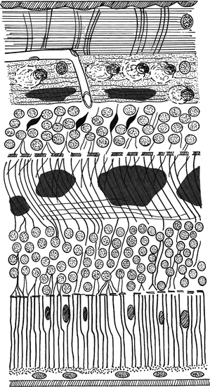
FIGURE 2-1. Diagram of the retina.
Stay updated, free articles. Join our Telegram channel

Full access? Get Clinical Tree


