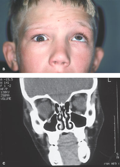
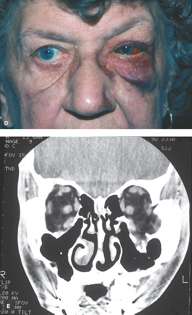
FIGURE 15-1. White-eyed blowout fracture. A and B. A 9-year-old boy with a history of being hit with an elbow 1 day prior. He has pain, worse with eye movement; double vision; and has had nausea and some vomiting. The photograph shows his lack of ecchymosis and swelling as well as severe restriction of upgaze. White-eyed blowout fracture. C. CT scan shows a small trap-door floor fracture with tissue entrapment. This fracture needs to be repaired promptly as the entrapped muscle may become ischemic. Orbital floor fracture. D. This 72-year-old woman fell and hit her eye on her bedpost. She has full motility but significant swelling, ecchymosis, and infraorbital hypesthesia. E. CT scan shows a large orbital floor fracture. The patient is at risk for development of enophthalmos from the large fracture.
MEDIAL WALL FRACTURE
Medial wall fractures can be isolated fractures of the medial wall only or they can be a part of larger fractures involving the nose and sinuses. Isolated fractures are treated much like orbital floor fractures (Fig. 15-2). Larger fractures usually involve a multidiscipline approach to the repair of the fractures.
Epidemiology and Etiology
• Age: Most common in second through fourth decades
• Gender: More common in males
• Etiology: Direct fractures occur from striking a solid object. Indirect (blow-out) fractures occur in association with and by similar mechanisms as orbital floor fractures.
History
• Trauma history is variable.
• Symptoms include diplopia and cosmetic deformities depending on the extent of the nasal fractures.
Examination
• Medial rectus entrapment with diplopia and eventual enophthalmos are the two ocular manifestations that may occur.
• Direct fractures often have significant damage to the nasal bridge and medial orbit. The nasal bridge may be depressed with telecanthus.
• Other findings that can occur include epistaxis, orbital hematoma, cerebral spinal fluid rhinorrhea, and damage to the lacrimal drainage system.
Imaging
• CT scanning will show the extent of the fracture and assist with potential planning of the repair.
• MRI does not image bone well and should not be used initially after trauma.
Special Considerations
• Medial wall fractures with entrapment of the medial rectus need to be repaired sooner than floor fractures (within 1 week) if possible.
Treatment
• If isolated, medial wall fractures often do not need repair.
• Medial rectus entrapment with diplopia is one indication for repair.
• If the fracture is large, enophthalmos can develop and require surgery to build up the orbit. Implants are sometimes placed.
• Larger fractures involving the nasal bridge and medial orbit require repair and plating, usually in conjunction with an otolaryngology specialist.
Prognosis
• Good. Larger fractures may require multiple surgeries and revisions.
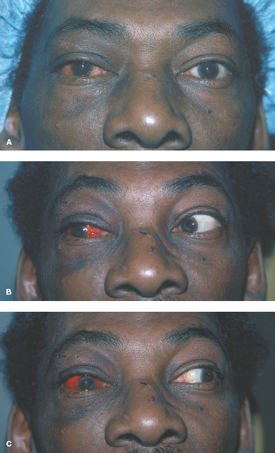
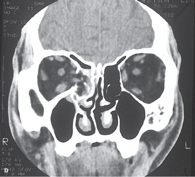
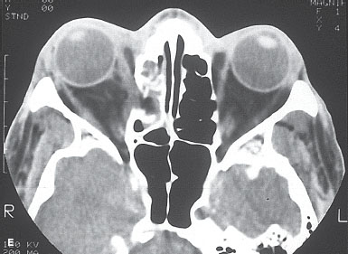
FIGURE 15-2. Medial wall fracture. A to C. A 55-year-old man struck in the face with an unknown object presented with horizontal diplopia. Motility is restricted in the right eye in both adduction and abduction. Medial wall fracture. D and E. CT scans show a medial wall orbital fracture with the medial rectus muscle pulled into the fracture.
ORBITAL ROOF FRACTURE
Orbital roof fractures (Fig. 15-3) are rare fractures that need to be recognized because of the potential for life-threatening neurologic sequelae. There may just be a small fracture with no neurologic problems or there may be significant intracranial air and bleeding. Treatment is in conjunction with neurosurgery.
Epidemiology and Etiology
• Age: Most common in second through fourth decades
• Gender: More common in males
• Etiology: Blunt trauma or direct injury by a thin object that goes above the globe under the superior orbital rim. An isolated roof fracture is rare.
History
• Trauma history will often suggest high-energy forces that caused the injury.
• These include hydraulic air hoses, a blunt object with high velocity, and so forth.
Examination
• Poor upgaze, supraorbital hypesthesia, and more swelling superiorly than inferiorly suggest an orbital roof fracture.
• Entrapment of the superior rectus or superior oblique muscle is extremely rare.
Imaging
• CT scanning will show the fracture usually just inside the orbital rim.
• MRI does not image bone well and should not be used initially after trauma.
• MRI can be of value to evaluate intracranial injury.
Special Considerations
• Important to consult neurosurgery for the potential of CNS complications with a roof fracture
Treatment
• Repair of a roof fracture is usually done for neurologic reasons rather than ocular.
• Any plating and repair is done via craniotomy.
• Nondisplaced fractures do not require repair.
Prognosis
• Variable depending on the extent of associated CNS injuries
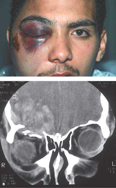
FIGURE 15-3. Orbital roof fracture. A. The patient was struck in the eye with a hydraulic air hose. Examination shows significant swelling, with decreased upgaze and supraorbital hypesthesia. B. CT scan shows an orbital roof fracture with intracranial hemorrhage.
ZYGOMATIC FRACTURE
Stay updated, free articles. Join our Telegram channel

Full access? Get Clinical Tree


