FIGURE 14.1 RetCam color photograph of type 1 ROP (stage 3, zone II, plus disease). Left eye temporal fundus with neovascular ridge, torturous arteries, and minimally dilated veins.
The RetCam is an integral tool for the documentation and management of fundus pathology in children with retinoblastoma (2). As with ROP, digital imaging of retinoblastoma lesions before and after treatment is superior to fundus drawing or relying on memory alone. An example of a pediatric patient with retinoblastoma is shown in Figure 14.2.
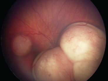
FIGURE 14.2 RetCam color photograph of retinoblastoma. Right eye with a medium-sized white retinoblastoma in the superotemporal macula and a large multilobulated retinal mass adjacent to the optic nerve in the inferonasal retina. (Courtesy of David H. Abramson.)
It is also important to document retinal findings when evaluating children for accidental and nonaccidental trauma. Figure 14.3 shows the fundus of a 13-month-old child who concurrently suffered from an acute subdural hematoma in the setting of a seizure.
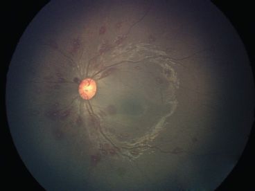
FIGURE 14.3 RetCam color photograph of a 13-month-old admitted for seizures and intracranial hemorrhage from accidental trauma. Left eye with multiple white-centered flame-shaped intraretinal hemorrhages centered around the optic nerve.
RetCam Review Software
The RetCam has proprietary software for image manipulation and review. The interface is user friendly and allows for image transfer to portable media.
One of the most important features of the RetCam software is the Compare function, whereby images from different dates can be reviewed side by side. This feature facilitates longitudinal review of fundus changes. A note entry feature includes input of additional data, which allows database search by date, physician, disease, and other parameters.
RetCam Imaging
All versions of the RetCam require the patient to be recumbent and cooperative, making it ideal in infants who can be swaddled and held down. When using the camera in older children, an examination under anesthesia is generally necessary.
After instilling topical anesthesia and placing an eyelid speculum, a viscous coupling agent is applied to the ocular surface, and the lens nosepiece is placed directly on the cornea with minimal pressure. The handpiece cord is directed superiorly (12 o’clock) by convention to preserve orientation of fundus images (i.e., macula to the left of the optic nerve for images of the right eye, and the converse for images of the left eye). Imaging should be performed with minimal pressure from the weight of the handpiece on the globe; otherwise vascular features may be altered as a consequence of the increase in ocular pressure. The computer screen is positioned to face the photographer during image acquisition. Adjusting the light intensity, focusing, and image capture can be performed with a foot pedal or by hand on the keyboard console. The latter method requires an assistant. The midperiphery is imaged with relative ease. Scleral indentation is useful to capture pathology in the far periphery.
RetCam Fluorescein Angiography
Performing FA with the RetCam requires three adjustments to the equipment. The light source, lens filter, and software each needs to be switched over to FA function. The fluorescein dye dosage is determined by weight (5 to 7.5 mg/kg body weight delivered intravenously). In premature infants in the perinatal period, the intravenous dose can be as low as 0.1 mg/kg and still provide angiographic images.
RetCam FA is useful in demonstrating otherwise subtle or inapparent vascular pathology. In ROP, flat areas of neovascularization are more easily detected (3), as are nonperfusion, vascular anomalies, and leakage in areas of vascularized retina (4).
RetCam Limitations
The optics of the camera demand clear media and good dilation for quality images. Small pupil size, media haze, and dark fundus pigmentation all impact on the ability to obtain quality images. Even relatively little nuclear sclerosis, as seen in patients only in their teens, may reduce contrast to a prohibitive degree when imaging low-contrast fundus pathology. The RetCam is also of limited utility in an outpatient setting because it requires a recumbent and still patient.
The Optos captures up to 200 degrees of fundus in one image as opposed to traditional cameras, which capture 30 or 50 degrees in a single image. Each picture is 3,900 × 3,072 pixels. The camera is a scanning laser ophthalmoscope, enabling visualization of the retina through moderate media opacities, such as cataract and vitreous hemorrhage. The technology uses an elliptical mirror and creates a virtual focal point inside the patient’s eye to image the far periphery. Besides wide-angle color fundus photographs, red-free, infrared, autofluorescence, and FA images can be taken with the Optos camera.
Optos was developed in Scotland specifically for the detection of peripheral retinal pathology through an undilated pupil in children. Figure 14.4 shows the fundus of a myopic child after retinopexy for an atrophic hole and lattice. Interestingly, however, a study of 205 eyes with 18 retinal holes/tears found that the sensitivity of Optos for detecting such lesions was only 33% when compared to a scleral depressed exam by a retinal specialist as the gold standard (5). Arguably, the major utility of the Optos camera to retinal specialists is the FA function.
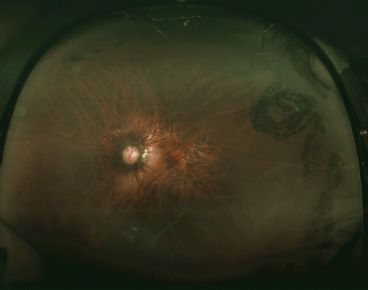
FIGURE 14.4 Optos color photograph of a 12-year-old myopic boy. Left eye with laser retinopexy scars superotemporally and temporally.
Optos Software
The viewing system that Optos uses is called Vantage V2. Color fundus images using Optos are taken with two channels only (633 nm red and 532 nm green). The absence of blue channel images results in a greendominant color aberration, which appears unnatural. Images can be processed either manually or automatically to appear more lifelike. Other software features include basic image processing capabilities such as brightness, contrast, zoom, and gamma manipulation. An interactive three-dimensional reconstruction feature called 3-D Wrap is useful for patient orientation and education.
Optos Imaging
The Optos is a nonmydriatic camera. The machine is large and not intended to be moved. The design of the legs and base makes the Optos wheelchair accessible. In the pediatric population, fluorescein dye is dosed by weight (5 to 7.5 mg/kg) and given intravenously. Because of the time it takes to console the child after an injection, later images are usually captured better. In theory, if early images are of interest, an intravenous line can be placed to allow the child time to calm down and position for the procedure. For some photographers, it is easier to capture images with an Optos machine compared to traditional fundus camera because the scanning laser ophthalmoscope technology is faster, it uses a less intense light source, and there is no ocular steering required to image the periphery.
There is no lower age limit for using Optos, and children as young as 5 years old may tolerate the procedure well. Patients can be positioned in several different ways, and the machine is adjustable vertically by 12 inches. Younger children can sit on a parent’s lap if desired, and older children can sit on their own. If their feet do not touch the ground, a step stool should be used for stability. Children approximately 3′10″?to 4′10″ tall have the option to stand up during the procedure instead of being seated.
Optos Fluorescein Angiography
In 2005, Optos FA became commercially available, and it has been most commonly used to evaluate diabetic retinopathy, retinal vein occlusions, sickle cell retinopathy, and other peripheral retinal pathology in adults (6–8). Its advantages over traditional FA include facile detection of peripheral pathology such as nonperfusion, neovascularization, and vascular leakage.
Optos Limitations
Two major disadvantages of the Optos are eyelash artifact and peripheral distortion. Eyelash artifact is typically more severe and noticeable inferiorly, because the superior eyelashes are longer and block the inferior fundus. When eyelash artifact is not confluent, information can be extracted from between the individual eyelashes. Significant blockage, defined as blockage located posterior to the equator, occurs in approximately 30% of pictures (Tsui, unpublished data), but this is somewhat variable depending on the experience of the photographer and patient’s eye. Adjustments to the procedure such as using a speculum or taping the eyelashes meet with limited success because of drying of the ocular surface.
Peripheral distortion comes from the fact that the Optos projects the round image of the retina onto a flat surface of an image. The problem is similar to the edges of a map being distorted when compared to a globe. Peripheral distortion with Optos appears as a mild ring of haze around the outer edges of the image. It is reportedly resolvable by capturing multiple images with ocular steering and then using a customized image processing method to montage the images (9).
EXAMPLES OF PEDIATRIC RETINAL DISEASES
The wide-field imaging techniques above can be useful to evaluate pediatric retinal diseases with macular or peripheral findings. Below are examples of diseases in which peripheral findings are important and wide-field imaging is of benefit.
Congenital Optic Nerve Anomalies
Congenital optic nerve anomalies vary from mild to severe in causing visual impairment and can involve the retina and choroid. They are oftentimes but not always autosomal dominant in inheritance. Examples include variants of morning glory (Fig. 14.5), optic nerve pits (Fig. 14.6), and retinal colobomas (Fig. 14.7). (Please also refer to Chapters 16 and 54 and the Atlas, Optic Nerve.)
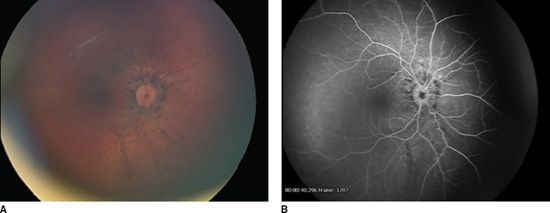
FIGURE 14.5 RetCam imaging of a 14-month-old girl with a congenital optic nerve anomaly. A: Color fundus photograph showing peripapillary pigmentation with linear atrophy radiating from the optic nerve. B: FA showing staining of the above and no choroidal neovascularization.
Stay updated, free articles. Join our Telegram channel

Full access? Get Clinical Tree


