Hyperglycemia affects 8% of all pregnancies in America.1 Preexisting diabetes affects 1% of all pregnancies. The hyperglycemia exists with Type 1 diabetes or gestational Type 2 diabetes. During the first trimester of pregnancy, hyperglycemia is associated with higher risk of spontaneous abortion and fetal malformations. In the third trimester, hyperglycemia increases the risk of macrosomia and metabolic complications at birth, increased incidence of caesarean section, and increased admission rates to newborn intensive care units. Risk of these birth complications is proportional to the degree of maternal hyperglycemia (Table 11-1).
TABLE 11-1 Risk Factors for Type 1 Pregnant Diabetic Patients
Acceleration of diabetic retinopathy
1. Elevated HbA1c
2. Longer duration of diabetes
3. Higher systolic blood pressure
Pathophysiology
Maternal Fetal Metabolism in Normal Pregnancy
With each meal, the pregnant woman experiences a complex set of maternal hormonal actions: rise in blood glucose, followed by secondary secretion of pancreatic insulin, glucagons, somatomedins, and adrenal catecholamines. Pregnant women develop hypoglycemia between meals and during sleep because the fetus continues to draw glucose across the placenta from the maternal bloodstream even during periods of fasting. Interprandial hypoglycemia increases as the pregnancy progresses and the glucose demand of the fetus increases.
Placental steroid and peptide hormones (estrogens, progesterone, and chorionic somatomammotropin) rise throughout the second and third trimesters. In the later stages of pregnancy, the levels of these hormones rise, creating increasing tissue insulin resistance despite increased insulin secretion, a response to the increased feeding. The mean insulin levels are 50% higher in the third trimester compared to the nonpregnant state2 (Tables 11-2 and 11-3).
TABLE 11-2 The Risk Factors for GDM
1. Previous history of GDM, prediabetes, impaired glucose tolerance, and impaired fasting glycemia.
2. Family history with first-degree relative with Type 2 diabetes
3. Maternal age (women over age 35)
4. Ethnic background: African Americans, Afro-Caribbeans, Native Americans, Hispanics, Asians, Pacific Islanders, and South Asian Indians of the India subcontinent
5. Overweight and obesity
6. Previous pregnancy which resulted in a child with high birth weight ≥4,000 g
7. Smokers
8. Polycystic ovarian syndrome
TABLE 11-3 Risk Factors for Pregnant Diabetic Patients
Acceleration of diabetic retinopathy
Lipid profiles (cholesterol and triglycerides)
Glycemic control over the course of her 9-month gestation
Level of diabetic retinopathy in the first trimester will progress into the third trimester. Hypertension status
Kidney status.
The Diabetic Pregnancy
If the maternal pancreatic insulin response cannot meet the needs of the increased feeding, then maternal and fetal hyperglycemia is the end result. The recurrent postprandial hyperglycemia is a cause for the increased growth of the fetus, leading to macrosomia. The episodes of maternal and fetal hyperglycemia are accompanied by fetal hyperinsulinemia. Fetal hyperinsulinemia promotes excessive nutrient storage, resulting in macrosomia. The fetal conversion of excess glucose into fat causes a depletion of fetal oxygen levels. The relative fetal hypoxia causes surges in adrenal catecholamines which in turn causes hypertension, cardiac hypertrophy, erythropoietin stimulation, red cell hyperplasia, and increased hematocrit. Polycythemia with a hematocrit over 65% occurs in 5% to 10% of newborns with diabetic mothers. This finding appears to be related to the level of glycemic control and level of fetal hypoxia. Elevated hematocrit in the neonate leads to poor circulation and hyperbilirubinemia.3
In the United States, 23.6 million people or approximately 7% of the population have diabetes. Another 6 to 7 million may be undiagnosed. An additional 57 million Americans are prediabetic and will become diabetic.4 While 3% to 10% of all pregnancies are complicated by diabetes, 90% of the diabetic pregnancies are due to gestational diabetes. The remaining 8% of the diabetic pregnancies are due to preexisting, insulin resistant or Type 2 diabetes.3
Diabetic Retinopathy in Pregnancy
The National Eye Institute (NEI), a division of the National Institutes of Health, states that “…during pregnancy, diabetic retinopathy may be a problem for women with diabetes. To protect vision, every pregnant woman with diabetes should have a comprehensive dilated eye exam as soon as possible.” In addition, the NEI also recommends that “your doctor may recommend additional exams during your pregnancy.”5 Thus, the official recommendations of National Institute of Health, an agency of the U.S. Department of Health and Human Services, suggest that diabetic pregnant women have dilated retinal exams. Studies involving diabetic retinopathy and pregnancy have shown that in Type 1 diabetic patients, elevated HbA1c, longer duration of diabetes, and higher systolic blood pressure were correlated with the progression of retinopathy5–7 (Table 11-4).
TABLE 11-4 Diabetic Pregnancy Guidelines
American association of clinical endocrinologists
HbA1c < 6%
FBS range: 60 and 90 mg/dL
1 hr postprandial plasma glucose ≤120 mg/dL.
Source: Moloney JB, Drury MI. The effect of pregnancy on the natural course of diabetic retinopathy. Am J Ophthalmol. 1982;93:745–756.
Gestational Diabetes Mellitus
Epidemiology
Gestational Diabetes Mellitus (GDM) is a condition in which women without previously diagnosed diabetes exhibit high blood glucose levels during pregnancy. Gestational diabetes has few symptoms, and is commonly diagnosed by fasting blood sugar (FBS) screening during pregnancy. Gestational diabetes affects 3% to 10% of pregnancies.
Factors in the apparent increase in GDM are varied. Prevalence of obesity in youth is thought to be due to low levels of physical activity and high levels of caloric intake. There is improved survival of female infants whose birth weights are at the extremes of the normal range. As adults, these infants will have altered insulin action and/or insulin secretory capacity that may predispose them to the development of GDM. Birth weight history may be helpful in the risk assessment of GDM.8
No specific cause has been identified, but it is believed that the hormones produced during pregnancy increase insulin resistance resulting in impaired glucose tolerance. The action of insulin as it binds to the insulin receptor is thought to be affected by pregnancy hormones.2 The interference of insulin receptor binding probably occurs at the level of the cells signaling pathway at the insulin receptor. Since insulin promotes the entry of glucose into the cell, insulin resistance prevents glucose from entering the cell and as a result, glucose remains in the blood stream. Plasma glucose levels rise and more insulin is needed to overcome this resistance. In a normal pregnancy, insulin is produced at 1.5 to 2.5 times the normal nonpregnant state.
Insulin resistance is a normal occurrence in the second trimester of pregnancy, which then progresses to levels seen in nonpregnant patients with Type 2 diabetes in the third trimester. Women with GDM have an insulin resistance that is not compensated with increased production of insulin in the beta cells of the pancreas.2 Placental hormones and to a lesser extent, increased fat deposits during pregnancy seem to mediate insulin resistance during pregnancy. Cortisol and progesterone are the main culprits, but human placental lactogen, prolactin, and estradiol are contributing factors to insulin resistance during pregnancy.2
It is postulated that there is a chronic beta-cell defect that is present in the prepregnancy and postpregnancy states and is accompanied by increasing blood glucose concentration. Thus, at the time of the diagnosis of GDM, some of the women may have had preexisting glucose intolerance that is demonstrated by glucose tolerance screening in pregnancy.
While it is unclear why some patients are unable to balance insulin needs and develop GDM, a number of explanations have been given, similar to those with Type 2 diabetes: obesity, autoimmunity, and gene mutations. In one study evaluating Pima Indian women with diabetes during pregnancy, the authors find that the 68 children of 49 mothers who had diabetes during pregnancy have a higher prevalence of obesity than the 541 control children born of 134 women who were prediabetic or than the 1,326 children of 446 mothers who remained nondiabetic. At 15 to 19 years of age, 58% of the offspring of diabetics weigh 140% or more of their desirable weight as compared with 25% of the of the prediabetics and 17% of the offspring of nondiabetics. Obesity in the children is directly related to maternal diabetes. The findings suggest that prenatal environment of the offspring of diabetic women results in the development of obesity in childhood and adolescence.9 Several humoral autoimmune markers have been found in GDM, islet cell antibodies (ICA) and ICA+ patients have poor glucose tolerance. Antibodies against glutamic acid decarboxylase (GAD) are detected in sera of Type 1 diabetics before and at the onset of the disease. In the study by Falluca et al.,10 GAD positivity was found in 3.6% of 83 GDM patients and 3.8% of 79 Type 2 diabetic pregnant women and none of the 64 pregnant nondiabetic women. These results suggest that the two diabetic pregnant populations have the same disposition to develop Type 1 diabetes and are likely to share the same disease.10
Genetic predisposition to GDM has been suggested since GDM clusters in families. Women with mutations in MODY (maturity onset diabetes of the young) genes often present with GDM. Common variants in several candidate genes (potassium inwardly rectifying channel subfamily J, member 11 (KCNJ11) glucokinase (GCK), hepatocyte nuclear factor-1 alpha (HNFIA) have been demonstrated to increase the risk of GDM.11
Risk Factors for Gestational Diabetes Mellitus
Prevalence of gestational diabetes is related to the patient’s race and culture. Prevalence rates are higher in African, Hispanic, Native American, and Asian women than in white women. Only 1.5% to 2% of Caucasian women develop GDM, whereas Native Americans from the Southwestern region of the United States have rates as high as 15%. In Hispanic, African American and Asian populations, the incidence is 5% to 8%. In these high-risk populations, the recurrence risk with future pregnancies has been reported as high as 68%. In patients with gestational diabetes, their risk of developing diabetes later in life is approximately 20% to 50%.5 In a study of Navajo women, the risk of diabetes after GDM was estimated to be 50% to 70% after 11 years.5,12,13
All patients with gestational Type 2 diabetes should be advised to have medical nutritional therapy at the time of diagnosis. Medical nutrition therapy should focus on food choices that allow for adequate energy intake to provide appropriate weight gain, normoglycemia, and the absence of ketones.
While weight loss during pregnancy is not recommended, obese or overweight women with gestational Type 2 diabetes may benefit from modest exercise and carbohydrate restriction. Given the stringent glycemic targets suggested during pregnancy, the patient should monitor blood glucose frequently and maintain a dietary log. The record of the patient’s meals will provide useful information about the patient’s dietary compliance to the obstetrician, endocrinologist, and nutritionist.
In the first trimester, the gestational diabetic patient may be sent to the ophthalmologist or retinal specialist for an evaluation. Baseline examination and fundus photographs are useful for documentation. While gestational diabetic patients usually do not have diabetic retinopathy in the first trimester, they can develop loss of vision as their diabetes worsens. At that point, additional ophthalmic follow-up is needed. Specifically, if the patient has symptoms such as decreased vision while reading or transient visual loss, increasing levels of HbA1c or worsening glycemic control, or elevated systolic blood pressure, she may be at risk for retinal changes. Thus, she may need more frequent follow-up with an ophthalmologist.
In a pregnant patient with existing diabetic retinopathy, her retinopathy frequently worsens during pregnancy.14 Even in prepregnancy planning, these women patients with known diabetic retinopathy should be seen by a retinal specialist. They should be counseled on their risk of developing diabetic retinopathy. They should also be told that their diabetic retinopathy can worsen during pregnancy unless they aim for glycemic control and good nutritional habits.
In a pregnant patient with a duration of diabetes of 15 years or more, they will need to see a retinal specialist.14 At the minimum, this type of patient will have mild nonproliferative diabetic retinopathy (NPDR) that will progress by the third trimester. For this group of patients, watch for clinically significant macular edema that may necessitate laser. By the time of delivery, these groups of patients require follow-up care until they stop breast feeding. Encourage them to do Amsler grid testing at home. They should be instructed to return to the retinal specialist’s office if there is deterioration of vision on Amsler grid testing in the postpartum period.
Women with diabetes who become pregnant should have a dilated fundus examination in the first trimester and followed as closely as needed. They should be seen in every trimester by a retinal specialist.
Ask the patient to send her HbA1c and FBSs to the ophthalmologist as well.14 If the HbA1c rises over 7.0 and then to 7.5, then with every 0.5% climb with each trimester, the patient will be at risk for acceleration of her diabetic retinopathy.14 It is important to be wary of new changes of her retinal status. She may be developing background diabetic retinopathy or mild NPDR with macular edema if her FBSs rise along with HbA1c.
During the first trimester of pregnancy, hyperglycemia increases the risk of spontaneous abortion and fetal malformation. Later in pregnancy, hyperglycemia increases the risk of macrosomia and metabolic complications at birth. According to the guidelines from the American Academy of Clinical Endocrinologists, the pregnant diabetic patient should ideally have a HbA1c ≤6%, fasting blood glucose <125 mg/dL, and nonfasting plasma glucose <200 mg/dL.15
The American Diabetes Association guidelines suggest FBS < 95 mg/dL, 1 hour postprandial <140 mg/dL, 2 hour postprandial glucose of <120 mg/dL. Blood pressure should be maintained <130/80 mm Hg.15,16
In addition, ask the patient for a copy of her lipid profiles. This information would allow the ophthalmologist to watch for any large increases in cholesterol and triglycerides between the first and third trimester. If the patient has diabetic retinopathy, the rise in lipid profiles will add to the hard exudates formation that can occur in the retina.
For the pregnant diabetic patient, the severity of her retinopathy will be dependent on her glycemic control over the course of her 9-month gestation. Whatever the level of diabetic retinopathy in the first trimester, there will be a progression into the third trimester. The progression can be mild or moderate or severe depending on the patient’s level of glycemic control and her overall health. Contributing factors are her hypertension status and kidney status.
Macular edema
Macular edema can present at any stage of the diabetic pregnancy. The edema can be quite severe and can require laser in the first, second, and third trimesters (Figs. 11-1–11-7).
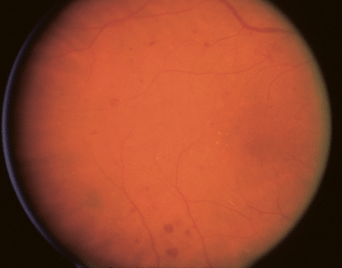
FIGURE 11-1. Pregnant Type 1 diabetic patient, 37-year-old female G0P1 for evaluation, Type 1 diabetes 10 years, first trimester vision 20/40 OU, Photo OD, dot blot hemorrhages, macular edema.
Stay updated, free articles. Join our Telegram channel

Full access? Get Clinical Tree


