Vitreous Anatomy
The vitreous is a translucent gel when it is in its normal state. The vitreous is a three-dimensional interactive matrix of collagen fibers with contractile properties and hyaluronic acid gel. The vitreous is in contact with the macula, pars plana, ciliary body, and retina in a sphere with a concavity for the lens. As aging occurs, the gellike structure becomes liquefied. In disease states such as diabetes, blood cells can be interspersed in the three-dimensional matrix, and the patient sees floaters. Or, in a dense vitreous hemorrhage, the patient experiences a loss of vision.
In visualizing the vitreous, the surgeon should start at the preoperative examination with dynamic ultrasound. Watch the movement of the vitreous with the epiretinal and periretinal membranes. Place the ultrasound probe at each clock hour of the retina to see the movement of the vitreous gel as the eye moves to the right, left, up, and down.
If there is a view of the vitreous and retina, the surgeon can use the Goldmann three-mirror lens to observe the peripheral vitreous, equator, midvitreous, and anterior vitreous. Consider the vitreous anatomy as one would enter the eye from anterior to posterior, using the Goldmann three-mirror lens. Take notes of various vitreous adhesions to retinal surfaces.
When examining the clear media in a patient with a traction retinal detachment from diabetic retinopathy, consider the anatomy of the vitreous and retina interface. Epiretinal membranes are white with a dull matte finish. The retina has a shiny, reflective sheen and is a slightly different shade of beige color than a white epiretinal membrane.
In cases of an incomplete posterior vitreous detachment, the epiretinal membrane will be attached at some places to the posterior hyaloid face. There will be areas where the tension of the vitreous gel pulls on the epiretinal membrane. This is an area of vitreous traction. It is at these “tension” points that microdissection and delamination can be initiated. These geometric forces can be seen by the preoperative examination.
The differences in the anterior-posterior direction of tension cause different types of epiretinal membrane formation. Tangential traction will cause starfolds and puckerlike folds. The most common type of epiretinal membrane in diabetic patients is anterior-posterior traction at sites of neovascularization and inflammation. The result is a plateau type of membrane structure.
In diabetic membranes, the epiretinal membrane may be pulling on the atrophic retina which has been thinned from lack of oxygen or chronic ischemia. These membranes look to be fully integrated with the retina. For these membranes, removing the anterior-posterior traction near the membrane or en bloc resection can be performed safely.
Membrane Peeling
The posterior hyaloid can be identified with a soft-tipped silicone suction cannula. When suction is lightly applied, the cannula becomes occluded by the vitreous gel as the cannula is brought close to the retinal surface. The cortical vitreous can be seen with the staining techniques of intravitreal injection of the dye such as fluorescein, indocyanine green, or trypan blue. Triamcinolone with its particulate matter may also be injected following core vitrectomy. The particles become entangled in the cortical vitreous, thus “labeling” the vitreous and its interface with epiretinal membranes. Residual triamcinolone appears to be well tolerated but can be associated with ocular hypertension, cataract, and sterile uveitis. The cortical vitreous is then peeled from the inner retinal surface, creating a posterior vitreous detachment. This can be done with the suction cutting instrument or cannula using active suction (100 to 200 mm Hg) at the area next to the optic nerve where the retina is thick and strong, able to tolerate some tissue manipulation in the posterior-anterior direction. Alternatively, the use of a bent myringotomy blade in the dominant hand, or of a light pick in the nondominant hand, may be useful in this maneuver. With the creation of a posterior vitreous detachment, the surgeon can visualize a floating Weiss’ ring. The vitrectomy is then completed, and removal of the posterior cortical vitreous is confirmed by the reaction of the brushing motion of the soft-tipped cannula.
Epiretinal membranes may be removed at this point using a bent myringotomy blade and intraocular forceps. Indirect ophthalmoscopy of the entire retina is performed to examine for iatrogenic retinal breaks and retinal detachments. Air-fluid exchange is then performed. Rapid accumulation of intravitreal fluid on the retinal surface usually occurs quickly, within the first 10 minutes after the air-fluid exchange. Therefore, many retinal surgeons allow at least 10 minutes to elapse before aspirating the accumulated intravitreal fluid. This technique would prepare for a maximal intravitreal gas bubble postoperatively. Face-down positioning is required postoperatively to provide retinal tamponade and repair of the diabetic traction retinal detachment.
A nonclearing vitreous hemorrhage in a diabetic patient is an indication for surgery. The timing of surgery is urgent if there is a traction retinal detachment threatening the macula. In the Diabetic Retinopathy Vitrectomy Study, vitreous hemorrhages were monitored for 6 months, however, that was in the era before optical coherence tomography (OCT) and before small incision vitrectomy surgery.
In many situations, vitrectomy for a diabetic patient is done within a few weeks of presentation to the ophthalmologist’s office. In these cases, the patient is a poor historian and is unable to say how long he has had the loss of vision. In other cases, the patient already has rubeosis of the iris and/or neovascular glaucoma or that his activities of daily living are severely hampered.
The surgical intervention begins with the preoperative consent. The team of physicians, consulting internist, endocrinologist, cardiologist, and nephrologist need to be consulted. In the cases of severe proliferative diabetic retinopathy, it is important to have medical clearance since the patient usually has other signs of systemic complications of diabetes. The most important risk factor for this patient is the cardiovascular risk factors. In fact, the need for diabetic vitrectomy surgery correlates with poor survival within 5 years of the surgery.1
In a core pars plana vitrectomy, where there is bright red, vitreous blood with ultrasonic evidence of attached retina, the light pipe can be used in the first part of the surgery to delineate the type of vitreous gel present. The vitreous can be dense clots or vitreous gel suffused with red blood cells. The light reflex shows the surgeon the texture of the vitreous (Fig. 10-1).
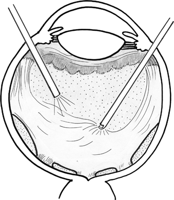
FIGURE 10-1. Core vitrectomy.
As the light pipe advances deeper into the posterior pole, the light reflections can help with the visualization of the vitreous gel and allow the surgeon to do en bloc dissection (Fig. 10-2). Using en bloc dissection, the vitreous gel, which is only partially detached, and the accompanying vitreoretinal membranes are removed as a single unit (see Fig. 10-2). Similarly, for new traction retinal detachment, membrane peeling and en bloc dissection techniques can be utilized (Fig. 10-3). While for some surgeons, the en bloc dissection technique serves to perform surgery with the minimal risk to the underlying retina (Fig. 10-4) because the countertraction of the vitreous gel enables the release of the adhesions of the membranes to the vitreous. All techniques utilize the fluidics of the vitrectomy probe, suction forces, and the response of the retina to the dissection techniques of the surgeon.
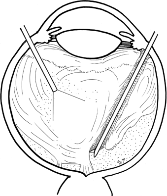
FIGURE 10-2. En bloc resection, step 1.
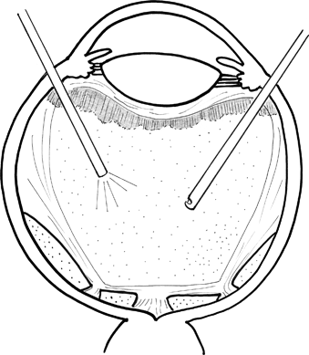
FIGURE 10-3. New traction retinal detachment, step 2.
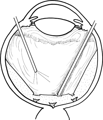
FIGURE 10-4. En bloc resection of traction retinal detachment.
Other techniques include delamination and methods of releasing the focal vitreoretinal adhesion with localized truncation of the anterior vitreoretinal forces of membranes. If there are tangential adhesions, membrane peeling is a challenge: care must be taken to create a space between the underlying retina and the overlying membrane. In that space, safe removal of the membrane can be performed.
In summary, all safe membrane peeling revolves around the creation of a space between the underlying retina and the membrane. If possible, the membrane can be removed by the vitrectomy probe with its cutting and suction action, thereby removing traction. For diabetic traction retinal detachments, membrane peeling techniques are based on the least trauma to the underlying atrophic or abnormally vascularized retina. In diabetic traction retinal detachments, the surgeon is concerned about bleeding from the fibrovascular membranes as well.
Using the smaller gauge instrumentation for pars plana vitrectomy, a 23- or 25-gauge system makes the vitrectomy very simple and accessible. Postoperatively, the patient has an excellent cosmetic result. The 23-gauge system has many compatible forceps and tools that can be used with the 23-gauge port of entry.
Of note, if the vitreous hemorrhage is dense, full of red clots of blood or old discolored heme, the 23-gauge system and 25-gauge system will take longer to remove the blood than the 20-gauge system. However, with practice and patience, the surgery results will be the same. There is precise control of the cutting action and the suction with the use of the small caliber instrumentation, thus, most retinal surgeons have converted to the smaller gauge systems.
Membrane Peeling for Traction Retinal Detachment
Once there is a traction retinal detachment of the macula, the clinician can measure the amount of traction with the OCT, fundus camera, and fluorescein angiography. There is usually a glial membrane that initiates the process of fibrovascular formation and consequent traction retinal detachment.
Traction retinal detachments occur after a vitreous hemorrhage or recurrent vitreous hemorrhage despite panretinal photocoagulation. These detachments are common and are common indications for pars plana vitrectomy with membrane peeling. Typically, the detachment is between the optic nerve and the macula.
There are two types of traction retinal detachment, (i) in the setting of active proliferative diabetic retinopathy, never having had panretinal laser photocoagulation and (ii) in the setting of multiple panretinal photocoagulation sessions. The first type is an active process where the fibrous bands have juicy red blood vessels and the retina that is being pulled upward and is encased in fresh, glistening glial tissue. This type of traction retinal detachment bleeds easily intraoperatively. Preoperatively, the retina surgeon could consider using intravitreal bevacizumab. The injection makes the activity of the blood vessels become less pronounced and hence less of an intraoperative bleeding hazard at the time of surgery.
The second type of traction retinal detachment, in the setting of old panretinal photocoagulation, is challenging because the surrounding tissue of the traction retinal detachment is thin and atrophic. Careful delamination is necessary rather than tissue manipulation of the atrophic retina. This type of thinned retina can lead to iatrogenic tears. Indirect membrane peeling using viscoelastic substances or silicon-tipped instruments are indicated for this type of traction retinal detachment repair.
At the time of vitrectomy surgery, while removing the blood, attention is paid to the anterior vitreous and then going more deeply into the posterior vitreous. At this point, the thickened vitreous at the periphery can be removed. For the peeling of the membrane, the process can start at the optic disc where the membrane is the thickest and then peeled across the papillomacular bundle, toward the fovea. Some surgeons like to start temporal to the macula and peel toward the fovea. The judgment of where to begin peeling depends on where the membrane is the thickest and where the surgeon can get the best control with the intraocular forceps to initiate the peeling. Bent 20-gauge needle is still a simple instrument to raise a membrane edge. There are diamond-tipped spatula needles as well.2
Delaminating scissors are useful for relieving the anterior-posterior traction. Various soft-tipped needles are useful in raising the edge in the membrane peeling process and continuing the peeling motion (Fig. 10-5).
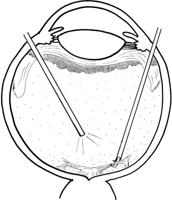
FIGURE 10-5. Membrane peeling with scissors.
Stay updated, free articles. Join our Telegram channel

Full access? Get Clinical Tree


