KENNETH C. CHERN AND MICHAEL A. SAIDEL
QUESTIONS
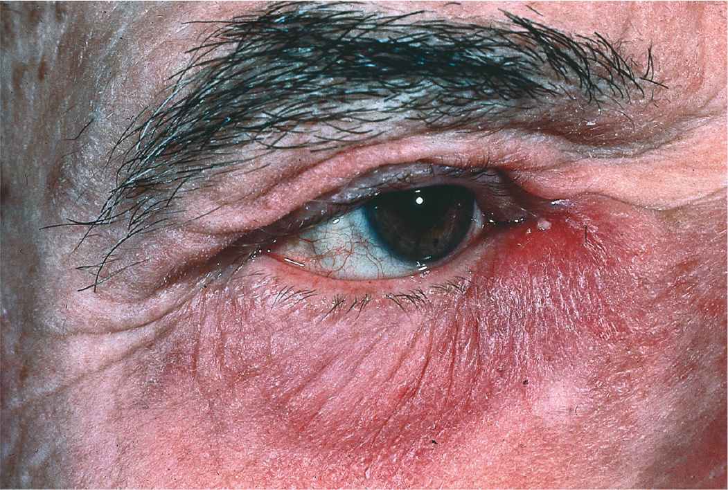
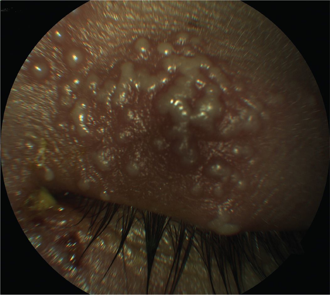
1 All of the following are included in the differential diagnosis of this condition shown in Figure 10-1 except:
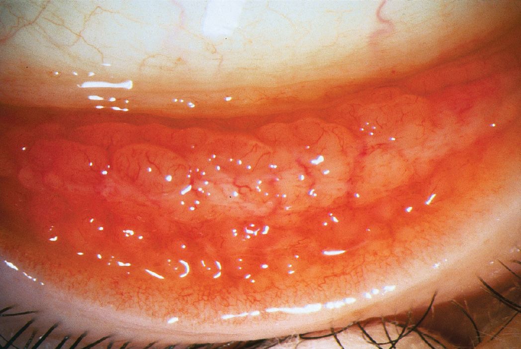
A) herpes simplex virus (HSV)
B) molluscum contagiosum
C) allergic drug reaction
D) Stevens–Johnson syndrome
2 Which disease is not caused by Chlamydia trachomatis?
A) Ligneous conjunctivitis
B) Adult inclusion conjunctivitis
C) Lymphogranuloma venereum
D) Trachoma
3 Following blunt trauma and the development of a hyphema, all of the following put a patient at higher risk for corneal blood staining except:
A) high intraocular pressure (IOP)
B) large blood clot
C) sickle cell trait or disease
D) Fuchs corneal dystrophy
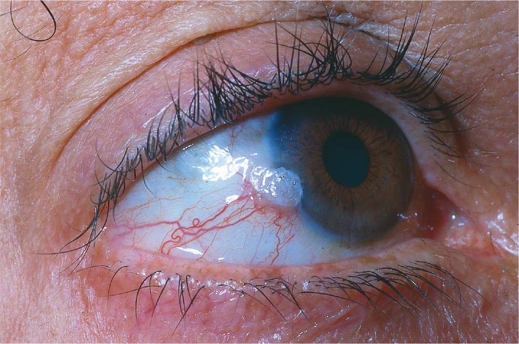
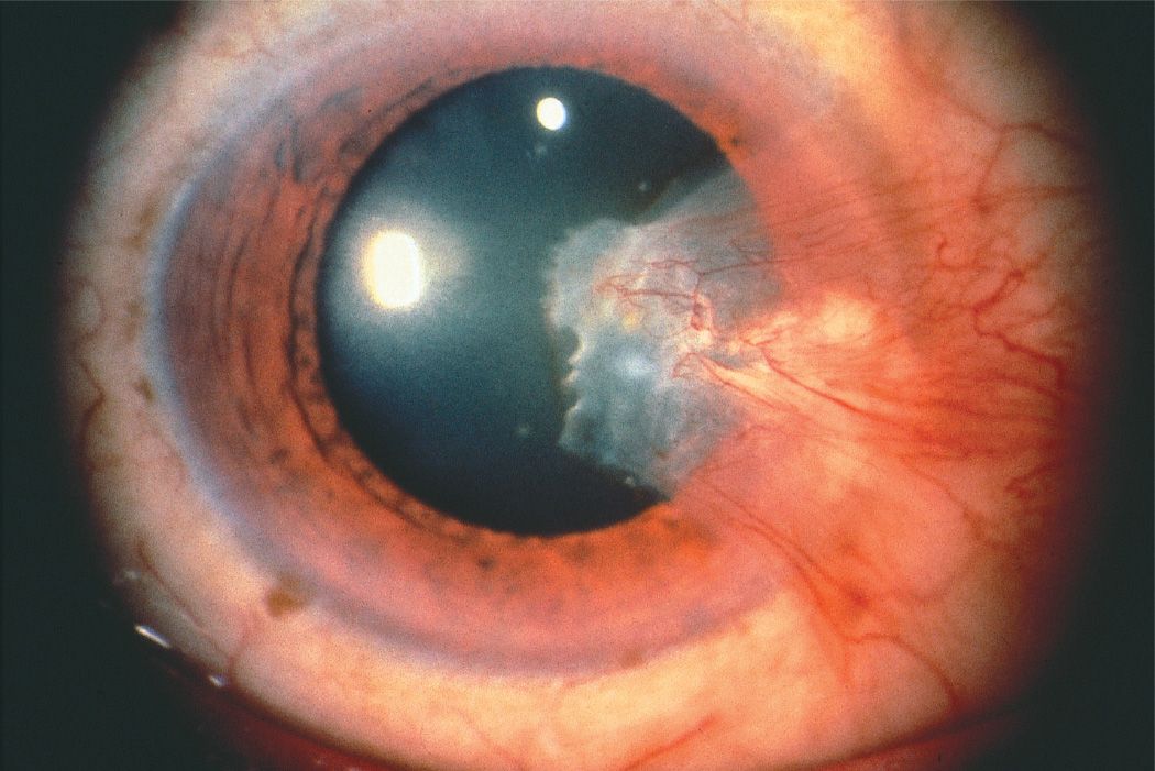
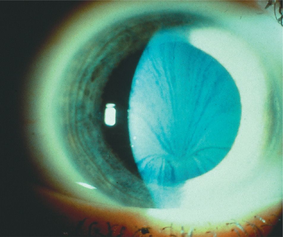
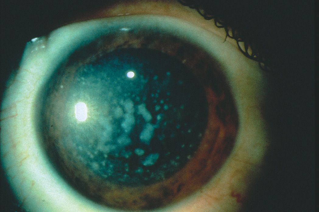
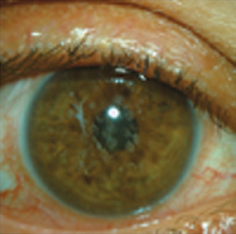
4 Based on the appearance of the corneal infiltrate in Figure 10-2, which laboratory test would be least helpful in aiding in the diagnosis?
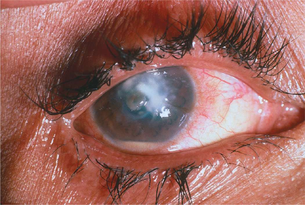
A) Culture on Sabouraud agar
B) Calcofluor white stain
C) Lowenstein–Jensen agar
D) Giemsa stain
5 Which one of the following associations between a microorganism and a useful medium for growth is correct?
A) Moraxella—blood agar in 5% to 10% carbon dioxide
B) Fungi—Sabouraud dextrose agar with cycloheximide
C) Mycobacterium tuberculosis—Loeffler medium
D) Haemophilus—blood agar
6 Which one of the following medications is commonly used to treat cases of filamentous fungal keratitis caused by Fusarium spp.?
A) Flucytosine
B) Natamycin
C) Amphotericin B
D) Miconazole
7 All of the following are appropriate therapy for primary HSV epithelial keratitis except:
A) ganciclovir 0.15% gel
B) difluprednate 0.05%
C) débridement of corneal lesions
D) valacyclovir
8 Which one of the following regarding megalocornea is true?
A) Most common inheritance is autosomal dominant
B) Associated with progressive corneal enlargement
C) Corneal diameter greater than 10 mm
D) Associated with Down syndrome
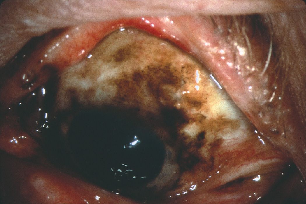
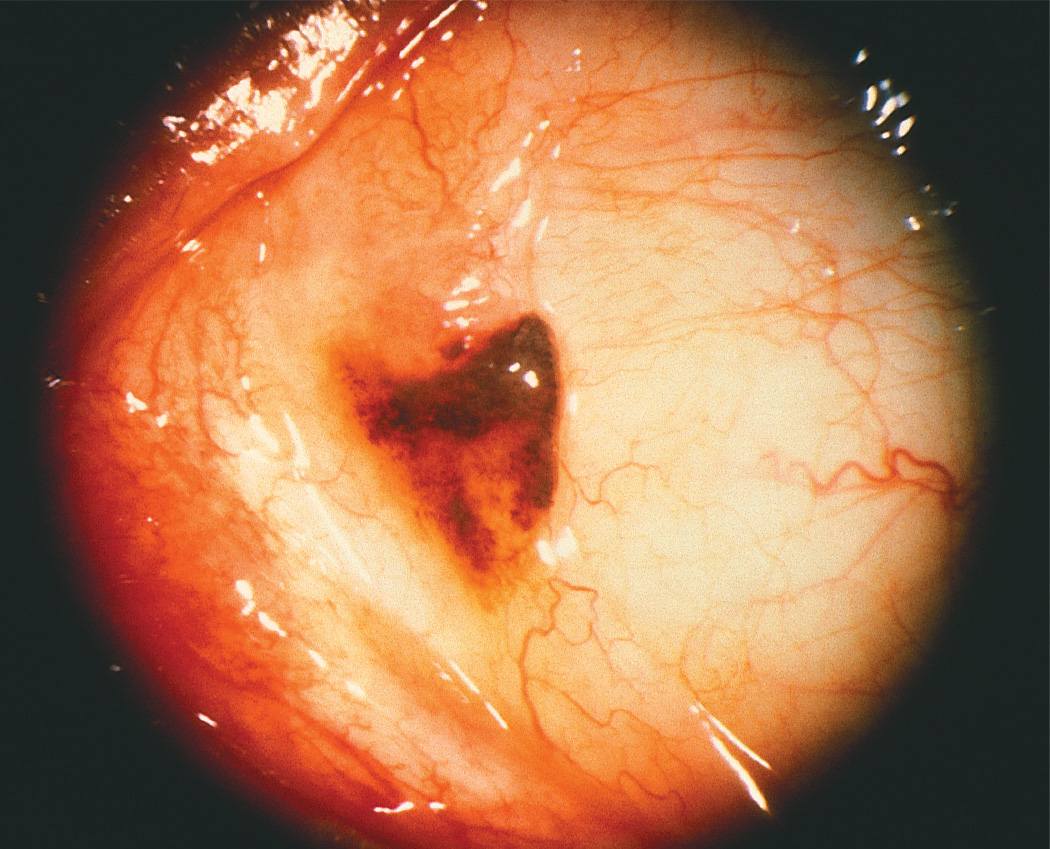
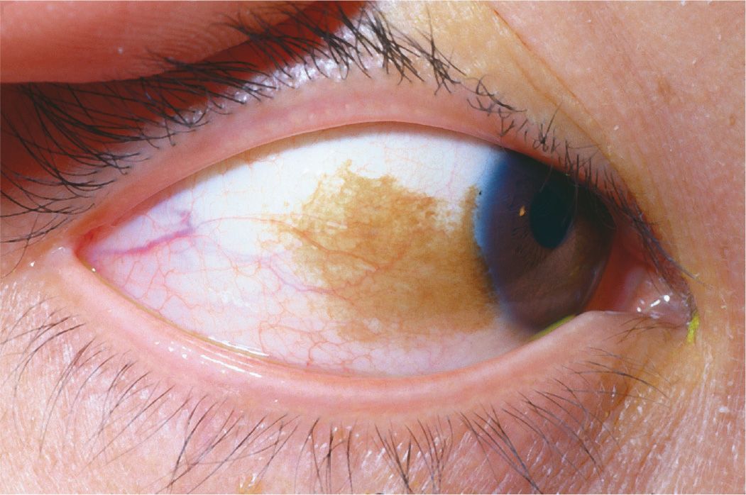
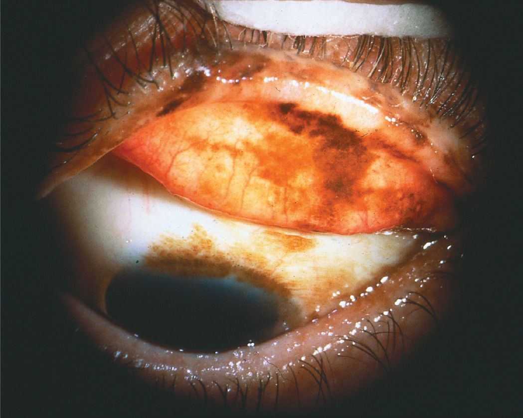
9 Which of the following conditions is not associated with rheumatoid arthritis?
A) Limbal follicles
B) Peripheral ulcerative keratitis
C) Necrotizing scleritis
D) Paracentral keratolysis
QUESTIONS 10 and 11 Select the condition(s) associated with the given finding.
10 Can result from mutation at the PAX6 gene:
A) Peters anomaly
B) Rieger syndrome
C) both
D) neither
11 Associated with glaucoma:
A) Axenfeld syndrome
B) Peters anomaly
C) both
D) neither
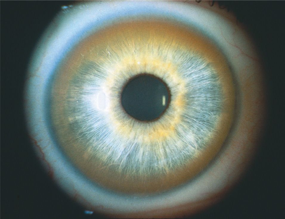
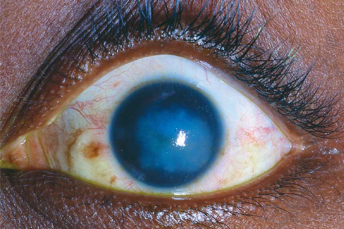
12 Which one of the following is not included in the differential diagnosis of blue sclera?
A) Hurler syndrome
B) Osteogenesis imperfecta
C) Turner syndrome
D) Marfan syndrome
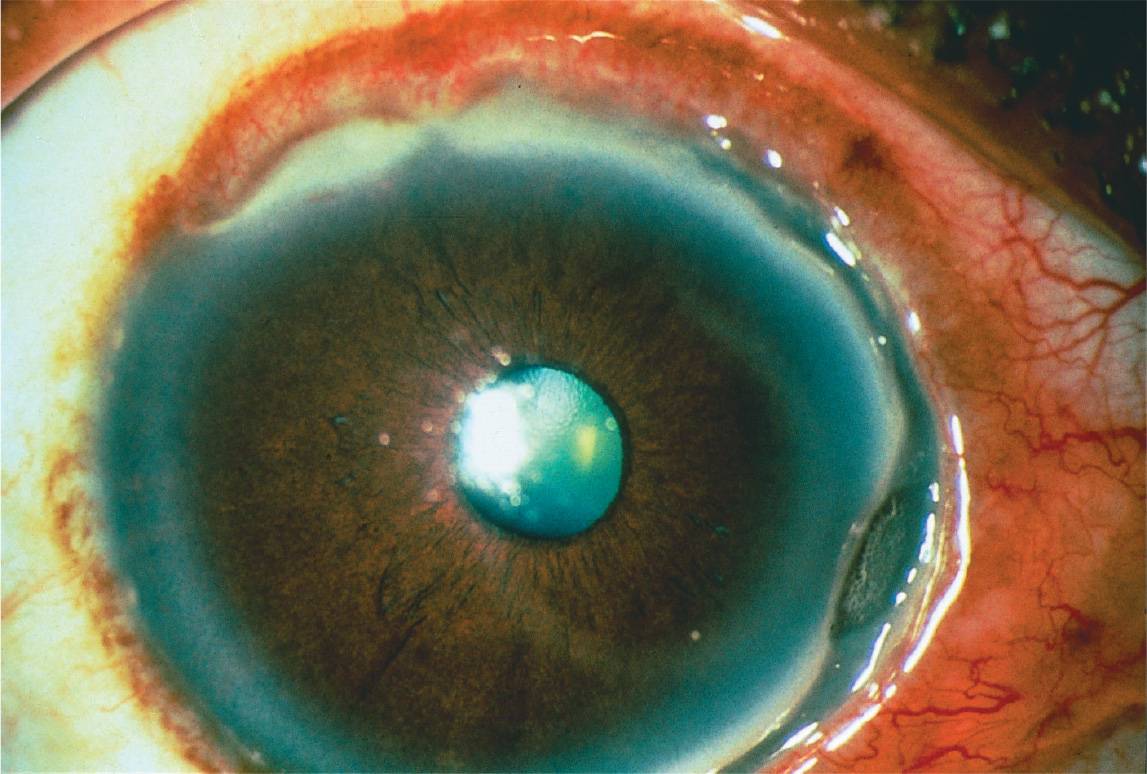
13 All of the following are associated with conjunctival cicatrization except:
A) atopic keratoconjunctivitis
B) ocular cicatricial pemphigoid
C) Stevens–Johnson syndrome
D) superior limbic keratoconjunctivitis
14 Which one of the following is a characteristic of sclerocornea?
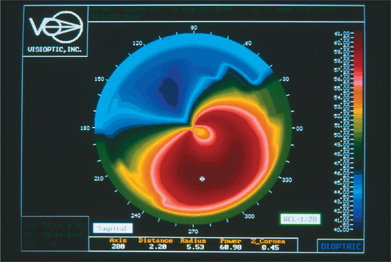
A) This disease is secondary to an inflammatory process.
B) Most cases are unilateral.
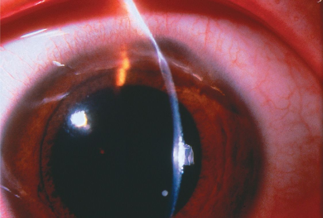
C) Most affected patients are women.
D) This process is nonprogressive.
15 All of the following may be considered part of the iridocorneal endothelial (ICE) syndrome except:
A) Chandler syndrome
B) essential iris atrophy
C) Cogan–Reese syndrome
D) posterior embryotoxon
16 Which one of the following is found with Rieger anomaly?
A) Autosomal dominant inheritance
B) Maxillary hypoplasia
C) Hypospadias
D) Peg-shaped teeth
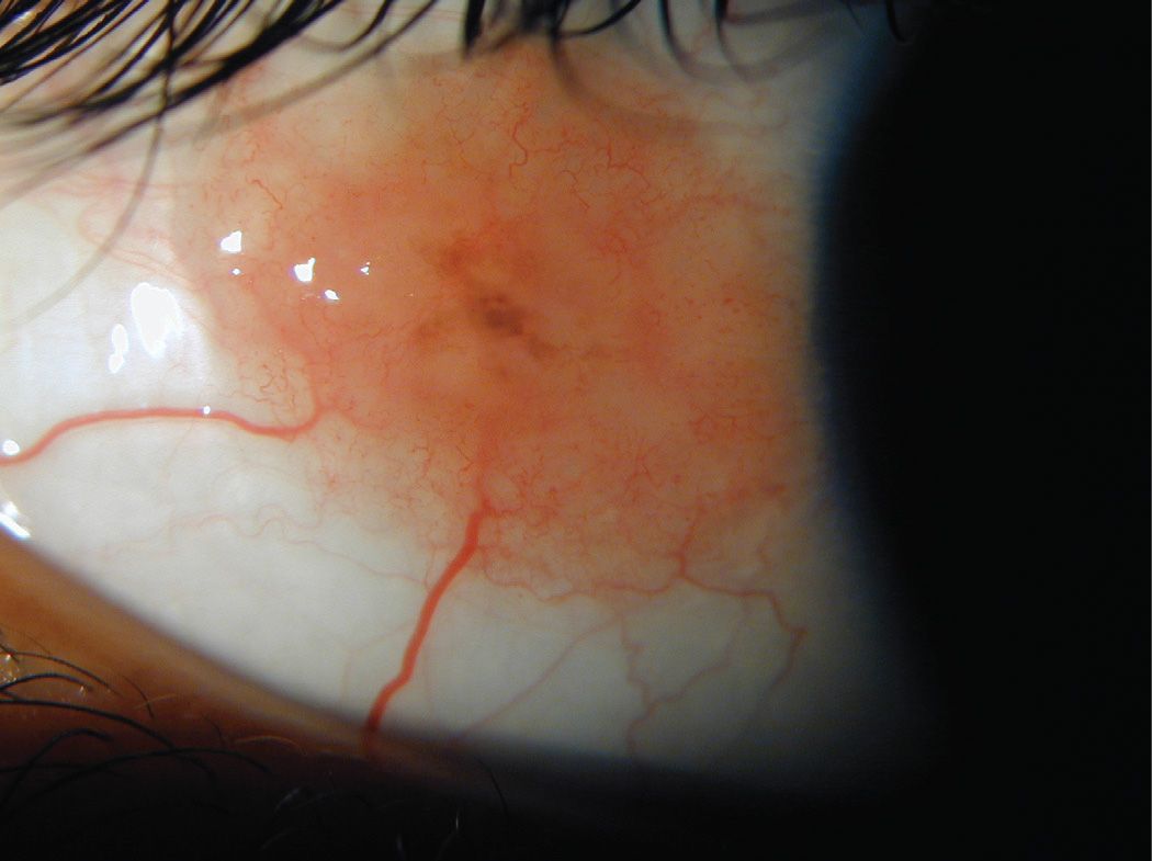
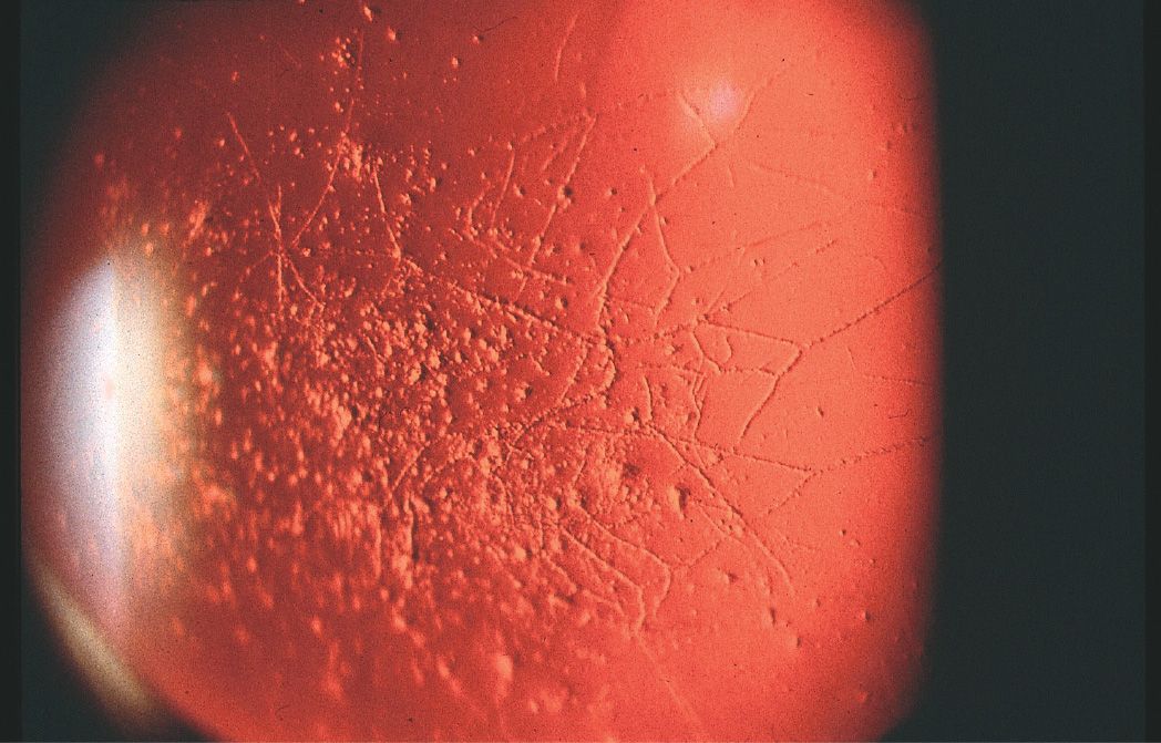
17 Which one of the following is not in the differential of a baby whose cornea is shown in Figure 10-3?
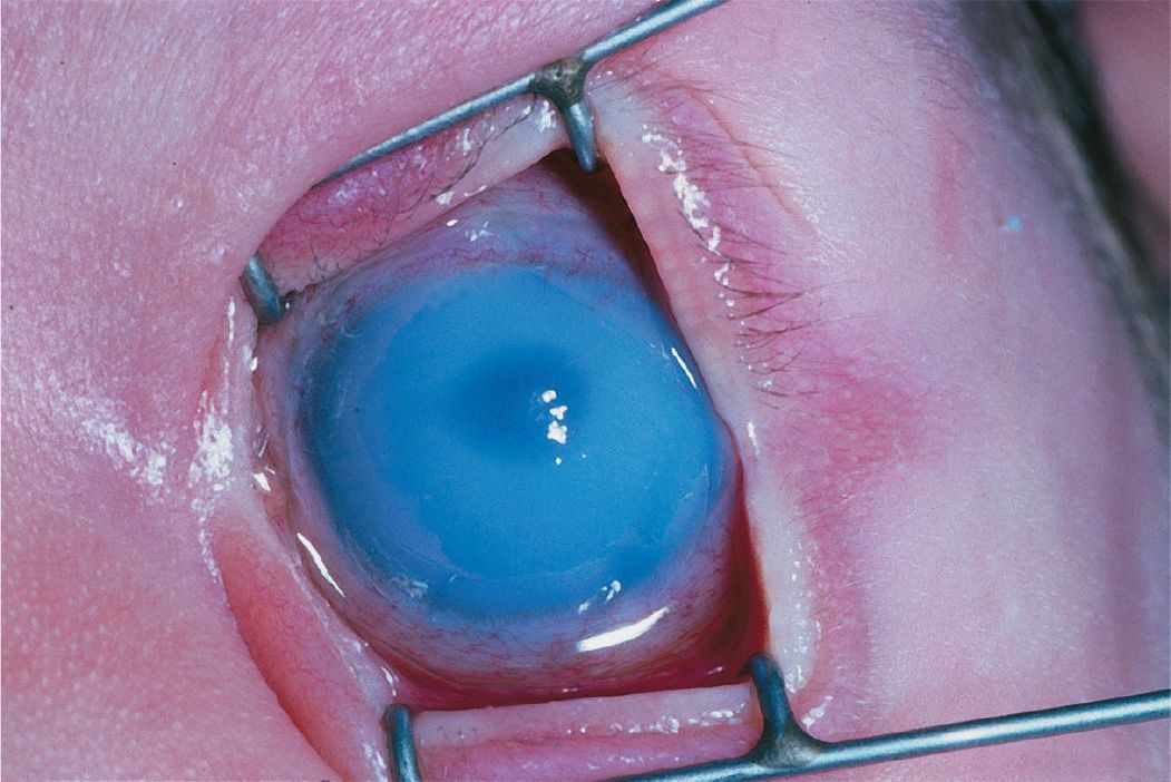
A) Congenital hereditary stromal dystrophy
B) Peters anomaly
C) Congenital glaucoma
D) ICE syndrome
18 Which one of the following represents a choristoma?
A) Dermolipoma
B) Keratoacanthoma
C) Hemangioma
D) Lymphangioma
19 Which one of the following would best be used to identify a corneal wound leak?
A) Gentian violet
B) Rose bengal
C) Fluorescein
D) Lissamine green
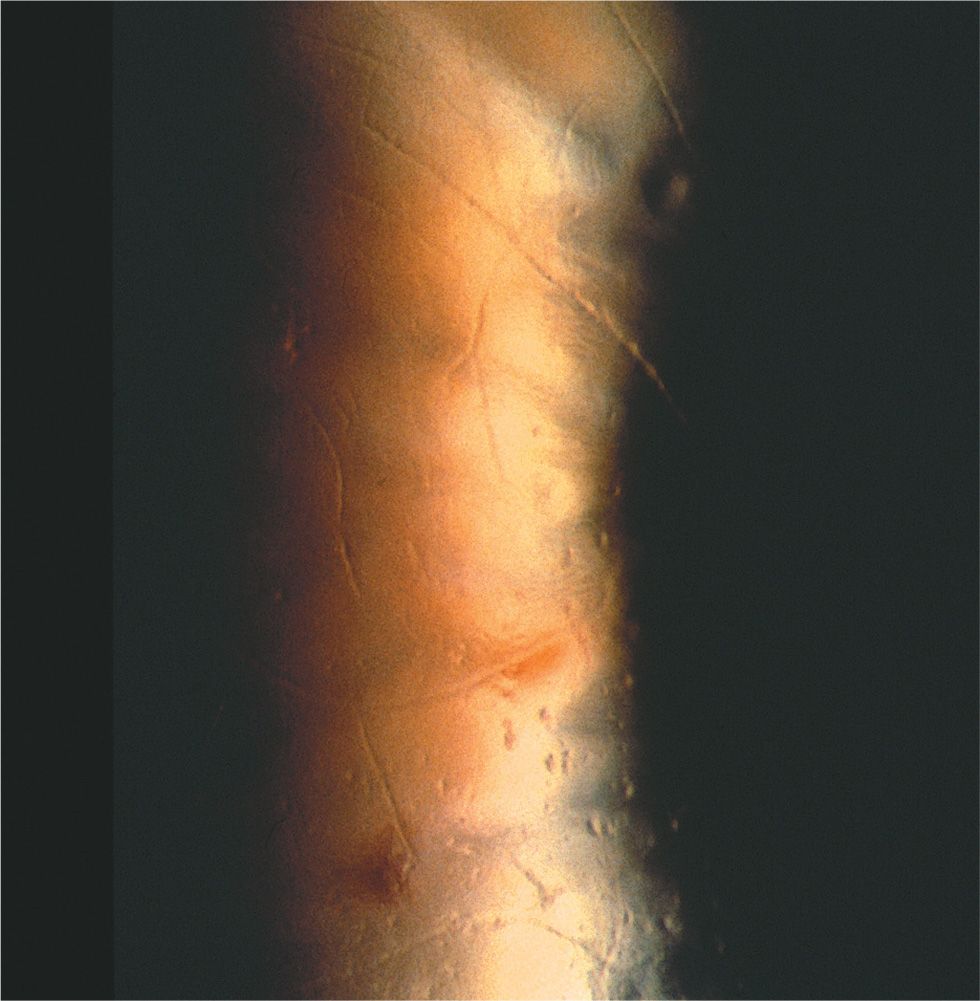
20 Which of the following occurs with the condition pictured in Figure 10-4?
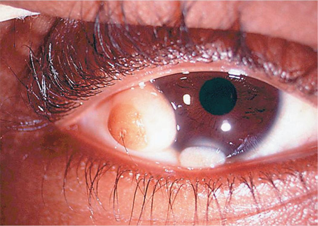
FIGURE 10-4 Courtesy of Helen K. Wu, MD.
A) The steepest meridian of the cornea is adjacent to this lesion.
B) These are benign with no malignant potential.
C) They can grow rapidly.
D) Patients may also have congenital cardiac defects.
21 Which structure is not found in the lesion in Figure 10-4?
A) Skin
B) Muscle
C) Sebaceous glands
D) Hair
22 All are true regarding Goldenhar syndrome except:
A) Iris colobomas may be present.
B) Upper eyelid colobomas may be present.
C) Preauricular skin tags may be present.
D) It can have an autosomal dominant inheritance.
23 Erythema multiforme major (Stevens–Johnson syndrome) is associated with which of the following etiologic factors?
A) Mycoplasma pneumonia
B) Sulfonamides
C) Coxsackievirus
D) All of the above
24 Which one of the following is not associated with enlarged corneal nerves?
A) Refsum disease
B) Congenital glaucoma
C) Ichthyosis
D) Multiple endocrine neoplasia (MEN), type I
QUESTIONS 25–27 (FIGS. 10-5A–D)
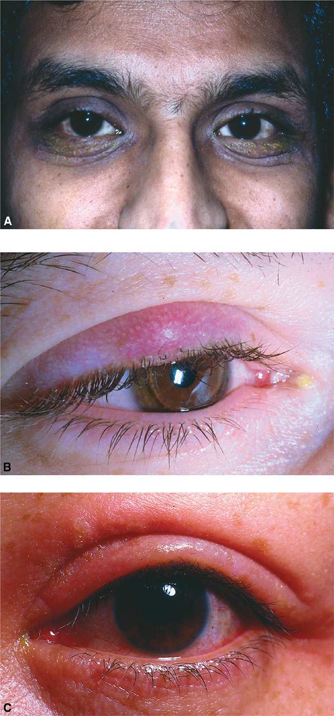
25 Which condition is caused by a virus?
A) Figure 10-5A
B) Figure 10-5B
C) Figure 10-5C
D) None of the above
26 Which treatment can be successful for the patient in Figure 10-5D?
A) Acyclovir
B) Steroid ointment
C) Incision
D) Doxycycline
27 All of the conditions are associated with the condition pictured in Figure 10-5A except:
A) rheumatoid arthritis
B) asthma
C) hay fever or seasonal allergies
D) eczema
28 Which organism is not a usual commensal found on the lids and lashes?
A) Moraxella catarrhalis
B) Haemophilus influenzae
C) Propionibacterium acnes
D) Staphylococcus epidermidis
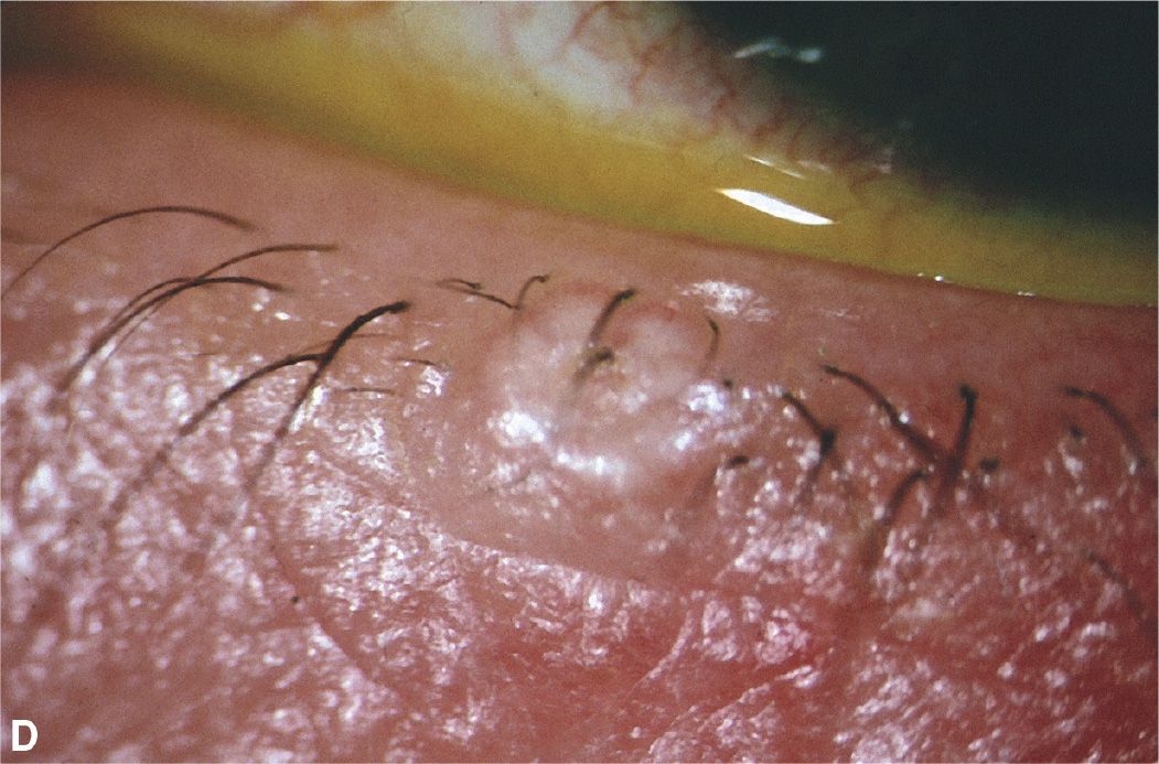
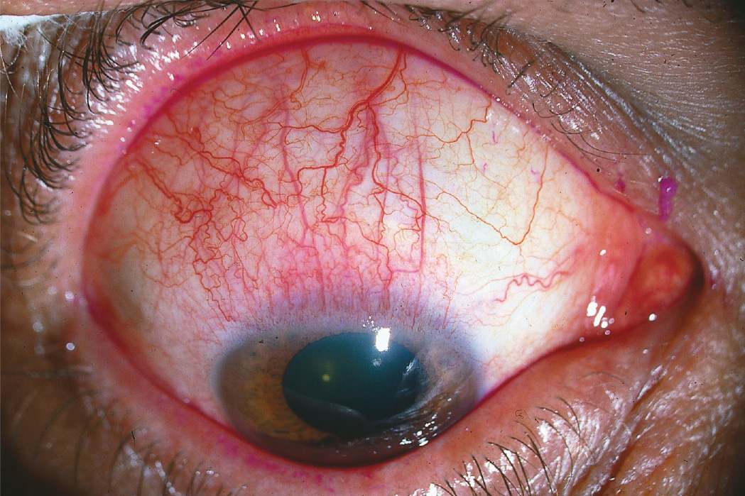
29 Which one of the following statements is correct for the condition in Figure 10-6?
A) Phthirus pubis is a normal commensal of adult meibomian glands.
B) Demodex folliculorum is transmitted by sexual contact.
C) Physostigmine acts as a respiratory poison to Phthirus pubis.
D) Demodex folliculorum is responsible for collarettes along the base of eyelashes.
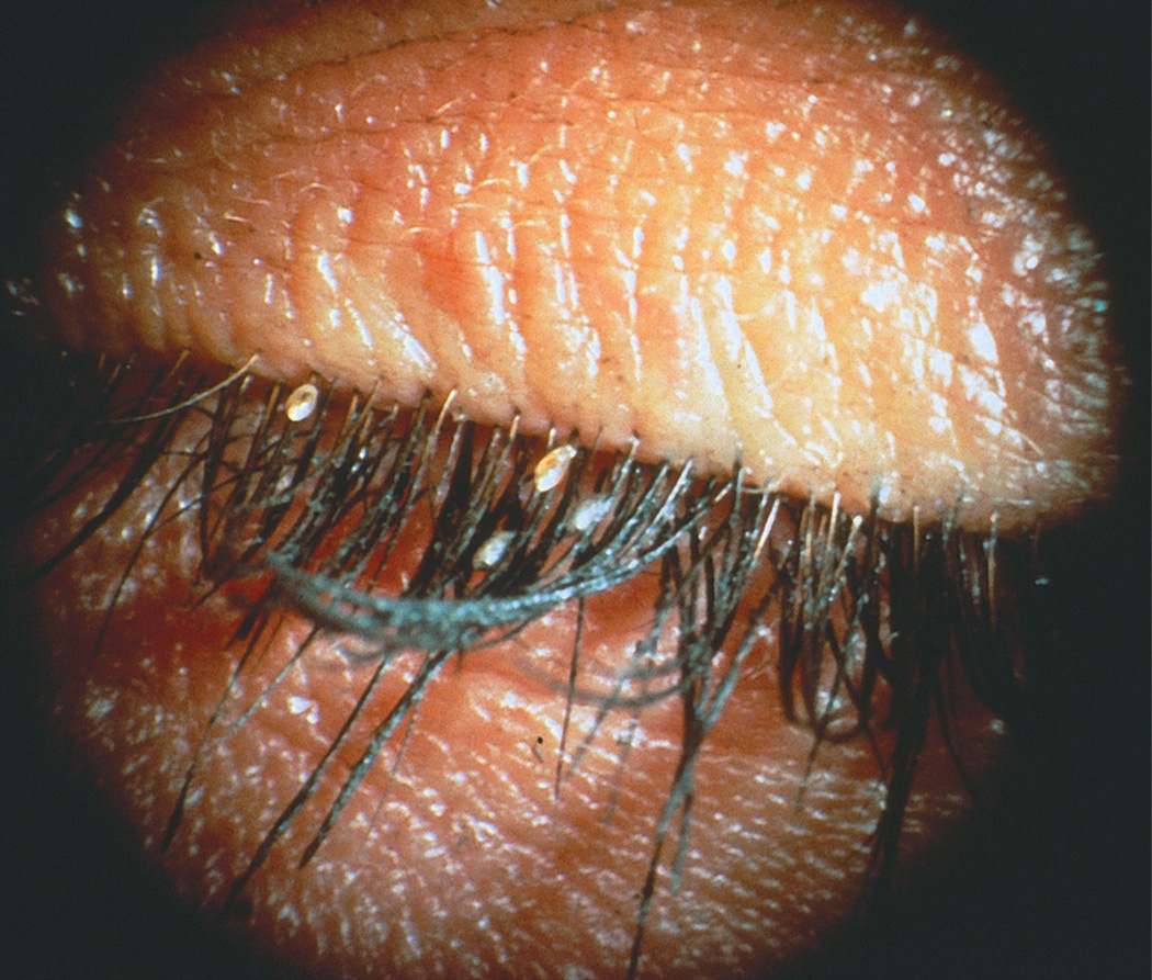
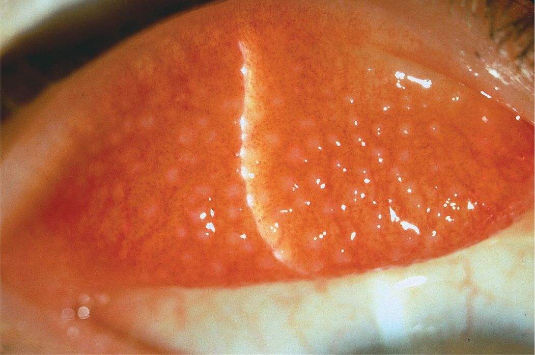
30 All of the following medications commonly cause the condition pictured in Figure 10-7 except:
A) atropine
B) neomycin
C) ketorolac
D) apraclonidine
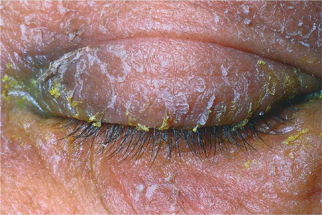
31 All of the following bacteria are commonly known to cause the clinical condition shown in Figure 10-8 except:
A) Streptococcus
B) Staphylococcus
C) Nocardia
D) Haemophilus
QUESTIONS 32 and 33 A 60-year-old man presents to your office complaining of a “spot” in his right eye, as shown in Figure 10-9.
32 Which of the following is not appropriate treatment for this lesion?
A) Local excision with wide margins followed by cryotherapy
B) Topical prednisolone drops
C) Local radiotherapy with a ruthenium-106 plaque sutured to the scleral bed following excision
D) Mitomycin C
33 The lesion pictured in Figure 10-9:
A) is always benign
B) is contagious and easily spread to others
C) affects only the superficial epithelium
D) can produce keratin
34 Which one of the following statements regarding the conjunctival lesion pictured in Figure 10-10 is true?
A) Microscopic examination without cellular atypia would confirm the benign nature of the lesion.
B) Growth and increased pigmentation can occur during puberty.
C) This lesion is confined to the conjunctiva and does not affect the underlying sclera.
D) Treatment with cyclosporin drops is needed since steroids are ineffective.
35 Which of the following conditions is thought to have a similar pathophysiology to staphylococcal marginal keratitis?
A) Phlectenulosis
B) Pterygium
C) Pseudogerontoxon
D) Pseudomembranes
36 Patients with ocular cicatricial pemphigoid:
A) have immunoglobulins bound to the conjunctival basement membrane
B) may have increased numbers of goblet cells
C) benefit from limbal stem cell transplants
D) frequently present with scleritis
QUESTIONS 37–39 A 24-year-old student presents after noticing a localized area of redness in his right eye near his caruncle as shown in Figure 10-11. He has had similar previous lesions that have resolved spontaneously.
37 What would the lesion likely show histopathologically?
A) Spindle-shaped atypical cells with dark nuclei
B) Acanthotic epithelium over fibrovascular cores
C) Small caliber vascular channels in collagenase stroma
D) Lined nonkeratinizing stratified squamous epithelium
38 Which therapy might be most appropriate at this time?
A) Observation
B) Simple excision
C) Photocoagulation
D) Excision with frozen section controls
39 Which virus has been implicated in causing this lesion?
A) Epstein–Barr virus
B) Human papilloma virus
C) HSV
D) Molluscum contagiosum virus
40 What is the most common malignant epithelial tumor of the conjunctiva?
A) Basal cell carcinoma
B) Squamous cell carcinoma
C) Malignant melanoma
D) Squamous papilloma
41 Regarding conjunctival intraepithelial neoplasia, which one of the following is true?
A) It rarely occurs in the interpalpebral zone.
B) Treatment is by enucleation.
C) Abnormal vascularization is rare.
D) The entire thickness of the epithelium may be involved.
42 Which one of the following is not a cause of secondary acquired conjunctival melanosis?
A) Pregnancy
B) Topical epinephrine drops
C) Addison disease
D) Scleritis
QUESTIONS 43–46 (Figs. 10-12 to 10-15)
A) Figure 10-12
B) Figure 10-13
C) Figure 10-14
D) Figure 10-15
43 Which has the lowest neoplastic potential?
44 Which often occurs bilaterally?
45 Which may enlarge during adolescence or with pregnancy?
46 Which requires a systemic disease workup?
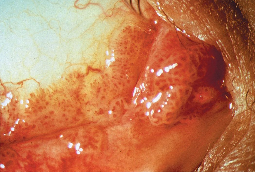
QUESTIONS 47–49 A 47-year-old farmer presents with the lesion pictured in Figure 10-16. He states it has been present for at least 2 years, and it has been gradually increasing in size.
47 This lesion may lead to all of the following except:
A) an adjacent dellen
B) flattening in the involved meridian with change in central astigmatism
C) destruction of Bowman layer
D) distant metastasis
48 Possible treatment options, if this were a recurrent lesion, would likely include all of the following except:
A) simple excision, leaving bare sclera
B) excision with amniotic membrane graft
C) excision with mitomycin C application and conjunctival autograft
D) excision with conjunctival autograft
49 Which is correct regarding the lesion pictured in Figure 10-16?
A) Histopathology shows fibrovascular ingrowth just beneath Bowman layer.
B) A pigmented iron line (Ferry line) is found at the leading corneal edge of the lesion.
C) Prolonged actinic exposure is a risk factor.
D) Recurrence after treatment is rare.
50 Which one of the following regarding the condition shown in Figure 10-17 is true?
A) Corneal sensation is reduced.
B) Resection of the adjacent conjunctiva may be indicated.
C) Treatment with a silver nitrate stick is beneficial.
D) A Fox shield at bedtime is helpful.
51 Which systemic finding might be found in association with this condition?
A) Increased urine catecholamines
B) Macular skin rash
C) Hyperextensible joints
D) Decreased thyroid-stimulating hormone levels
52 Which of the following is appropriate treatment of the condition shown in Figure 10-18?
A) Topical moxifloxacin
B) Oral doxycycline
C) Change from hard contact lenses to soft contact lenses
D) Topical mast cell stabilizers or corticosteroid drops
QUESTIONS 53–55 Corneal staining patterns in Figure 10-19. The cornea is stained with fluorescein or a vital dye.
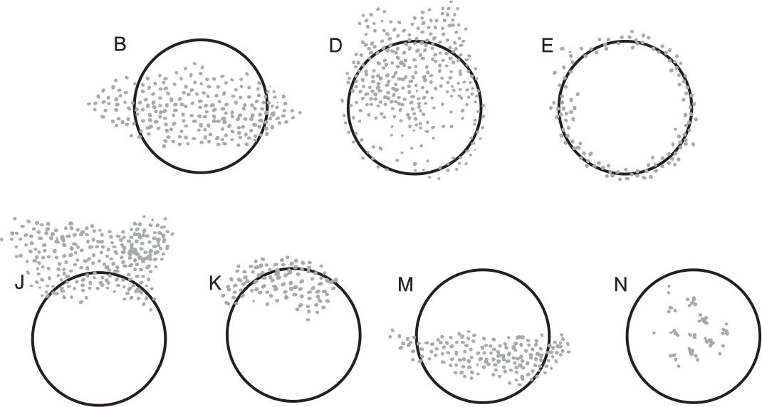
53 Which pattern can be found in a patient with thyroid eye disease and proptosis?
A) Figure 10-19M
B) Figure 10-19D
C) Figure 10-19N
D) Figure 10-19E
54 Which pattern is most typical of Thygeson keratopathy?
A) Figure 10-19K
B) Figure 10-19E
C) Figure 10-19N
D) Figure 10-19B
55 Figure 10-19J corresponds to which condition below?
A) Superior limbic keratitis
B) Epidemic keratoconjunctivitis (EKC)
C) Rosacea keratoconjunctivitis
D) Exposure keratopathy
56 All of the following drugs can cause a corneal appearance as in Figure 10-20 except:
A) amiodarone
B) lithium
C) chloroquine
D) indomethacin
57 Which metabolic disease can manifest as shown in Figure 10-20?
A) Fabry disease
B) Tay–Sachs disease
C) Alport syndrome
D) Refsum disease
58 Which one of the following is most accurate regarding a 54-year-old man with cystinosis?
A) All other siblings would have similar findings.
B) He is likely of short stature and has renal dysfunction.
C) He is unlikely to develop epithelial erosions.
D) The cystine deposits begin centrally within the anterior stroma and progress to involve the entire cornea.
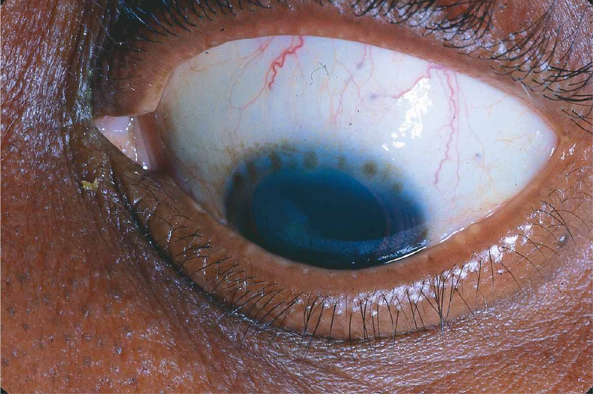
QUESTIONS 59 and 60 A 15-year-old girl presents with muscle tremors and a brownish ring near the limbus shown in Figure 10-21.
59 The deposits shown in the photograph are localized to which layer of the cornea?
A) Epithelium
B) Bowman layer
C) Posterior stroma
D) Descemet membrane
60 Which one of the following statements regarding this disease is true?
A) It is an isolated, nonhereditary disease.
B) The corneal findings can be used to monitor therapy.
C) It is caused by a defect in the kidneys.
D) The corneal findings are pathognomonic for this patient’s disease.
61 The findings in Figure 10-22 can be found in which condition?
A) Adult inclusion conjunctivitis
B) Vernal keratoconjunctivitis
C) Trachoma
D) Staphylococcal marginal keratitis
62 All of the following ocular findings may accompany this condition except:
A) superior corneal pannus
B) conjunctival scarring
C) superior conjunctival follicles
D) leukoplakia
63 Which one of the following statements regarding the disease process shown in Figure 10-23 is false?
A) The deposits consist of calcium hydroxyapatite and are found mainly in Bowman layer.
B) This patient may have deposition of copper in the liver, kidneys, and brain.
C) Patients with this disease may be on long-term topical steroids.
D) This disease may be associated with chronic mercurial exposure.
64 Which topical agent might be used to treat this condition?
A) Ethylenediaminetetraacetic acid (EDTA)
B) Penicillamine
C) Corticosteroids
D) Acetylcysteine
65 Which one of the following statements regarding spheroidal degeneration of the cornea is false?
A) It is usually bilateral.
B) Pathologically, it appears as lipid deposition in the cornea.
C) Patients usually remain asymptomatic.
D) Actinic exposure is implicated in the pathogenesis of spheroidal degeneration.
66 Which one of the following is the most accurate statement regarding the condition shown in the Figure 10-24?
A) It is often associated with a systemic autoimmune disease.
B) With-the-rule astigmatism may be induced.
C) Thinning is more apparent than real.
D) Epithelium remains intact.
67 Which one of the following statements regarding the clinical condition found in this 73-year-old woman (Fig. 10-25) is true?
A) Biopsy of the adjacent conjunctiva may show increased plasma cells.
B) Corneal perforation will occur rapidly.
C) Systemic immunosuppressives will be necessary.
D) It is a painless, slowly progressive process.
68 Which is true of pellucid marginal degeneration?
A) Decreased vision results from lipid deposition.
B) Protrusion of the cornea is at the point of maximal thinning.
C) This is a bilateral condition.
D) Best surgically treated with PRK.
69 Which one of the following is not characteristic of the condition shown on corneal topography (Fig. 10-26)?
A) Apical scarring
B) Scissoring of the red reflex on retinoscopy
C) Spontaneous perforation
D) Fleischer ring
70 All of the following are accepted treatment measures for visual rehabilitation in this condition except:
A) riboflavin and corneal cross-linking
B) hard contact lens fitting
C) penetrating keratoplasty
D) photorefractive keratectomy (PRK)
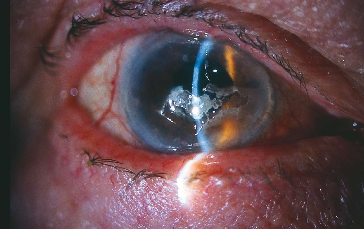
71 This same patient comes back with a dramatic decrease in vision and this corneal appearance (Fig. 10-27). What is the best initial treatment?
A) Hypertonic saline
B) Antibiotic drops
C) Corneal transplantation
D) Descemet stripping endothelial keratoplasty (DSEK)
QUESTIONS 72 TO 73 PERTAIN TO THE FOLLOWING FIGURE (FIG. 10-28). A 25-year-old woman presents with 2 days of eyelid redness, swelling, and discomfort. She has been using cold compresses with no improvement.
72 What diagnostic tests, if any, would be helpful in confirming the diagnosis?
A) Gram stain looking for diplococci
B) Adenovirus DFA
C) Conjunctival biopsy looking for eosinophils
D) No tests needed, diagnosis based on clinical appearance
73 What other findings would be commonly seen with this eyelid appearance?
A) Conjunctival membranes
B) Epithelial dendrites
C) Shield ulcer
D) Hypopyon
74 Which one of the following is true regarding congenital hereditary endothelial dystrophy?
A) Nystagmus is absent in the recessive form of the disease.
B) There are usually associated systemic abnormalities.
C) The recessive form is nonprogressive, whereas the dominantly inherited form is slowly progressive.
D) Corneal clouding is present at birth in both forms of the disease.
75 Which one of the following concerning congenital hereditary stromal dystrophy is false?
A) Autosomal dominant inheritance
B) Symptoms of pain, tearing and photophobia
C) Central anterior stromal flaky, feathery opacity
D) Cornea of normal thickness
76 Of the corneal dystrophies below, which is the most common?
A) Lattice stromal dystrophy
B) Macular stromal dystrophy
C) Granular stromal dystrophy
D) Meesmann dystrophy
QUESTIONS 76 and 77 A 24-year-old woman presents with ocular irritation, foreign body sensation, decreased vision in the right eye, and the findings shown in Figure 10-29.
77 What would histopathologic examination of the corneal specimen show?
A) Amyloid deposits
B) Cholesterol and neutral fats
C) Acid mucopolysaccharides
D) Hyaline
78 Which one of the following statements regarding the disease shown in Figure 10-29 is true?
A) Both of the patient’s siblings are likely affected.
B) The disease is caused by a defect in the synthesis of keratan sulfate.
C) Epithelial erosions are a frequent, recurring problem.
D) In the majority of cases, only the central cornea is affected.
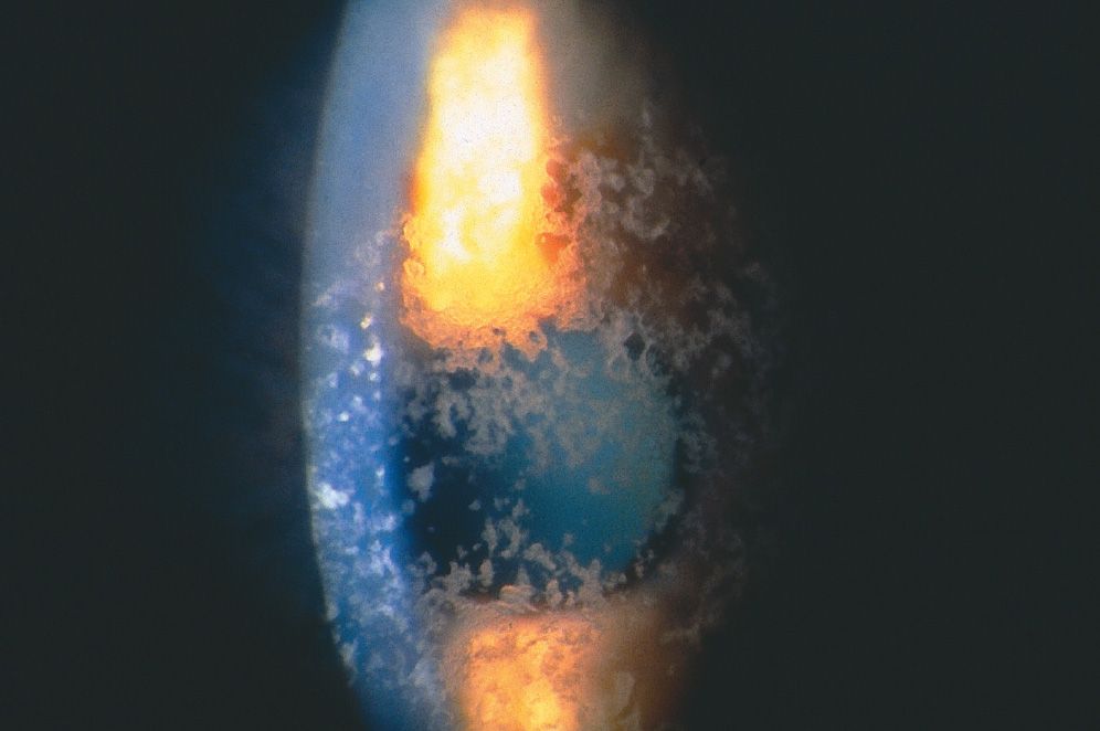
QUESTIONS 79 and 80 A 27-year-old woman presents with foreign body sensation in her eye and has the findings shown in Figure 10-30.
79 Which of the following is true?
A) Initially, deposits are concentrated in the periphery.
B) The corneal findings are best shown by specular reflection.
C) The deposits are found in the posterior stroma.
D) Skin findings and nerve palsies are manifestations of systemic involvement.
80 All of the following stains will highlight the deposits seen in this disease except:
A) thioflavin T
B) Congo red
C) alcian blue
D) crystal violet
81 Each of the following statements regarding granular corneal dystrophy is true except:
A) the corneal findings precede symptoms by several years.
B) the intervening cornea is characteristically clear between lesions.
C) recurrent erosions are common.
D) the deposits consist of hyaline, which stains with Masson trichrome.
QUESTIONS 82–86 Match the condition(s) with the associated finding.
A) Figure 10-31
B) Figure 10-32
C) Both
D) Neither
82 Progressive disorder?
83 Recurrence of disease in grafts?
84 Autosomal recessive inheritance?
85 Epithelial erosions occur frequently?
86 Vision severely affected in most cases?
QUESTIONS 87 and 88 A 34-year-old man presents with a report of decreased visual acuity in both eyes. He has had many episodes of pain and redness that last several days. His father has the same condition. His right eye is shown in Figure 10-33.
87 Which one of the following statements regarding these findings is true?
A) The opacities are at the level of the stroma.
B) The lesions are among the most common to recur after penetrating keratoplasty.
C) This condition is not progressive.
D) Recurrent erosions are rare.
88 Histopathology would show:
A) disruption and absence of Bowman layer
B) “peculiar substance” replacing Bowman layer
C) birefringence and dichroism
D) staining of these lesions with Oil Red O
89 All of the following statements regarding the condition pictured in Figure 10-34 are true except:
Stay updated, free articles. Join our Telegram channel

Full access? Get Clinical Tree


