David T. Tse
Classically, patients present with temporal headaches, visual symptoms, jaw claudication, and temporal artery tenderness. Constitutional complaints of fever, anorexia, weight loss, and fatigue are also common. Polymyalgia rheumatica is frequently associated with temporal arteritis, and most now believe that these two disorders represent a spectrum of disease.
In temporal arteritis, the erythrocyte sedimentation rate (ESR) is usually higher than 50 mm/h and commonly exceeds 100 mm/h. A normal ESR, however, does not rule out temporal arteritis. The Westergren method of performing the sedimentation rate is preferred to the Wintrobe method, because of its increased accuracy at the higher test values.
The term cranial arteritis has been used to describe this disorder because of the frequent involvement of the superficial temporal, vertebral, ophthalmic, and posterior ciliary arteries. Temporal arteritis has also been referred to as granulomatous arteritis and giant cell arteritis, as a result of the characteristic granulomatous inflammation with giant cells seen on histopathologic examination.
The exact etiology of temporal arteritis is unknown. It is believed, however, to be related to an autoimmune reaction directed against the elastic tissue within the media and adventitia of the arteries of the head and neck.
The superficial temporal artery (STA) is chosen for biopsy almost exclusively, because of its readily accessible location and its high frequency of involvement. The rich arterial anastomosis within the forehead allows the anterior branch of the STA to be excised without a significant risk of vascular compromise. Additionally, “skip” areas of involvement have been demonstrated within a single arterial specimen; therefore, a large biopsy (more than 2 cm) is recommended. A positive arterial biopsy is required to conclusively establish the diagnosis of temporal arteritis.
The surgical technique presented here is based on the common anatomic branching patterns of the STA, the various fascial layers, and the location of the temporal branches of the facial nerve. Proper surgical technique, combined with a working knowledge of the anatomy of the temporalis region, adds a new dimension to the safety of the temporal artery biopsy procedure.
Superficial temporal artery
The superficial temporal and maxillary arteries are the two terminal branches of the external carotid artery. The STA arises from within the parotid gland and ascends superiorly to cross the zygomatic process of the temporal bone, approximately 1 cm anterior to the tragus.
The transverse facial artery (zygomaticomalar) runs in a medial direction either along the surface, or immediately inferior to the zygomatic arch. The middle temporal artery arises from the undersurface of the STA and travels superiorly over the anterior surface of the zygomatic arch. The artery then penetrates the superficial fascial planes to supply the underlying deep temporalis fascia (DTF).
The distal STA divides into an anterior frontal and a posterior parietal branch. There are five major branching patterns described for the distal STA; however, in 80% of cases the bifurcation of the frontal and parietal branches occurs above the zygomatic arch. The frontal and parietal branches, along with the transverse facial, supraorbital, and supratrochlear arteries, arborize extensively to form a rich arterial plexus within this region. Collectively, these vessels supply the superficial temporal fascia (STF) and the overlying skin.
The STF is also referred to as the temporoparietal fascia and epicranial aponeurosis. The STF is bounded by and contiguous with the galea superiorly, the frontalis anteriorly, and occipitalis posteriorly, and the subcutaneous musculoaponeurotic system inferiorly. As such, there are no clearly defined boundaries to this fascial plane.
Immediately deep to the STF is a loose avascular areolar layer, separating the STF from the DTF. The DTF covers the temporalis muscle superiorly and splits into a superficial and deep layer below the level of the superior orbital rim. The point at which the deep temporal fascial layers unite together as a single unit has been referred to as the temporal line of fusion. The superficial and deep temporal fat pads are separated by the superficial and deep layers of the DTF, respectively. The DTF vasculature is supplied by the middle temporal artery without contributions from the frontal or parietal branches of the STA. The intervening loose avascular areolar layer separates the vasculature of the STF and the DTF.
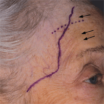
The temporal branches of the facial nerve supply motor innervation to the frontalis, upper half of the orbicularis oculi, and corrugator supercilii. These branches initially emerge from the parotid gland just above the zygomatic arch and arborize extensively. Despite variable branching patterns, all nerve fibers travel within the same plane as they traverse the zygomatic arch. Branches of this nerve run along the undersurface of the STF just within the loose avascular areolar layer (Fig. 35-1B). While maintaining this constant position immediately below the STF, these branches proceed in a superomedial direction to innervate the facial musculature from the undersurface. The zone where the temporal branches of the facial nerve are most vulnerable to injury during a temporal artery biopsy is outlined by the following anatomic landmarks: (1) the tragus of the ear, (2) the junction of the zygomatic arch and the lateral orbital rim, (3) 2 cm above the level of the superior orbital rim and in a line directly superior to (2), (4) superior to the tragus and in horizontal alignment with (3) (Fig. 35-1A). (The vertical dotted line defines the site of the cross-sectional diagram [Fig. 35-1B] and is not an incision line.) This “danger zone” delineates the area where the facial nerve is superficial in location and in close proximity to the frontal branch of the STA.

Figure 35-1A. The proximal STA gives off four main branches: the transverse facial, middle temporal, parietal, and frontal.
Stay updated, free articles. Join our Telegram channel

Full access? Get Clinical Tree


