Osseous Approaches to the Temporal Bone


Osseous Approaches to the Temporal Bone


Joseph B. Nadol, Jr.


A variety of osseous approaches are used in surgery of the ear and temporal bone. Presented in this chapter are procedures and their modifications for special surgical problems, the relevant surgical anatomy and landmarks, the factors that must be considered in selecting an appropriate surgical approach, and a uniform method of nomenclature to describe the surgical procedure. Details of the soft tissue incisions are discussed in Chapter 9, and details of each procedure when applied to specific disease processes are described in subsequent chapters.


SURGICAL ANATOMY

Important surgical anatomy of the lateral and basal aspects of the temporal bone is shown in Figure 11.1A,B. The temporal bone is divided into six parts: tympanic, mastoid, squamous, zygomatic, styloid, and petrous. The osseous structures of the lateral aspect of the temporal bone, which are of particular importance as surgical landmarks, are shown in Table 11.1.


PNEUMATIZATION OF THE TEMPORAL BONE

An understanding of the development and variability of pneumatization of the temporal bone is essential for the mastoid surgeon. The pneumatized spaces or potential pneumatized spaces serve as important internal landmarks, guiding the surgeon to the middle and posterior fossa bony plates, facial nerve, jugular vein, carotid artery, and labyrinth. The more medially located tracts may serve as surgical pathways to the petrous apex.

The surgical anatomy of the pneumatized regions of the temporal bone have been described by Allam (1) and Schuknecht (2). Five pneumatized regions of the temporal bone are recognized: middle ear, mastoid, perilabyrinthine cells, petrous apex, and accessory cells (Figs. 11.2 and 11.3; Table 11.2).


Region 1: Middle Ear

The pneumatized middle ear and its extensions are larger than those defined by the bony tympanic ring. Lines drawn tangent to the tympanic ring (Fig. 11.3) help define the approximate limits of the epitympanum, protympanum, hypotympanum, and posterior tympanum.


Clinical Significance

Removal of part of the bony tympanic ring may be necessary for surgical exposure of the middle ear. For example, the transcanal approach to the stapes requires curettage of the posterior tympanic annulus. Similarly, full exposure of the hypotympanum may require drilling of the inferior tympanic annulus in cases of chronic otitis media or glomus tumors. Extension of chronic otitis media to the facial recess and sinus tympani requires surgical access by careful bone removal anterior and anteromedial to the facial nerve (Chapter 17).

In canal wall-up tympanomastoidectomy for chronic otitis media or cochlear implantation, access to the middle ear from the mastoid is achieved through the posterior tympanic (facial recess) cells and the posterior tympanum. For procedures requiring thorough obliteration of the eustachian tube, such as tympanomastoid obliteration for chronic otitis media or for persistent spinal fluid leak, access to the eustachian tube and the peritubal cells, which may enter the eustachian tube several millimeters anterior to the tympanic end of the tube, requires exposure of the protympanum. Symptomatic retraction pockets and early cholesteatoma may be managed by exteriorization of the epitympanum through the ear canal.


Region 2: Mastoid Cells

The growth of the mastoid with age and variations in normal development in the adult have been documented by Eby and Nadol (3). Shortly after birth, only the middle ear and aditus are pneumatized. The central mastoid tract becomes pneumatized in two growth spurts: from 0 to 2 years and again at puberty.


FIG. 11.1 A: Osseous landmarks of the lateral surface of the left temporal bone. B: Osseous landmarks of the base view of the left temporal bone.


TABLE 11.1 Osseous structures of the lateral aspect of the temporal bone that are of particular importance as surgical landmarks


Structure


Surgical structure


Temporal line


Serves as an approximate guide to the level of the tegmen mastoidea and temporal lobe


Suprameatal spine of Henle


Classically described as a landmark to the location of the antrum


Cribriform area


1.


Small foramina that perforate the lateral mastoid cortex in the fossa mastoidea. Particularly in the young, these foramina may provide a pathway for purulence to spread from the pneumatized mastoid to the subperiosteal space.


2.


Also used as an approximate guide to the position of the antrum.


Mastoid process


The lateral mastoid aspect of the bone, which serves as an attachment of the sternocleidomastoid muscle and covers the pneumatized central mastoid tract.


Mastoid foramen


The vascular foramen for the emissary vein to the lateral venous sinus and the mastoid branch of the occipital artery. May be encountered in a wide-field mastoidectomy where vigorous bleeding may be controlled by bone wax or Gelfoam pledgets and pressure.


Tympanomastoid fissure


1.


Anatomic division between tympanic and mastoid bone.


2.


Serves as a conduit for Arnold’s branch of the tenth cranial nerve.


3.


May provide a pathway for infection from the external ear canal to the skull base, as in malignant external otitis.


4.


Posteriorly based canal skin flaps generally require sharp dissection over this suture.


Digastric groove


1.


Origin of digastric muscle.


2.


This groove externally and the digastric ridge within the mastoid end anteriorly at the stylomastoid foramen and hence serve a useful surgical landmark to the end of the mastoid segment of the facial nerve.


Notch of Rivinus


The tympanic incisure represents a developmental dehiscence in the tympanic ring. On its posterior side the fibrous annulus of the tympanic membrane leaves the bony tympanic annulus and is hence easily identified for elevation of an anteriorly based tympanomeatal flap.


Tympanosquamous fissure


Elevation of a laterally based anterior canal skin flap will commonly be adherent at this suture line between tympanic and squamous bone and require sharp dissection. Troublesome bleeding vessels within the suture can easily be cauterized.


Clinical Significance

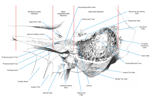
The facial nerve exits the stylomastoid foramen on the lateral surface of the mastoid bone and is not covered by the enlarging mastoid process until approximately age 1½ years; hence it is susceptible to injury with a standard postauricular incision.

The tegmental and sinodural areas within the mastoid are common sites of recurrent, chronic, suppurative otitis media. The posterior fossa plate overlying the lateral venous sinus and the middle fossa plate overlying the temporal lobe define the sinodural angle of Citelli (Fig. 11.4A). It is important to remember in the positioning of the patient and in exenterating the mastoid that the tegmental cells may lie medial to the most lateral overhanging extent of the temporal lobe (Fig. 4B,C). In a well-pneumatized mastoid the sinal cells may extend both lateral and medial to the sinus. Exenteration of these cells will more fully expose the posterior fossa (cerebellar) plate, providing better exposure in the transmastoid approach to the cerebellopontine angle. Exenteration of the lateral and medial tip cells will define the digastric ridge between them, which followed anteriorly is an excellent landmark for the stylomastoid foramen. The retrofacial cells extend from the central mastoid tract medial to the descending segment of the facial nerve to the infralabyrinthine and hypotympanic cells. In a well-pneumatized mastoid the inexperienced surgeon may follow a well-aerated central mastoid tract into the retrofacial cell tract, not recognizing that the facial nerve is lateral to these cells. Because the retrofacial cells drain into the infralabyrinthine cell tract, complete exenteration of the retrofacial cells in chronic otitis media is not as essential as it is for the central mastoid, sinodural, and tegmental cells. Conversely, exenteration of the retrofacial cell tract provides added exposure to the sinus tympani area for removal of hidden cholesteatoma.


FIG. 11.2 Pneumatization of the temporal bone can be divided into five regions, as presented in Table 11.2. Three of these—the mastoid, perilabyrinthine, and petrous apex regions—are illustrated here.


FIG. 11.3 Pneumatization of the middle ear region can be divided into five areas: the epitympanum, hypotympanum, protympanum, posterior tympanum, and mesotympanum. The boundaries of these areas can be roughly delineated by creating horizontal and vertical tangents to the margins of the osseous tympanic ring as indicated.


Regions 3 and 4: Perilabyrinthine Cells and Petrous Apex

The perilabyrinthine region is anatomically divided into supralabyrinthine and infralabyrinthine areas (Table 11.2). The petrous apex may be divided into peritubal and apical areas. Chole (4) has divided the petrous apex into anterior and posterior portions defined by a line drawn through the internal auditory canal in the coronal plane.


Clinical Significance

The clinical relevance of the perilabyrinthine cells relates to their role as a route of communication among the central mastoid tract, middle ear, and petrous apex. Thus suppuration within the mastoid may extend through these pneumatized spaces to the petrous apex, and drainage or access to the petrous apex may be achieved in chronic otitis media and other lesions of the petrous apex without destruction of the membranous labyrinth through the perilabyrinthine cell tracts. Conversely, a spinal fluid leak from the middle or posterior fossae may access the eustachian tube via perilabyrinthine cells. It is important to recognize that the peritubal cells may drain directly into the eustachian tube several millimeters medial to the tympanic orifice (5).

Only gold members can continue reading. Log In or Register to continue

Stay updated, free articles. Join our Telegram channel

Sep 23, 2016 | Posted by in OTOLARYNGOLOGY | Comments Off on Osseous Approaches to the Temporal Bone

Full access? Get Clinical Tree

Get Clinical Tree app for offline access