Purpose
To demonstrate the correlation of ultrasound biomicroscopy (UBM) features of iris melanoma with histopathology.
Design
Retrospective analysis of medical records.
Methods
The medical records of patients that underwent surgery for iris melanoma at the Princess Margaret Hospital, University of Toronto, from June 1990 to October 1998 were reviewed. The clinical features, as well as the UBM findings prior to surgical intervention, were evaluated. The anatomic features noted on UBM were correlated with histopathologic features seen in the surgical specimens.
Results
Fourteen cases met the inclusion criteria and were included in the final analysis. The ultrasound acoustic characteristics showed a broad spectrum of findings among iris melanomas. Tumor acoustic parameters correlated well with histologic features, including tumor vascularity, surface plaque, extrascleral extension, ciliary body involvement, and integrity of iris pigment epithelium.
Conclusions
UBM is a useful imaging technique for the in vivo assessment of primary iris melanoma and can provide detailed imaging of the tumor’s interface with the angle structures. The preoperative assessment of these tumors by UBM may aid the surgeon in choosing the most appropriate technique to ensure total removal.
Primary iris melanomas are rare tumors of the uveal tract that generally have a more benign course than choroidal or ciliary body melanomas. Growth is typically slower and metastases infrequent. The nonaggressive nature of these lesions should obviate surgical intervention unless the risk of progression is high. Morbidity from these lesions is usually related to local effects such as glaucoma, cataract, hyphema, or direct invasion of the angle.
The diagnosis of iris melanomas is difficult on clinical grounds alone as no specific feature is pathognomonic. Suspicion arises from the growth pattern, color, vascularity, and presence of sectoral cataract or glaucoma; ultimately, pathologic evaluation of the lesion is required. Shields’ clinical diagnostic criteria include melanotic and amelanotic lesions of at least 1 mm in height and 3 mm in base diameter that replace the iris stroma. Three of 5 clinical features are also required for the diagnosis: prominent vasculature, ectropion iridis, secondary cataract, secondary glaucoma (intraocular pressure >24), and documented growth. The correlation of these clinical findings with melanoma of the iris is uncertain. Documented growth seems to be the most widely accepted clinical feature for the diagnosis. Ancillary diagnostic testing has not proven very useful. There is no specific fluorescein angiographic pattern diagnostic of melanoma, and standard water bath ultrasound has lacked the resolution to differentiate melanoma from simulating lesions. Certain high-risk characteristics suggesting metastatic potential include diffuse iris involvement, epithelioid cell type, and direct invasion of the ciliary body, but these features are difficult to assess clinically.
Ultrasound biomicroscopy (UBM) has been used to define and follow iris tumors in previous studies. Serial examinations allow for objective documentation of growth or changes in ultrastructure. Its high resolution allows identification of the anterior margin of peripheral choroidal melanoma. It has also been useful for differentiating ciliary body melanomas from primary iris tumors and for planning surgical management.
Methods
A retrospective review was performed of the medical charts of proven cases of primary iris melanoma assessed pathologically by light microscopy, and in which preoperative UBM was carried out.
Clinical Characteristics
Clinical characteristics such as the largest dimension of the tumor, as well as pigmentation, location, presence of intrinsic vascularity, ectropion iridis, sectoral cataracts, documented growth, and increased intraocular pressure, were reported.
Ultrasound Biomicroscopy Findings
Ultrasound biomicroscopic examinations were carried out with a 50-MHz transducer through the plane of the tumor that best showed the relationship of the tumor to anterior segment structures. Tumor thickness was measured on a radial ultrasound section through the thickest part of the tumor. The lesion internal reflectivity was determined by comparison to the reflectivity of the iris stroma, which was considered as medium.
The following features were assessed on UBM and pathology: 1) presence of a surface plaque; 2) tumor vascularity; 3) iris pigment epithelium integrity; 4) interface with the ciliary body; and 4) evidence of extrascleral extension. All UBM evaluations were performed by the same experienced physician (C.J.P.).
Histopathologic Findings
The pathologic specimens were fixed in 10% formalin and underwent routine processing. Appropriate sections were evaluated under high-power light microscopy with hematoxylin-eosin stain. Some specimens removed by iridectomy or iridocyclectomy were fragmented, making pathologic measurement impossible.
The diagnosis of an iris melanoma, in contradistinction to a nevus, was based on the criteria of Jakobiec and Silbert except that spindle cells of type A were not considered as indicative of a nevus. Particular attention was paid to the cellularity of the lesion, the degree of expansion of the iris leaflet, and the extent of stromal involvement (90% or more of the thickness of the expanded leaflet).
Results
Fourteen eyes of 14 patients met the inclusion criteria and were included in the final analysis. The average age was 50 years (range 16-72 years). All patients were white. Ten patients (71%) were female. Eleven patients (79%) had the left eye involved, and all but 1 patient had blue irides. Tumor location was inferonasal in 6 eyes (43%) and inferotemporal in 8 eyes (57%). Four tumors (29%) were amelanotic and 13 (93%) displayed obvious intrinsic vascularity. All of the lesions had an irregular profile. The median larger basal diameter was 5.7 mm (range 3-12 mm). The average tumor height was 1.6 mm (range 0.9-2.3 mm). Ectropion iridis was present in 9 eyes (64%). Secondary cataract was seen in only 1 eye (7%). Six eyes (43%) presented with secondary glaucoma. In 5 cases (36%) there was involvement of the ciliary body.
A pathologic specimen was obtained by iridocyclectomy in 7 eyes (50%), enucleation in 4 eyes (29%), excisional iridectomy in 2 eyes (14%), and incisional biopsy in the remaining eye (7%). All operations were carried out by the same ophthalmic surgeon (H.D.M.). The iris melanomas were classified pathologically as spindle cell melanoma in 7 cases and mixed spindle and epithelioid cell melanoma in 7 cases. Table 1 summarizes the diagnostic features, pathologic type of the tumor, and type of surgery performed for the 14 cases.
| Case No. | Diagnostic Criteria | Surgery | Pathology Cell Type | |||||||
|---|---|---|---|---|---|---|---|---|---|---|
| BD, mm | Thickness, mm | CB Involvement | Vascularity (0 – 4+) | Glaucoma | EI | Documented Growth | Sectorial Cataract | |||
| 1 | 6 | 2.2 | No | 4+ | Yes | Yes | Yes | No | Iridocyclectomy | Spindle cell |
| 2 | 4 | 1.2 | No | 2+ | No | Yes | Yes | No | Iridectomy | Mixed cell |
| 3 | 8 | 1.3 | No | 4+ | No | Yes | Yes | No | Iridocyclectomy | Spindle cell |
| 4 | 5 | 0.9 | Yes | 3+ | Yes | Yes | No | No | Iridocyclectomy | Mixed cell |
| 5 | 6.3 | 2 | Yes | 2+ | No | No | Yes | Yes | Iridocyclectomy | Spindle cell |
| 6 | 6.5 | 2.3 | Yes | 1+ | No | No | Yes | No | Enucleation | Mixed cell |
| 7 | 3.5 | 2 | No | 1+ | Yes | No | No | No | Enucleation | Mixed cell |
| 8 | 3 | 0.9 | No | 3+ | No | Yes | Yes | No | Iridectomy | Spindle cell |
| 9 | 12 | 1.5 | No | 1+ | Yes | Yes | Yes | No | Enucleation | Mixed cell |
| 10 | 6 | 2.1 | No | 4+ | No | Yes | No | No | Incisional biopsy | Spindle cell |
| 11 | 7.5 | 1.6 | Yes | 2+ | No | Yes | Yes | No | Enucleation | Mixed cell |
| 12 | 4.5 | 2.1 | Yes | 4+ | No | No | Yes | No | Iridocyclectomy | Spindle cell |
| 13 | 4.5 | 1.5 | No | 1+ | Yes | Yes | Yes | No | Iridocyclectomy | Spindle cell |
| 14 | 3.5 | 1.6 | No | 0 | No | No | Yes | No | Iridocyclectomy | Mixed cell |
Five tumor characteristics were seen commonly on UBM and evaluated pathologically.
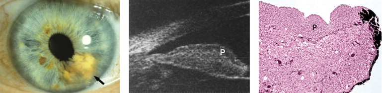
Surface Plaque
Five eyes (36%) had evidence of a surface plaque. The plaque was hyporeflective in 3 cases: 2 mixed epithelioid and spindle cell melanomas and 1 spindle cell melanoma. The plaque was hyperreflective in 2 mixed cell melanomas with a predominance of epithelioid cells. Figure 1 represents an example of a mixed cell melanoma with a hyporeflective plaque on UBM, which corresponded on histopathology to a layer of tightly packed tumor cells of mainly spindle type.
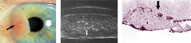
Tumor Vascularity
Tumor vascularity showed 2 patterns on UBM: 1 with small hyperechoic dots relating to small-caliber vessels in the iris stroma and the other with large hypoechoic spaces relating to larger vascular spaces ( Figure 2 ). All cases had small hyperreflective blood vessels and 3 spindle cell melanomas had large hyporeflective ones.

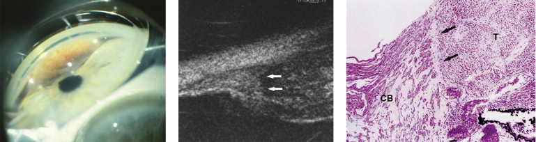
Tumor/Ciliary Body Interface
The tumor interface with the ciliary body was seen on UBM as a line of change in the internal reflectivity and correlated with the pathologic interface between tumor cells and the normal ciliary muscles. In 5 cases (36%) UBM was able to demonstrate the interface between the tumor and ciliary body. This finding was confirmed on histopathology after the tumor had been removed ( Figure 3 ).
Integrity of the Iris Pigment Epithelium
In 1 spindle cell melanoma, UBM was able to demonstrate a focal area of disruption of the highly reflective iris pigment epithelium, suggesting tumor breakthrough and posterior growth. Histopathologic examination confirmed this finding and showed splitting of the iris pigment epithelium by tumor cells ( Figure 4 ).
Extrascleral Extension
In 1 melanoma case, the patient had been followed for 12 years for a diffuse pigmented iris lesion and developed 2 episcleral pigmented nodules. UBM demonstrated an intrascleral emissary canal connecting the tumor with 1 of the nodules, suggesting extrascleral extension ( Figure 5 ). Histopathologically, the canal was found to contain melanoma cells and these cells were intrascleral and extravascular.
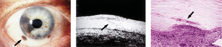
Table 2 compares the UBM appearances and histopathologic findings.
| Case Number | UBM Features | Pathologic Features |
|---|---|---|
| 1 |
|
|
| 2 |
|
|
| 3 |
|
|
| 4 |
|
|
| 5 |
|
|
| 6 |
|
|
| 7 |
|
|
| 8 |
|
|
| 9 |
|
|
| 10 |
|
|
| 11 |
|
|
| 12 |
|
|
| 13 |
|
|
| 14 |
|
|
Stay updated, free articles. Join our Telegram channel

Full access? Get Clinical Tree


