Thyroid-Associated Orbitopathy: An Evidence-Based Approach to Diagnosis and Management


Thyroid-Associated Orbitopathy: An Evidence-Based Approach to Diagnosis and Management


Harmeet S. Gill MD, FRCSC


I. MANAGEMENT OF GRAVES’ DISEASE


Overview

Graves’ disease (GD) is a systemic, organ-specific, autoimmune condition that primarily affects the thyroid gland, pretibial skin, and the orbit. The endocrinologic manifestations should be distinguished from the ophthalmologic manifestations. Although they tend to be closely associated and typically present within months of one another, each appear to run an independent clinical course. In this chapter, the endocrinologic manifestations are referred to as dysthyroidism and the ophthalmologic manifestations as thyroid-associated orbitopathy (TAO). About 30% to 50% of GD patients will demonstrate signs of TAO during their course of disease.


Demographics

GD is typically diagnosed between the third to fifth decades of life, with a possible second peak incidence during the seventh decade of life. Women are four to eight times more likely than men to be affected.1,2 The incidence of TAO is 14 per 100,000 in adults,3 0.1 per 100,000 in prepubescent children, and 3.0 per 100,000 in postpubescent children.4 There is no known pattern of inheritance, but a genetic predisposition to autoimmune disease triggered by environmental factors is likely.5 The most significant environmental factor is cigarette smoking. Genes such as CTLA4, TNF, CD40, PTPN22, and ICAM1 may contribute to disease susceptibility.6,7


Thyroid Physiology

The thyroid gland produces hormones including thyroxine (T4) and triiodothyronine (T3), which help regulate body temperature, energy levels, sleep, appetite, metabolic rate, and many other bodily functions. The pituitary gland produces thyrotropin, also called thyroid-stimulating hormone (TSH), when serum concentrations of T4 and T3 are low and decreases TSH production when levels are high. The thyroid gland has receptors for TSH (TSH-R) and produces T4 and T3 in response to serum TSH concentration.


Graves’ Pathophysiology

Thyroid gland follicular cells and orbital fibroblasts share a common antigen, the TSH-R.8,9,10 Autoantibodies known as thyroid-stimulating immunoglobulins (TSI) or thyrotropin receptor antibodies bind TSH-Rs. “Activating” antibodies are more prevalent and result in T4 and T3 overproduction (hyperthyroidism), while “blocking” antibodies found in up to 15% of subjects can result in hypothyroidism. Some patients will develop goiter or thyroid gland enlargement. TAO occurs because an immune response, consisting of both humoral and cell-mediated pathways, is triggered. Autoreactive T-lymphocytes target orbital fibroblasts and then antigen-presenting cells, including B-lymphocytes, release cytokines and chemokines that activate cell-surface receptors like CD-40. This results in overproduction of hyaluronan, a glycosaminoglycan (GAG) secreted by the connective tissue network
surrounding extraocular muscles.11 The hydrophilic GAGs cause edematous expansion of eye muscles and other orbital tissues. Some orbital fibroblasts and muscle cells will differentiate into adipocytes (adipogenesis), further increasing the orbital volume.12 A heterogeneous population of orbital fibroblasts among patients may provide the basis for variability in clinical activity. In addition to their function as antigen-presenting cells, B-cells are also precursors to antibody-producing plasma cells.

Histological findings of TAO include fibroblast proliferation, lymphocyte and plasma cell infiltration, GAG accumulation, edema, and fibrosis.13,14 The most common immune cells that infiltrate orbital tissue are T-lymphocytes, B-lymphocytes, and mast cells.14 A second orbital antigen implicated in TAO is insulin-like growth factor 1 receptor (IGF-I-R).15 Autoantibodies against IGF-I-Rs may be responsible for the chemokine release that triggers an orbit-specific homing signal to the rest of the immune system.16 Bone marrow-derived fibrocytes are fibroblast-like cells detectable in orbital tissue of patients with GD that recruit various inflammatory mediators.17 They are absent in healthy subjects. Some in vitro studies suggest that oxidative stress and high concentrations of oxygen-free radicals are also involved in TAO pathogenesis.18,19 The underlying molecular and immune pathways that eventually lead to spontaneous resolution of disease remain unknown.


Clinical Features of Systemic Disease

Symptoms from GD can present suddenly or progress over longer periods of time delaying diagnosis. Dysthyroidism most commonly manifests as hyperthyroidism (90% or greater), but some patients will be euthyroid or hypothyroid or have Hashimoto’s thyroiditis.3 Signs and symptoms of hyperthyroidism are summarized in Table 17.1. Pretibial myxedema presents as a nonpainful erythematous and indurated dermatopathy over the shins.


TABLE 17.1 Clinical features of hyperthyroidism


Tachycardia


Palpitations


Anxiety


Tremulousness


Thyroid gland enlargement


Sensitivity to heat, diaphoresis


Hair thinning or loss


Weight loss with increased appetite


Diarrhea


Insomnia


Difficulty concentrating


Fatigue and muscle weakness


Brittle nails


Menstrual irregularity in women


Breast enlargement in men


Thyroid storm (rare but fatal)


Clinical Features of Thyroid-Associated Orbitopathy

Orbital inflammation and fibrosis can cause significant disfigurement of the eye, vision loss, and decreased quality of life. Most patients (90%) will demonstrate upper eyelid retraction, many (70%) will demonstrate periorbital edema, some (30%) will demonstrate restrictive strabismus and diplopia, and few (5% or less) will suffer decreased visual function from compressive optic neuropathy.2,3 The common manifestations of TAO are summarized in Table 17.2. Bilateral symmetrical or asymmetrical disease can occur (see Figs. 17.1, 17.2 and 17.3A). Upper eyelid retraction is initially caused by beta-adrenergic stimulation from thyrotoxicosis but persists because of inflammation and scarring of the levator palpebrae superioris muscle. Inflammation of the conjunctiva, extraocular muscles, and orbital fat results in chemosis, diplopia, and exophthalmos, respectively. Exophthalmos exacerbates upper and lower eyelid retraction. Upper eyelid lag on downgaze, lagophthalmos, and exposure keratopathy are common findings. Enlargement of multiple extraocular muscles with relative
sparing of muscle insertions is typical.20 The inferior and medial recti tend to be enlarged more than the superior and lateral recti. The enlarged recti muscle bellies are best visualized on a coronal orbital image (see Fig. 17.4). Abnormally enlarged extraocular muscles are seen in up to 90% of patients radiographically, but diplopia occurs in only one-third.21 Imaging is helpful to distinguish fat-predominant versus muscle-predominant orbital expansion. Significant crowding at the orbital apex can result in congestive and compressive optic neuropathy. This is particularly dangerous in the absence of exophthalmos. Signs of compressive optic neuropathy include decreased visual function, optic disc edema or pallor, color desaturation, or an afferent pupillary defect. TAO is more severe in older patients and in males.22 Compressive optic neuropathy does not occur in children.4 Rarely, patients suffer concomitant autoimmune myasthenia gravis, which presents as variable blepharoptosis, diplopia, or saccadic fatigue.


TABLE 17.2 Manifestations of thyroid-associated orbitopathy


Anterior segment


Exposure keratopathy


Conjunctival hyperemia


Chemosis


Elevated intraocular pressure


Posterior segment


Optic disc edema or pallor (rare)


Eyelids


Upper eyelid retraction (most common)


Lower eyelid retraction


Eyelid lag on downgaze


Eyelid edema/fullness


Lagophthalmos


Orbit


Restrictive strabismus and diplopia


Exophthalmos


Globe subluxation


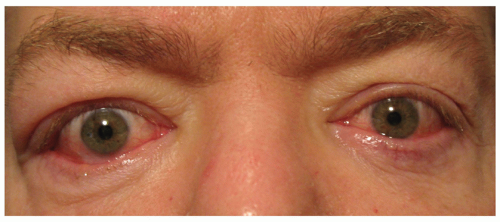
FIGURE 17.1 Mild-to-moderate asymmetrical disease. Bilateral upper eyelid swelling, conjunctival injection, and chemosis. Right upper and lower eyelid retraction.


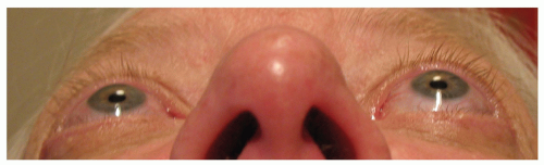
FIGURE 17.2 Moderate-to-severe asymmetrical disease. Submental view of left exophthalmos.


Natural History of Disease

A period of worsening signs and symptoms lasts for 6 to 12 months followed by a plateau and burning-out period over 18 to 24 months.23 The “inflammatory” or “active” phase represents maximum activation of the immune system, which is followed by a period of fibrosis and scarring. After the disease has burnt out, the term “inactive” or “quiescent” disease may be used. There are some rare reports of a chronic or relapsing disease course.24 Cigarette smoking, including second-hand exposure, increases the risk, severity, and duration of TAO by tenfold.25,26


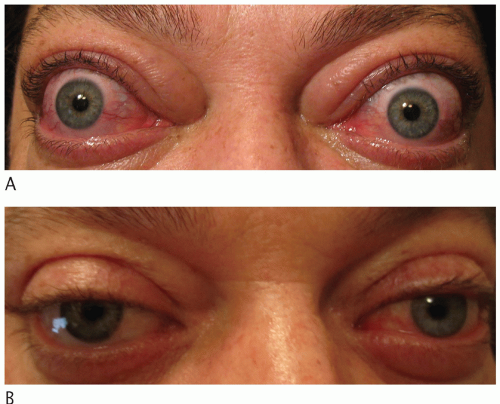
FIGURE 17.3 (A) Vision-threatening symmetrical disease. Upper and lower eyelid swelling and retraction, conjunctival injection and chemosis, restrictive strabismus and exophthalmos, and compressive optic neuropathy. (B) Six months after high-dose steroids, surgical decompression, strabismus repair, and eyelid retraction repair.


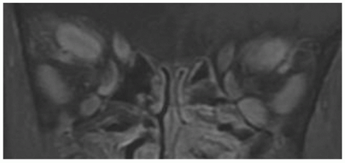
FIGURE 17.4 Coronal T1-weighted, fat-suppressed, gadolinium-enhanced magnetic resonance image of enlarged extraocular muscle bellies from vision-threatening thyroid-associated orbitopathy.


Ophthalmologic Examination

A comprehensive evaluation includes testing visual function, pupillary responses, intraocular pressure, optic discs, extraocular motility, exophthalmometry, cornea, conjunctiva, margin-to-reflex distances, and determination of lagophthalmos for each eye. Visual function tests include best-corrected visual acuity, color testing (red saturation or Ishihara plates), and perimetric testing. An important objective of this evaluation is to determine both “activity” and “severity” of disease.

Disease activity refers to the degree of inflammatory reaction to autoantigen presently taking place. This was quantified in the late 1970s using an ordinal score called the NOSPECS index (N, no signs or symptoms; O, only signs, no symptoms; S, soft-tissue involvement; P, proptosis; E, extraocular muscle involvement; C, corneal involvement; S, sight loss).27 This index was later modified to a continuous total eye score.28 Since the late 1990s, the clinical activity score (CAS) developed by Mourits et al. has become popular.29 The European Group on Graves’ Orbitopathy (EUGOGO) consensus statement recommends a CAS of three or more out of seven (the first seven clinical parameters) as a threshold for active disease (see Table 17.3).30

Disease severity describes the actual physical sequelae (i.e., optic neuropathy, exophthalmos, and eyelid retraction) that occur and can be categorized as vision-threatening, severe, moderate, or mild. These sequelae are present during active or inactive disease.


Work-Up and Treatment for Dysthyroidism

From the panel of possible thyroid function tests, the most valuable for diagnosing and monitoring dysthyroidism are serum-free T4 and TSH. Most patients with GD are
comanaged with internists or endocrinologists whose goals are to stabilize thyroid function with antithyroid drugs (ATDs) and to help in the decision-making process regarding if and when thyroid ablation is required. Oral thioamides (methimazole, carbimazole, or propylthiouracil) are commonly used, and randomized, controlled studies support initial therapy with methimazole for efficacy and tolerability.31 The ATD administration regimens are either titration or a block-and-replace strategy. Thyroid ablation, whether by surgical thyroidectomy or radioiodine (I-131), is typically used when thyroid levels are unstable on ATDs. Thyroidectomy (total, near total, or subtotal) may be preferable for patients that exhibit a goiter or thyroid gland enlargement, but risks include hypoparathyroidism and permanent damage to the recurrent laryngeal nerve. The main disadvantage of radioiodine ablation is the increased risk and severity of TAO in susceptible patients.32 This phenomenon is worse in smokers and likely related to the high concentration of thyroid antigens released during necrosis of thyroid follicular cells. An oral administration of 5 to 15 mCi results in an absorbed radiation dose of 50 to 100 Gy. A study of 450 patients shows that the rate for TAO progression during radioiodine ablation is 15% but can be mitigated with a short course (1 to 3 months) of oral steroids.33 Posttreatment hypothyroidism should also be avoided.34 Patients who are nonsmokers and lack evidence of TAO can probably undergo radioiodine ablation without steroid prophylaxis. A recent randomized, placebo-controlled study found that Ginkgo biloba extract decreases radiation-induced genotoxic damage during radioiodine therapy.35


TABLE 17.3 Clinical activity score (CAS) to quantify disease activity


PAIN


1.


Retrobulbar pain


2.


Pain on attempted up- or downgaze


REDNESS


3.


Redness of the eyelids


4.


Redness of the conjunctiva


SWELLING


5.


Swelling of the eyelids


6.


Chemosis


7.


Swollen caruncle or plica


IMPAIRMENT


8.


Increase in exophthalmos by ≥2 mm over 1-3 mo


9.


Decrease in eye movement in any direction by > five degrees over 1-3 mo


10.


Decrease in pinhole visual acuity by 1 or more lines on the Snellen chart over 1-3 mo


The European Group on Graves’ Orbitopathy (EUGOGO) recommends a CAS of three or more out of seven (the first seven clinical parameters) as a threshold for active disease. Bartalena et al.30 [www.eugogo.eu].


Currently, the choice of treatment for dysthyroidism is based on expert opinion rather than level 1 evidence.36 There is an ongoing debate regarding whether or not thyroid ablation has any effect on TAO progression. Removing the bulk of shared antigens should theoretically reduce the severity of disease.37 Total thyroid ablation (TTA) is a combined strategy in which surgical thyroidectomy is followed immediately by radioiodine ablation for remnant tissue. The long-term follow-up data from a randomized study comparing thyroidectomy alone versus TTA showed that TAO symptoms resolved more quickly in the TTA group, but overall outcomes were comparable.38 Euthyroid maintenance seems to prevent TAO progression.39 However, based on the current evidence, thyroid ablation is believed to treat dysthyroidism alone and has little to no effect on the course of ophthalmologic disease.33


Work-Up for Thyroid-Associated Orbitopathy

Antibodies generated against thyroid peroxidase (TPO) and thyroglobulin are detectable in patients with GD. However, the blood test most valuable for monitoring TAO activity is TSI.40 This antibody titer correlates strongly with CAS but does not correlate with thyroid function tests (T4, TSH) or TPO antibody.41 A recent cross-sectional study showed that 98% of 108 patients with active TAO had a positive TSI assay.42 Furthermore, TSI levels were higher in moderate-to-severe versus mild disease (p < 0.001) in this study. A trend-based
analysis of TSI is preferable to using specific values as thresholds for intervention. When the TSI levels begin to decrease and plateau, this indicates disease stabilization. The values typically never return to normal levels. Neuroimaging with computed tomography or magnetic resonance imaging can help confirm the diagnosis of TAO, especially for euthyroid patients (see Fig. 17.4).


Differential Diagnosis

It is not uncommon for patients with dry eye symptoms alone and no prior history of GD or other autoimmune disease to have occult TAO.43 Although the diagnosis of TAO is often apparent, a differential diagnosis includes myasthenia gravis, orbital myositis, orbital tumors, chronic progressive external ophthalmoplegia, and orbital arteriovenous fistulas. Once the diagnosis has been confirmed, TAO activity and severity will determine whether observation alone or treatment with medication, radiation, or surgery is required.


II. TREATMENT OF THYROID-ASSOCIATED ORBITOPATHY


Overview

The purpose of this section is to provide guidelines regarding the treatment of TAO based on the best available evidence. The information has been organized by treatment modality and the implications for practice are highlighted prior to a detailed review of the relevant studies. Areas of future research are summarized at the end of each subsection. (See Appendix A for a summary of clinically relevant studies of treatment for TAO.)


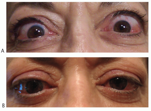
FIGURE 17.5 (A) Bilateral upper eyelid retraction left greater than right. (B) Two months after bilateral upper eyelid recession (posterior transconjunctival mullerectomy).

Smoking cessation is the single most important intervention for every patient regardless of disease severity.44 The majority of patients can be managed conservatively with close monitoring and reassurance while awaiting spontaneous recovery. Some patients will require an immunosuppressive or surgical intervention for vision-threatening disease, eyelid swelling, exophthalmos, diplopia, or exposure. The order of surgical intervention is to perform orbital decompression first, followed by strabismus surgery, followed by eyelid surgery as needed (see Figs. 17.3 and 17.5). Some patients will eventually require a combination of these procedures for functional and/or aesthetic rehabilitation.45 Any surgical intervention performed during the active phase will worsen orbital inflammation, and so rehabilitative surgery is considered after quiescence. Vision-threatening cases are initially managed with high-dose steroid (HDS) therapy with some requiring adjunctive surgical decompression of bony orbital walls and intraorbital fat. Retrobulbar irradiation (RI) is less effective as a first-line intervention but may be useful in refractory cases. One approach to quantification of treatment response is presented by Bartalena et al.
(EUGOGO) using major and minor criteria.33 (See Table 17.4 for an overview of management options based on disease severity.)


TABLE 17.4 Management of thyroid-associated orbitopathy based on disease severity


Vision-threatening disease (compressive optic neuropathy or severe corneal exposure)


High-dose steroid therapy


– intravenous > oral


Surgical orbit decompression


– bony walls (1 -, 2-, or 3-wall)


– intraorbital fat


Retrobulbar irradiation


Newer biologics (rituximab)


Severe disease (restrictive strabismus)


Prismatic correction


Botulinum A toxin injection


Strabismus surgery


Moderate disease (eyelid retraction with exposure keratopathy)


Botulinum A toxin injection


Upper eyelid recession


Lower eyelid elevation (± spacer graft, ± midface elevation)


Mild disease (dry eyes, mild keratopathy, chemosis, eyelid edema, mild eyelid retraction)


Ocular lubricants (tears, ointments)


Selenium supplementation


Immunosuppressive Therapy

The primary goal of TAO management is to minimize inflammatory damage while awaiting disease resolution. Systemic glucocorticoid therapy is the most effective intervention to accomplish this goal. Steroids directly inhibit the proinflammatory cytokines and other inflammatory mediators released during the active disease phase, but confer no benefit during the inactive fibrotic phase. They improve soft-tissue swelling, motility disturbance, and compressive optic neuropathy but do not significantly decrease exophthalmos. Both orally administered and intravenous (IV) pulses of HDSs can be used, but the latter seems to be better tolerated and more effective.46 Many retrospective and nonrandomized studies found IV more effective than oral administration, but had drawbacks such as use of oral agents during the interpulse period or concomitant radiotherapy.47,48,49,50 A prospective, randomized, controlled study comparing IV and oral HDS over 12 weeks for moderate-to-severe TAO found that IV administration resulted in a more rapid and significant improvement in CAS (p < 0.01), exophthalmos (p < 0.038), extraocular muscle changes (p < 0.02), optic neuropathy (p < 0.001), intraocular pressure (p < 0.04), visual acuity (p < 0.03), quality of life (p < 0.0001), and overall treatment response (72% versus 49%) (p < 0.001).51 In another single-blind, prospective, randomized, controlled study, 70 patients with active and severe TAO received either 0.5 g of methylprednisolone IV once weekly for 6 weeks then 0.25 g for 6 weeks (cumulative dose 4.5 g) or 100 mg daily of oral prednisone for 1 week tapered by 10 mg per week for a total of 12 weeks.52 The IV group had a 77% response rate compared with 51% for oral HDS (p < 0.01). One biological explanation for this finding is that IV therapy more effectively reduces circulating dendritic cells, which are potent antigen-presenting cells involved in the primary immune response of TAO.53 IV HDSs also significantly decrease serum TSH-R autoantibody levels in patients with GD.54 The response rate achieved using IV pulse therapy is about 80% compared with 50% for oral HDSs.30 However, the specific dosing strategies for IV administration are variable.51,55 Weekly pulse therapy is probably preferable to consecutive daily administration in order to minimize the risk of adverse events, the most significant being hepatic failure.56 Hepatic failure occurs when the cumulative dose of IV methylprednisolone exceeds 8 g.57 The morbidity and mortality of IV HDSs are 6.5 and 0.6%, respectively.56 Patients should be followed closely for adverse events, and any indication of hepatic dysfunction demands liver enzyme testing. Less serious and more common side effects include weight gain, cushingoid features, hypertension, hirsutism, psychosis, osteoporosis, and gastrointestinal (GI) problems.28 These are all worse with oral compared with IV administration.51 Prophylactic H2 receptor blockers are helpful to
reduce GI symptoms. Overall, HDSs decrease the quality of life for most patients.58 Contraindications to HDSs include uncontrolled diabetes, severe hypertension, recent hepatitis, and pregnancy. As the disease stabilizes and the dose is tapered, rebound inflammation can occur.30

Only gold members can continue reading. Log In or Register to continue

Stay updated, free articles. Join our Telegram channel

Aug 2, 2016 | Posted by in OPHTHALMOLOGY | Comments Off on Thyroid-Associated Orbitopathy: An Evidence-Based Approach to Diagnosis and Management

Full access? Get Clinical Tree

Get Clinical Tree app for offline access