Swallowing is a complex physiologic function that involves precisely coordinated movements within the oral cavity, pharynx, larynx, and esophagus. This article reviews the anatomy, muscular control, and neurophysiological control of normal, healthy swallowing.
Key points
- •
Swallowing is a rapid yet complex sequence of movements with 4 primary phases: oral preparatory, oral transport, pharyngeal, and esophageal.
- •
These phases are primarily coordinated by 5 cranial nerves and 3 peripheral nerves, which are mediated centrally via the medulla oblongata of the brain stem.
- •
In addition to the brain stem, the cortical and subcortical regions of the brain play an integral part in mediating the swallow, especially the oral preparatory and oral transport phases.
- •
Impaired sensory or motor ability at any level impairs the efficiency of swallow physiology; impairment that affects the mechanisms responsible for airway protection also affects swallow safety.
Introduction
Swallowing is a normal physiologic function that is often taken for granted. As humans, we swallow an average of 500 times per day. A normal swallow requires the precise coordination of more than 30 muscles located within the oral cavity, pharynx, larynx, and esophagus ( Table 1 ). Muscle movements are controlled by several cranial (CN V, VII, and IX-XII) and peripheral (C1-C3) nerves and are coordinated within the brain stem (mainly medulla oblongata, where a network of sensory nuclei, motor nuclei, and interneurons form what is known as the “swallowing center” ). Often, individuals with neurologic and/or structural abnormalities of the head and neck due to causes such as stroke, Parkinson’s disease, or cancer, experience problems with swallowing, or dysphagia. Dysphagia can lead to several negative outcomes, including pneumonia, malnutrition, dehydration, and reduced quality of life. As a result, rehabilitation is often required to prevent negative outcomes and ensure, where possible, safe and adequate oral intake. To understand the processes involved in rehabilitating a disordered swallow, it is imperative to first understand the anatomy, muscular control, and neurophysiological control of a normal, healthy swallow.
| CN | Cranial nerve |
| DSG | Dorsal swallowing group |
| FEESST | Fiberoptic endoscopic evaluation of swallowing with sensory testing |
| ISLN | Internal branch of the superior laryngeal nerve |
| LAR | Laryngeal adductor response |
| LCA | Lateral cricoarytenoid |
| LCR | Laryngeal cough reflex |
| LES | Lower esophageal sphincter |
| SLN | Superior laryngeal nerve |
| UES | Upper esophageal sphincter |
| VSG | Ventral swallowing group |
| Category | Muscle Name | Innervation | Attachments | Function |
|---|---|---|---|---|
| Muscles of the face | Orbicularis oris | CN VII | Maxilla; mandible; mucous membrane of lips | Closes and protrudes lips |
| Buccinator | CN VII | Maxilla and mandible (alveolar process); pterygomandibular raphe; orbicularis oris | Flattens and compresses cheek | |
| Muscles of mastication | Temporalis | CN V | Temporal fossa of the parietal bone; mandible (coronoid process) | Elevates mandible |
| Masseter | CN V | Zygomatic bone; zygomatic arch; lateral surface of ramus of mandible | Elevates mandible | |
| Medial pterygoid | CN V | Medial surface of lateral plate (pterygoid process); palatine bone (pyramidal process); maxilla (tuberosity and pyramidal process) | Elevates mandible | |
| Lateral pterygoid | CN V | Greater wing of sphenoid bone; lateral surface of lateral plate (pterygoid process); mandible (condolyoid process) | Moves mandible laterally (rotary chew); depresses and protrudes mandible | |
| Intrinsic muscles of the tongue | Superior longitudinal | CN XII | Median septum of tongue; submucosal connective tissue of tongue; mucous membrane of tongue | Shortens tongue; raises tip and lateral margins of tongue |
| Inferior longitudinal | CN XII | Root of tongue; hyoid bone; apex of tongue | Shortens tongue; pulls tongue tip down | |
| Transverse | CN XII | Median septum of tongue; submucosal connective tissue on lateral margins of tongue | Narrows and elongates tongue | |
| Verticalis | CN XII | Submucosal connective tissue on dorsal surface and ventral regions of tongue | Flattens and widens tongue | |
| Extrinsic muscles of the tongue | Genioglossus | CN XII | Mandible (superior mental spine); hyoid bone; dorsum of tongue | Depresses center of tongue (groove); protrudes tongue |
| Hyoglossus | CN XII | Hyoid bone; lateral aspect of tongue | Depresses and retrudes tongue | |
| Styloglossus | CN XII | Styloid process; styloid ligament; lateral surface of tongue | Elevates and retracts tongue | |
| Palatoglossus | CN X | Palatine aponeurosis; lateral margin of tongue | Depresses soft palate; moves palatoglossal fold toward midline; elevates back of tongue | |
| Muscles of the soft palate | Levator veli palatini | CN X | Temporal bone; palatine aponeurosis | Elevates soft palate |
| Musculus uvulae | CN X | Posterior nasal spine; palatine aponeurosis; mucosa of uvula | Elevates and retracts uvula | |
| Tensor veli palatini | CN V | Medial pterygoid plate (sphenoid bone); palatine aponeurosis | Tenses soft palate; opens pharyngotympanic tube | |
| Pharyngeal musculature | Superior pharyngeal constrictor | CN X | Pharyngeal raphe; medial pterygoid plate (pterygoid hamulus); pterygomandibular raphe; mandible | Constricts pharynx |
| Middle pharyngeal constrictor | CN X | Pharyngeal raphe; hyoid bone; stylohyoid ligament | Constricts pharynx | |
| Inferior pharyngeal constrictor | CN X | Pharyngeal raphe; cricoid cartilage; thyroid cartilage; crosses cricothyroid muscle | Constricts pharynx | |
| Stylopharyngeus | CN IX | Styloid process (temporal bone); pharyngeal wall | Elevates pharynx | |
| Salpingopharyngeus | CN X | Pharyngotympanic tube; pharyngeal wall | Elevates pharynx | |
| Palatopharyngeus | CN X | Palatine aponeurosis; lateral pharyngeal wall | Elevates pharynx; moves posterior pharyngeal wall toward midline | |
| Suprahyoid muscles | Mylohyoid | CN V | Medial body of mandible; hyoid bone | Elevates hyoid, floor of mouth |
| Geniohyoid | CN XII; C1-2 | Anterior body of mandible; hyoid bone | Fixed mandible, pulls hyoid forward; fixed hyoid, depresses and retracts mandible | |
| Digastric (anterior) | CN V | Anterior body of mandible; intermediate tendon; hyoid bone | Fixed mandible, elevates hyoid; fixed hyoid, depresses mandible | |
| Digastric (posterior) | CN VII | Temporal bone (mastoid process); intermediate tendon; hyoid bone | Elevates and retracts hyoid bone | |
| Stylohyoid | CN VII | Temporal bone (styloid process); hyoid bone | Elevates hyoid | |
| Muscles of the larynx | Lateral cricoarytenoid | CN X | Arch of cricoid cartilage; vocal process of arytenoid cartilage | Adducts vocal folds and closes off/protects airway |
| Transverse arytenoid | CN X | Arytenoid cartilage on one side; contralateral arytenoid cartilage | Adducts vocal folds and closes off/protects airway (especially at posterior commissure) | |
| Thyroarytenoid | CN X | Inner surface of thyroid cartilage (anterior); arytenoid cartilage (anterior surface) | Helps to close off airway by narrowing laryngeal inlet | |
| Infrahyoid muscles | Sternothyroid | Ansa cervicalis (C1-C3) | Manubrium of sternum; thyroid cartilage | Depresses larynx (and hyoid) |
| Sternohyoid | Ansa cervicalis (C1-C3) | Manubrium of sternum; hyoid bone | Depresses hyoid | |
| Thyrohyoid | CN XII; C1 | Thyroid cartilage; hyoid bone | Depresses hyoid; elevates larynx | |
| Omohyoid (superior and inferior bellies) | Ansa cervicalis (C1-C3) | Scapula; hyoid bone | Depresses and retracts hyoid | |
| Muscles of the upper esophagus | Inferior fibers of the inferior pharyngeal constrictor | CN X | Cricoid cartilage; pharyngeal raphe | — |
| Cricopharyngeus | CN IX, X | Lateral aspects of cricoid cartilage | Contracted at rest to prevent reflux; relaxes during swallow to allow bolus to pass from pharynx into esophagus | |
| Upper fibers of the esophagus | CN X | Lower borders of cricoid cartilage | — |
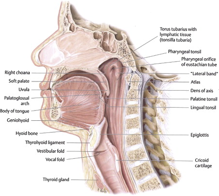
Anatomy and contributing landmarks
Many structures within the oral cavity, pharynx, and esophagus are involved in swallowing, including several bones, cartilages, teeth, spaces, salivary glands, and muscles. Familiarity with these structures is a necessary first step to differentiate between normal and disordered swallowing ( Fig. 1 ).
Bones and Cartilages
Bones such as the mandible, maxilla, hard palate, hyoid bone, cervical vertebrae (C1-C7), and skull (specifically the styloid and mastoid processes of the temporal bone) are critical during swallowing in that they support and stabilize the involved muscles and aid in mastication. Similarly, cartilages such as the thyroid cartilage, cricoid cartilage, arytenoids, and epiglottis are important during swallowing. Cartilages, like bones, provide support for several of the muscles that are involved in mastication as well as lingual and pharyngeal bolus transport. They anchor the muscles that protect the airway as the liquid or food bolus traverses the pharynx. In particular, the epiglottis deflects downward, thereby directing the oncoming bolus away from the airway and into the esophagus.
Teeth
A healthy adult has 32 permanent teeth, all of which are critical to bolus preparation. The incisors are used for cutting and biting, and the molars are used for grinding solid food. Any disruption to an individual’s dentition can affect his/her ability to consume a typical adult diet.
Spaces
As a point of reference when describing the swallow, the upper aerodigestive tract is divided into 4 main areas or spaces: the oral cavity, nasopharynx, oropharynx, and hypopharynx ( Fig. 2 ). Within each of these main spaces, there are several smaller spaces through which fluids and foods pass during a normal swallow, namely, the valleculae and pyriform sinuses. There are other spaces that are effectively sealed during the swallow and hence make no contact with the ingested bolus. These include the lateral and anterior sulci, laryngeal vestibule, and laryngeal ventricle. After the completion of the swallow, residue of the liquid or solid bolus in any of these spaces indicates dysphagia.


