The First Anti-VEGF Therapy: Pegaptanib Sodium
Anthony P. Adamis
INTRODUCTION
Until recently, the treatment of ocular neovascular diseases such as age-related macular degeneration (AMD), diabetic retinopathy (DR), and retinal vein occlusion (RVO) has been restricted to ablative laser-based procedures and surgical interventions. More than a decade of research has identified a central role for vascular endothelial growth factor (VEGF) in the etiology of these diseases, resulting in revolutionary new treatments based on inhibiting VEGF. Two anti-VEGF agents are approved as intravitreal therapies for neovascular AMD. Pegaptanib sodium, the first approved agent, is an RNA aptamer that binds the VEGF165 isoform while ranibizumab is a monoclonal antibody antigen-binding fragment that binds all VEGF isoforms. Bevacizumab, a related monoclonal antibody, also is being evaluated for the treatment of ocular neovascular diseases in off-label studies. This chapter will focus on the development and clinical application of pegaptanib.
VEGF AS A THERAPEUTIC TARGET IN OCULAR NEOVASCULAR DISEASE
The importance of VEGF in ocular neovascular diseases stems from its key properties as a regulator of physiological and pathological angiogenesis (1) and as a potent promoter of vascular permeability (2). As such, it contributes to two proximate causes of vision loss: the growth of aberrant retinal vasculature and macular edema. Clinical studies established that vitreous levels of VEGF were elevated in a number of ocular neovascular conditions (3, 4, 5 and 6) while preclinical work with several animal model systems demonstrated that such elevations were both necessary and sufficient for the development of ocular neovascularization (7,8). Studies using animal models of diabetes also were important in establishing the role of VEGF in promoting the increased retinal leukostasis characteristic of DR believed to mediate much of the damage to the retinal vasculature associated with this disease (9).
Alternative splicing of the VEGF gene generates six principal isoforms (1,10) with VEGF165 and VEGF121 being the most common variants in normal eyes (11). Preclinical evidence suggests that VEGF165 is especially pathogenic in that it was dramatically upregulated in a rodent model of ischemic neovascularization (12) and had more potent proinflammatory properties than VEGF121 (13). Such properties included chemoattraction of monocytes (13), which amplifies neovascularization induced by ischemia (12) or laser wounding (14, 15 and 16), and upregulation of retinal expression of intercellular cell adhesion molecule-1 (13), a mediator of retinal leukostasis in DR (9). These data suggested that specific inactivation of VEGF165 could significantly ameliorate the effects of VEGF in ocular neovascular diseases. This concept was supported by two key preclinical observations in rodent models: not only was intravitreal pegaptanib as effective as a VEGF-receptor (R)-Fc fusion protein that binds all VEGF isoforms in suppressing ischemic ocular neovascularization (12) but it was also capable of reversing the diabetes-induced breakdown of the blood-retinal barrier (17).
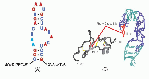 Figure 37-1. A: The sequence and predicted secondary structure of pegaptanib with 2′-O-methylated purines shown in red, 2′-fluorine-modified pyrimidines shown in blue, and unmodified ribonucleotides shown in black. The nucleotide modifications were made to increase bioavailability. The site of attachment of a 40 kDa polyethylene glycol (PEG) moiety is shown. B: The interaction between the 55 amino acid heparin-binding domain of VEGF165 and pegaptanib. The free heparin-binding domain of VEGF165 is shown in gray with disulfide bonds in yellow. Pegaptanib is shown in teal, with the interaction between cysteine-137 of VEGF165 and uridine-14 of the aptamer indicated in red. (Reproduced from Ng EW, Shima DT, Calias P, et al. Pegaptanib, a targeted anti-VEGF aptamer for ocular vascular disease. Nat Rev Drug Discov 2006;5:123-132, with permission.). |
VEGF exerts a wide range of actions both in the eye, where it is important in the maintenance of retinal neurons (18) and the choriocapillaris (19), and in the nervous system, kidney, bone, and liver (9,20 and 21). Furthermore, the aberrant neovasculature found in AMD and DR is especially permeable, increasing the potential for systemic exposure to intravitreal anti-VEGF agents (22), as evidenced by the detection of therapeutic effects in the fellow eyes following anti-VEGF therapies in DR (23) and AMD (24). The safety of nonselective versus selective anti-VEGF therapies will be explored in the clinical section of this review.
DEVELOPMENT OF PEGAPTANIB AS AN ANTI-VEGF OCULAR THERAPEUTIC AGENT
Pegaptanib is an RNA aptamer that was developed through three applications (25, 26 and 27) of the SELEX [systematic evolution of ligands by exponential enrichment] procedure (28,29) using VEGF165 as a target. Chemical modifications of the constituent nucleotides were used to improve its nuclease resistance and affinity, and two 5′ polyethylene glycol substituents were added to increase bioavailability (30). Pegaptanib was found to block the binding of VEGF165 to endothelial cell receptors, to inhibit VEGF165-mediated cell signaling and proliferation (31), and to reduce VEGF165-induced vascular permeability (27,32) while not affecting responses to VEGF121 (31). Pegaptanib’s selectivity for VEGF165 derives from its interaction with the heparin-binding domain of VEGF165 (33), which is not present in VEGF121 (10). The predicted secondary structure of pegaptanib and its interaction with the heparin-binding domain of VEGF165 are shown in Figure 37-1 (30).
Initial pharmacokinetic studies of pegaptanib performed in monkeys found mean half-lives after intravitreal and intravenous administration of 94 hours and 9.3 hours, respectively, suggesting that clearance from the eye is rate-limiting; pegaptanib remained fully active in VEGF-binding assays 28 days after intravitreal injection (34). A subsequent clinical study of pegaptanib pharmacokinetics following intravitreal injection evaluated 147 patients with neovascular AMD who received either 1 mg or 3 mg every 6 weeks for 54 weeks (35). Mean maximal plasma concentrations in the 1 mg group ranged from 20 to 24 ng/ml; pegaptanib remained above the level of detection (8 ng/ml) for 1 week with a plasma mean terminal half-life of 10 days. Repeat injections did not result in any plasma accumulations nor were there detectable serum antibodies to pegaptanib (35).
Efficacy and Safety of Pegaptanib in the Treatment of Neovascular AMD
The efficacy and safety of pegaptanib for the treatment of neovascular AMD was established in the pivotal V.I.S.I.O.N. (VEGF Inhibition Study in Ocular Neovascularization) phase 2/3 trials (36). These two concurrent, multicenter, randomized, shamcontrolled, dose-ranging trials enrolled subjects with all angiographic subtypes of neovascular AMD and lesions ≥12 disk areas. Subjects were randomized to receive intravitreal pegaptanib (0.3, 1, or 3 mg) or sham injections every 6 weeks for 54 weeks. For subjects with predominantly classic lesions, photodynamic therapy (PDT) with verteporfin was administered at the investigator’s discretion. The primary endpoint was the proportion of subjects losing >15 letters of visual acuity (VA); secondary endpoints included proportions gaining ≥0, ≥5, ≥10, or ≥15 letters, proportions losing ≥30 letters, mean changes in VA, and proportions progressing to legal blindness (VA 20/200 or worse) in the study eye. Of 1208 subjects enrolled, 1186 (98%) received at least one treatment (mean: 8.5 of a possible 9 injections) and were evaluated at week 54.
Efficacy of Pegaptanib
All pegaptanib doses proved superior to sham according to the primary endpoint. Compared to 55% of sham subjects, proportions losing ≥15 letters for the 0.3 mg, 1 mg, and 3 mg groups were 70% (p < .001), 71% (p < .001), and 65% (p < .03), respectively (36). As no further benefit accrued to higher doses, further analysis was restricted to the 0.3 mg dose. Pegaptanib was superior to sham for all secondary endpoints as well, including proportions losing ≥30 letters (10% vs. 22%, respectively; p < .001) or progressing to legal blindness (38% vs. 56%, respectively; p < .001), mean change in VA (-7.95 vs. -15.05 letters, respectively; p < .05), and gaining ≥0, ≥5, ≥10, or ≥15 letters (p


Stay updated, free articles. Join our Telegram channel
Full access? Get Clinical Tree
