Purpose
To assess the efficacy of a novel ultraviolet (UV) lens device on the killing of Acanthamoeba cysts and the impact of efficacious doses of UV upon soft contact lens parameter and material characteristics.
Design
Prospective, in vitro, experimental study of a device.
Methods
A UV lens device was constructed and used to expose Acanthamoeba cysts to various levels of UV irradiation. Once an efficacious dose, as defined by a greater than 3 log reduction, was determined (130 mJ/cm 2 ), 6 soft contact lens materials (etafilcon A, senofilcon A, galyfilcon A, lotrafilcon A, polymacon, and comfilcon A) were exposed to that dose for 30 cycles and tested for visual parameters, mechanical parameters, and cytotoxicity.
Results
The UV device produced an average log reduction of over 3.5 log of Acanthamoeba cysts when the lens and solution inside of the inset case was irradiated with 130 mJ per cm 2 of UV or greater. After 30 cycles of 130 mJ per cm 2 UV dose each, no gross changes were observed in mechanical properties or cytotoxicity tests in any soft contact lenses tested. In visual parameters, polymacon and lotrafilcon A exhibited a shift in sphere power and diameter, respectively.
Conclusions
The novel UV lens device was able to provide a marked log reduction to Acanthamoeba cysts, one of the most resistant ocular disease–causing organisms found in lens cases, without a detrimental effect on many lens materials.
Contact lens wear is the most common risk factor for the development of microbial keratitis (MK). Microbial contamination of contact lens cases by bacteria, fungi, and amoebae is common. These microbes can adhere to the contact lens and are then transported onto the surface of the cornea when the user inserts his or her lens. Studies have shown that microorganisms isolated from the corneal ulcers of contact lens wearers were the same strains as those isolated from the patients’ lens case, strongly suggesting that contact lens cases are a source of potential pathogenic microbes.
Most cases of contact lens–related MK are attributable to bacteria, with Pseudomonas and Staphylococcus strains being the most common microbes involved. In recent years, however, there has been an increase in the number of cases of keratitis caused by the free-living amoeba Acanthamoeba in the United States. A lthough relatively rare, Acanthamoeba keratitis (AK) is potentially blinding, and approximately 90% of AK patients are contact lens wearers. Acanthamoeba are virtually ubiquitous in nature and are commonly found in dust, soil, swimming pools, air conditioning systems, and domestic tap water. Acanthamoeba exist as a dimorphic organism: the trophozoite, which is the motile, feeding, and replicating form, and the cyst, which is the dormant, resistant form. Acanthamoeba transform into the cyst form (encyst) in unfavorable conditions, such as starvation, exposure to biocides, and changes in osmolarity. Cysts are highly resistant to chemical disinfection, desiccation, and extremes of temperature and can remain viable and pathogenic after decades in storage.
Following an outbreak of AK in soft contact lens wearers in the United States starting in 2004, epidemiology studies found that 21 of the 39 AK cases were found to be associated with the use of a single multipurpose solution (MPS), which resulted in the manufacturer’s voluntary global recall of the product in May 2007. Since the 2007 product recall, the incidence of AK in the United States still remains elevated above previous baseline levels, indicating that additional risk factors are involved in the recent increase in cases of AK, such as users having poor lens care practices (Brown AC. Elevated Acanthamoeba keratitis incidence despite a 2007 outbreak-associated product Recall—A multi-state investigation, 2008–2011. Presented at CDC EIS Conference. April 16, 2012; Atlanta, Georgia.)
Although compliance in lens case hygiene is clearly important, the recent outbreak of AK highlights the need for improved contact lens disinfection systems, in particular against the more resistant cyst form of the organism. MPSs are currently the most common systems used for the care of soft contact lenses, being complex formulations of buffering, chelating, surfactant, wetting, and antimicrobial agents that can vary significantly from one MPS to another. Studies have shown that MPSs can vary in their disinfection efficacy against Acanthamoeba , and this efficacy also varies depending on the strain tested. Some of this variation could, however, be attributable to significant interlaboratory variation, as the current microbiological requirements for contact lens care products do not include efficacy against Acanthamoeba and there are currently no standardized test methods for the evaluation of contact lens disinfectant solutions against Acanthamoeba .
Ultraviolet (UV) light has been proposed as a method for disinfecting contact lenses. Although reports have shown successful UV disinfection of bacteria-contaminated contact lenses, lenses directly exposed to disinfecting levels of UV often exhibit changes in their properties. To remedy the issue of direct UV-induced changes, a commercially available UV device that exposes only the solution to UV while protecting the lens from direct irradiation has been created. The efficacy of this device was shown to be high for bacteria; however, the device exhibited low efficacy against Acanthamoeba cysts. Striking a balance between the germicidal dose of UV light and its ability to alter the polymer of soft contact lenses is a great challenge to the successful use of UV as a contact lens disinfecting agent.
The purpose of this study was to assess the disinfection efficacy of a novel directly irradiating germicidal UV device against Acanthamoeba castellanii ATCC 50370 cysts, while in the presence of a contact lens. A secondary objective was to assess the impact of efficacious (greater than 3 log reduction) doses of UV on soft contact lens parameters and material characteristics. Previous studies have shown that UV treatment on its own is ineffective in killing all Acanthamoeba cysts in a single exposure and that cysts are resistant to UVB up to 800 mJ per cm 2 . The results of this study showed that the novel UV device produced over 3 log reduction of Acanthamoeba cysts when the lens and solution inside of the inset case was irradiated with 130 mJ per cm 2 of UV or greater. This study also highlights methodology that can be used to assess the efficacy of contact lens disinfecting solutions or devices in a regimen assay against Acanthamoeba .
Methods
This study was a prospective, in vitro, experimental study of a novel UV contact lens disinfection device.
Materials
For the construction and testing of the novel UV device, National Institute of Standards and Technology (NIST)-certified UV germicidal detector PMA2122 and data logger PMA2100 were purchased from Solar (Glenside, Pennsylvania, USA). UV bulbs were purchased from LCD Lighting (Orange, Connecticut, USA). Teflon was purchased from Industrial Plastics and Machine (Houston, Texas, USA) and Achieve 1605 polypropylene homopolymer was purchased from ExxonMobil Chemical (Irving, Texas, USA).
For contact lens testing, Table 1 shows the lenses that were used.
| Material | Power (Diopter) | Diameter (mm) | Base Curve (mm) | Purchased From |
|---|---|---|---|---|
| etafilcon A | −1.00 | 14.0 | 8.7 | Johnson & Johnson Vision Care (Jacksonville, Florida, USA) |
| senofilcon A | −1.00 | 14.0 | 8.4 | Johnson & Johnson Vision Care (Jacksonville, Florida, USA) |
| galyfilcon A | −1.00 | 14.0 | 8.3 | Johnson & Johnson Vision Care (Jacksonville, Florida, USA) |
| lotrafilcon A | −1.00 | 13.8 | 8.4 | Ciba Vision (Duluth, Georgia, USA) |
| polymacon | −1.00 a | 14.0 | 8.4 | Bausch & Lomb (Rochester, New York, USA) |
| comfilcon A | −1.00 | 14.0 | 8.6 | Cooper Vision (Fairport, New York, USA) |
a −3.00 diopter lenses were used for parameters testing for polymacon only, owing to lack of availability of -1.00 diopter lenses at the time of testing.
Phosphate-buffered saline (PBS), Eagle’s minimal essential medium (EMEM), and fetal bovine serum (FBS) were purchased from Mediatech (Manassas, Virginia, USA). V79-4 cells (CCL-93), Escherichia coli (8739), and Acanthamoeba castellanii (50370) were purchased from ATCC (Manassas, Virginia, USA). TableCurve2D was purchased from Systat Software (San Jose, California, USA). Flat-bottomed 24- and 96-well tissue culture treated microtiter plates were purchased from Helena Biosciences (Tyne and Wear, UK). Hemocytometers were purchased from Hawksley (Sussex, UK) and all other materials were purchased from VWR (Atlanta, Georgia, USA) or Fisher (Leicestershire, UK).
Novel Device Construction
A circular custom-made germicidal UV bulb was made by LCD Lighting (Orange, Connecticut, USA) to allow for a uniform distribution of UV light to both surfaces of the lenses. A Teflon piece was machined by First Cut (Maple Plain, Minnesota, USA) to hold the inset case and UV bulb. A piece of aluminum was placed beneath the Teflon and UV bulb. Electronic components to power the UV bulbs were created and purchased from Turnkey Electronics (Melbourne, Florida, USA). Inset cases were injection molded by Tool Room Express (Binghamton, New York, USA) using polypropylene.
Ultraviolet Dose Quantitation and Verification
The UV dose within the inset case was calculated as described in Putt and associates. Briefly, UV energy from the bulbs was measured using a UV germicidal detector. A photodegradable dye, erythrosine B (10 μg/mL), was added to the wells of the inset case and irradiated. The percent absorbance remaining of the dye after exposure to several different doses of UV light as measured by the UV sensor was determined. A standard curve was generated correlating the external UV dose measured via the sensor to the actual UV dose present within the inset case.
Acanthamoeba Strains and Preparation of Cysts
Acanthamoeba castellanii ATCC 50370 cysts were prepared as described previously in Neff’s constant pH encystment medium at 28 C, 100 rpm for 7 days, then counted using a modified Fuchs-Rosenthal hemocytometer, adjusted to a concentration of 2 × 10 6 cysts/mL, stored at 4 C, and used within 14 days.
Acanthamoeba Cyst Kill Assay
Lenses were subjected to UV doses in the inset cases at 7 different challenge time periods (Groups 1–7), ranging from 6 minutes exposure to 24 minutes exposure. A no-rub, no-rinse regimen was used. At the end of each challenge time period, surviving organisms both on the lens and in the soaking solution were then enumerated, to maximize recovery of the cysts.
For each challenge time period, 2 groups of 4 senofilcon A lenses (8 lenses in total) were inoculated with organisms in accordance with the ISO 14729 protocol. Immediately following the UV challenge, surviving Acanthamoeba both on the lens and in the Unisol 4 (Alcon; Alcon Laboratories Inc, Fort Worth, Texas, USA) soak solution were enumerated. The lenses were removed from the inset cases and added to the outer 4 wells of 2 24-well microtiter plates; each well contained 1 mL of one-quarter-strength Ringer’s solution. The rest of the wells on the plates contained 0.9 mL of one-quarter-strength Ringer’s solution. Each lens was then rinsed thoroughly in situ with a pipette to remove adherent amoebae and serial 10-fold dilutions were made across the wells of the plate. Live Escherichia coli (ATCC 8739) were then added to the wells and the plates incubated at 28 C for 14 days. The most probable number of surviving Acanthamoeba across the 2 groups of 4 lenses was then calculated using trimmed Spearman-Karber computation. Surviving Acanthamoeba in the individual soak solutions were enumerated using both the trimmed Spearman-Karber most probable number method, as described previously (for enumeration of high numbers of surviving amoebae), and the plaque agar overlay technique, as described previously. All plates were incubated at 28 C for up to 14 days.
Lens Parameters and Mechanicals
In accordance with ISO 11981 (Ophthalmic optics–Contact lenses and contact lens care products–Determination of physical compatibility of contact lens care products with contact lenses), lenses in Unisol 4 saline solution were irradiated with 130 mJ/cm 2 of UV energy per cycle for 30 cycles in the novel UV device. Cycle times were dependent on individual bulb energy measurements, and were 12 minutes in duration on average (range 10–15 minutes). The saline solution was replaced after each irradiation cycle.
Twelve lenses were treated for parameter testing (diameter, center thickness, sphere power, and base curve) while 8 lenses were treated for mechanical testing (tensile strength, modulus, elongation, and toughness). For lens diameter, each lens was imaged and automated software determined several points on the edge of the lens from which the diameter was calculated. For center thickness, the lens was cut through the center and placed on a Rehder electronic thickness gauge (Rehder Development Co, Castro Valley, California, USA), where the center thickness was measured. For sphere power, a custom-built interferometer was used to measure the interference pattern generated from each lens, which is used to calculate the sphere power. For base curve, the sagittal depth of the lens was measured acoustically using a Panametric system (Olympus, Waltham, Massachusetts, USA). For mechanical testing, a portion of each lens was placed in an Instron 1122 tensile tester instrument (Instron, Norwood, Massachusetts, USA), where the lens was stretched until breakage. From the force data collected, a stress–strain curve was generated from which tensile strength (peak load/original cross-sectional area), modulus (change in stress/change in strain), elongation ((sample length at break–initial length of sample)/initial length of sample), and toughness (energy break/original sample volume) were calculated.
Direct Contact Cytotoxicity
In accordance with ISO 10993-5 (Biological evaluation of medical devices–Part 5: Tests for in vitro cytotoxicity), Chinese hamster V79 cells were grown in EMEM media supplemented with 5% FBS and incubated at 37 C in a 5% CO 2 , 95% air atmosphere. Upon reaching confluence, cells were split at a 1:6 ratio. Cells were resuspended in EMEM media supplemented with 10% FBS at a concentration of 100 cells/mL. Then 0.5 mL of the cell suspension was added to each well of a 24-well plate. Control lenses or lenses that had been irradiated with 130 mJ/cm 2 of UV energy per cycle for 30 cycles in the novel UV device were added to the wells containing cells. The plates were incubated for 6 days in the same growing conditions as described above. Following incubation, the media were removed from each well and the cells were fixed using 100% methanol. The cells were then stained with Giemsa (10% vol/vol in PBS) and the number of cell colonies in each well was counted. The number of remaining cells was then compared between the UV-treated lens group and the untreated lens group and the untreated lens group and the non-lens controls.
Results
Novel Device Construction
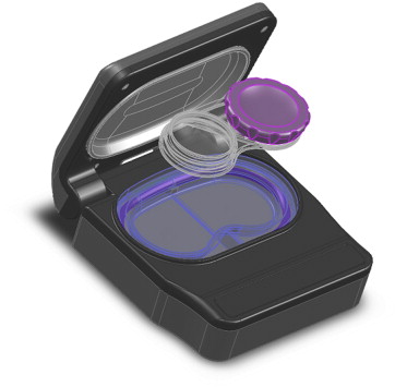
First, various plastics were screened for UV transmission and degradation to be used as an inset case (data not shown). It was found that a polypropylene exhibited limited degradation (as defined by minimal changes to UV transmission, visible color change, and leachables present in solution after irradiation) while maintaining a reasonable rate of UV transmission. Therefore, the inset cases shown in Figure 1 were injection molded from this plastic. To construct the unit responsible for delivering the UV dose, a curved germicidal UV bulb was created that wrapped horizontally around the entire inset case ( Figure 1 ). A fluoropolymer that diffuses the UV light was placed between the UV bulb and the inset case. The shape of the cavity in which the inset case sits was designed with reflecting surfaces to further provide a more even dose of light. This cavity was designed to completely separate the inset case from the bulb and other electronic components in the case of liquid spills. Over 70 different inset case and UV device iterations were pursued before an optimal configuration for even distribution of the UV light was determined. Once the optimal configuration was identified, a plastic flip case was designed and built to house all of the components.
Ultraviolet Dose Quantitation and Verification
To determine the actual amount of ultraviolet energy present within the inset case, a standard curve was generated to correlate the energy from a direct measure of the UV bulb to the energy present within the inset case (data not shown). This was accomplished by adding a photodegradable dye to the inset case and monitoring the dye’s percent change in absorbance while being exposed to UV light while simultaneously directly measuring the power and energy of the UV bulb, as detailed in Putt and associates.
Ultraviolet Acanthamoeba Cyst Kill
To determine the effect of UV irradiation on Acanthamoeba cysts, 4 × 10 4 A. castellanii ATCC 50370 cysts were inoculated onto senofilcon A lenses and placed in the inset cases (n = 8) containing 1 mL of Unisol saline solution. Organism kill was evaluated in the presence of lenses, as the UV absorbance of the lenses themselves can greatly affect the efficacy of the UV light (data not shown). For each challenge time period, the inset cases were divided into 2 groups of 4 and irradiated for a specific time. The power of the bulb was recorded and the energy present within the inset case was calculated using the method described above. The number of viable Acanthamoeba were then determined as described in the methods section. As outlined in Table 2 , the average of each treatment group showed a greater than 1.0 log reduction in viable cysts. Once the average dose reached a value of ∼130 mJ/cm 2 and above (Challenge Group 4 and above), the log reduction was greater than 3.0. As the dose reached ∼200 mJ/cm 2 (Challenge Group 6 and above) the log reduction was greater than 4.0. When each lens is shown individually ( Figure 2 ), a strong dose-dependent effect between the UV dose and the number of viable cysts is apparent, with nearly complete kill observed at UV doses greater than 250 mJ/cm 2 .
Stay updated, free articles. Join our Telegram channel

Full access? Get Clinical Tree


