Purpose
To characterize the microscopic relationships between the lower eyelid retractors and the lacrimal caruncle and to define the possible role of the caruncle in the lacrimal drainage process.
Design
Observational anatomic study.
Methods
Twelve eyelids with their orbital tissues (6 right and 6 left), fixed in 10% buffered formalin, were studied in 10 Asian cadavers (mean age at death: 77.1 years, age range: 62–92 years). An oblique incision in the harvested tissue specimen passed through the lower eyelid retractors and the lacrimal caruncle. The sliced specimens were stained with Masson’s trichrome.
Results
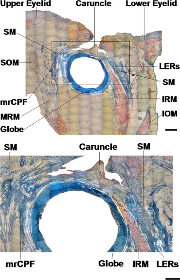
In all specimens, the lower eyelid retractors were shown to have direct connections to the lacrimal caruncle at the level of the medial horn of the lower eyelid retractors and to reach the medial rectus capsulopalpebral fascia. At this level, smooth muscle fibers were abundant both in the lower eyelid retractors and in the medial rectus capsulopalpebral fascia, which were symmetrically situated to the caruncle.
Conclusions
The lower eyelid retractors have direct connections to the lacrimal caruncle, at a level that is rich in smooth muscle fibers. These anatomic findings may confer functional advantages to the lacrimal drainage system.
The lower eyelid retractors are composed of the capsulopalpebral fascia, the capsulopalpebral head, and smooth muscle fibers. The retractors originate from the inferior rectus muscle fascia, surround the inferior oblique muscle, and reach the Lockwood ligament. Up to the point of the Lockwood ligament, the retractors are named “capsulopalpebral head,” whereas further distally they are named “capsulopalpebral fascia.” The retractors contain a large amount of smooth muscle fibers in the inferior conjunctival fornix, and they were also shown to have a medial and a lateral horn, similar to the retractors of the upper eyelid. The medial horn is situated in close proximity to the lacrimal caruncle.
The lacrimal caruncle is situated in the medial canthal area, nasal to the lacrimal punctum and the lacus lacrimalis. It was shown to be a component of the medial rectus capsulopalpebral fascia, which originates from the medial rectus muscle fascia; contains the medial rectus muscle pulley, the medial check ligament, and the lacrimal caruncle; and finally reaches the medial aspect of the tarsal plate. The lacrimal caruncle is embryologically derived from the lower eyelid, and that is why it contains sebaceous glands, as well as hair and other cutaneous appendages. Based on these anatomic characteristics, the lacrimal caruncle is situated both in the medial canthus and in the lower eyelid.
Although the proximal part of the lower eyelid retractors and the medial rectus muscle pulley (which is part of the medial rectus capsulopalpebral fascia) were shown to have direct connections around the globe equator, the anatomic relationships between the distal part of the lower eyelid retractors and the lacrimal caruncle (which is also part of the medial rectus capsulopalpebral fascia) have not been investigated so far. Additionally, although the lacrimal caruncle is positioned in close proximity with the lacrimal drainage system, its functional role in this system is still obscure.
The purpose of the present study is to characterize the microscopic relationships between the lower eyelid retractors and the lacrimal caruncle and to define the possible role of the caruncle in the lacrimal drainage process.
Materials and Methods
The method of removing and preparing the specimens has been previously described. Twelve eyelids with their orbital contents (6 right; 6 left), fixed in 10% buffered formalin, were studied in 10 Asian cadavers (mean age at death: 77.1 years, age range: 62–92 years). None of the cadavers had any history or clinical evidence of previous eyelid or orbital trauma, surgery, tumor, Graves’ orbitopathy, cerebral nerve palsy, strabismus, or any other periocular pathology. In addition, there was no relevant history of systemic diabetes, neurologic disease, or other vascular or cardiac pathologies that may have had an effect on the anatomic findings in the cadavers.
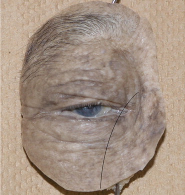
After an exenteration of the eyelid and orbital contents, an oblique incision in the harvested tissue specimen passed through the lower eyelid retractors and the lacrimal caruncle ( Figure 1 ). The temporal part of the incised specimen was used for analysis. These were stained with Masson’s trichrome and examined microscopically. Photographs were taken with a digital camera system attached to a microscope (Moticam 2000; Shimadzu Rika Kikai, Tokyo, Japan). Adobe Photoshop CS2 software (Adobe Systems Co Ltd, Tokyo, Japan) was used for photomerging.
Results
In all specimens, the lower eyelid retractors were shown to have direct connections to the lacrimal caruncle ( Figure 2 , A and B) at the level of the medial horn of the lower eyelid retractors and to reach the medial rectus capsulopalpebral fascia. At this level, smooth muscle fibers were abundant both in the lower eyelid retractors and in the medial rectus capsulopalpebral fascia, which were symmetrically situated to the caruncle.
Stay updated, free articles. Join our Telegram channel

Full access? Get Clinical Tree


