This article discusses the types of telemedicine technologies that are currently in place and being used successfully in otolaryngology. It examines how these technologies have been applied in several different otolaryngology telemedicine programs and discusses their relative merits and successes.
Telemedicine refers to the use of telecommunications technology to provide remote access to medical diagnosis and patient care. As technology has improved and the costs of using that technology have decreased, the promise of telemedicine and its potential to enhance the quality of care, improve efficiency, and reduce costs has grown significantly. The use of the Digital Imaging and Communications in Medicine (DICOM) imaging standard in radiology, for example, led to the widespread adoption of a telemedical approach to radiologic imaging and interpretation that has now become the standard of care. Historically, radiology, dermatology, psychiatry, and cardiology account for the most widespread use of telemedicine in the United States, but otolaryngology remains uniquely suited to the use of telemedicine. In addition to patient history and physical examination, many otolaryngologic diagnoses reflect information obtained from objective sources, such as tympanograms, audiometry, and telescopic and diagnostic imaging. These sources can be easily transmitted to allow for remote interpretation. Like many medical specialists, otolaryngologists are usually found in urban settings, making access to specialty care in remote and rural settings challenging. One promising use of telemedicine involves providing care to rural settings that would otherwise be difficult to provide in person, and this is a particular need that telemedicine is uniquely positioned to provide for.
Telemedicine: store and forward versus live feeds
Telemedicine consultations commonly take one of 2 forms:
- 1.
Live and interactive
- 2.
Delayed
The live and interactive method is intuitively appealing because it most closely approximates a real-life patient encounter; however, the use of a live consultation requires a level of coordination between the patient, referring physician, and specialist physician that makes this method of teleconsultation both more expensive and more challenging logistically.
The second form of telemedicine consultation is commonly known as store and forward. This involves the referring physician collecting and forwarding all of the relevant patient information, including history and imaging, to the consulting physician who then can review the data at a later time. One advantage of this type of consultation is that it does not require the physical presence of the referring physician or the patient ( Fig. 1 ).

In 1997, Sclafani and colleagues at the New York Eye and Ear Infirmary presented a study at the annual meeting of the American Academy of Otolaryngology – Head and Neck Surgery investigating the use of live and store-and-forward telemedicine in their otolaryngology practice. Patients were interviewed by a chief resident in otolaryngology who performed a relevant physical exam and flexible fiberoptic nasopharyngolaryngoscopy and who then presented his findings to 2 people: a locally available otolaryngologist and a remote otolaryngologist. Both the local and remote otolaryngologists were able to observe the interaction as well as a fiberoptic nasopharyngolaryngoscopy and to direct the chief resident. Afterward, another otolaryngologist, who was not present for the live encounter, was asked to review the electronic patient records. The investigators found concordance rates of 92% between the local and remote otolaryngologists (live videoconference) with a slightly lower concordance of 64% between the live physician and the delayed remote physician (store and forward). The investigators explain the diagnostic discordance seen with the store-and-forward remote physician as primarily the result of technical problems: color-shifting phenomenon, degraded video quality, and an insufficient quantity of high-quality images. Sclafani and colleagues state that these are problems that are easily remedied by the capability of new technology to provide a greater number of higher-quality still and video images. They conclude that remote interactive tele-otolaryngology can be used to evaluate a range of patient complaints with a high degree of diagnostic reliability.
Telemedicine and video otoscopic imaging
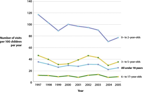
Otitis media is one of the most commonly encountered diagnoses in both pediatric and otolaryngologic practice, and the cost associated with the diagnosis and treatment of the disease is estimated to be in excess of 5.3 billion dollars per year. Acute otitis media is the most common bacterial infection in children and is the most frequent indication for antimicrobial therapy in the pediatric population ( Fig. 2 ). Patients who suffer from either recurrent acute otitis media or chronic otitis media are often treated with tympanostomy tube insertion, which is currently one of the most common procedures performed in children in the United States. After surgery, patients are seen in follow-up in the clinic to assess the patency of the tympanostomy tubes. This follow-up commonly occurs 1 month after surgery and at regular intervals thereafter. Because a significant proportion of the United States population lives in rural settings without nearby access to subspecialty surgical care, arranging follow-up for these patients can be challenging, which presents a unique opportunity for telemedicine to play a role in the postsurgical follow-up of patients with tympanostomy tubes.
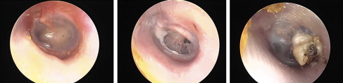
To establish a role for video otoscopy in providing telemedical care, it is important to ensure that video otoscopy is an accurate form of data gathering and that it provides reproducible results when images are reviewed by different practitioners. In 2003, Patricoski and colleagues of the Alaska Native Medical Center addressed this question in a study examining whether the results of still images of the tympanic membrane were comparable with in-person microscopic examination. They selected 40 patients who had previously undergone tympanostomy tube placement, had them independently examined by 2 otolaryngologists, and had imaging done using a video otoscope ( Fig. 3 ). The examining otolaryngologists reviewed the images 6 and 12 weeks later and the investigators compared their diagnostic concordance using κ statistics. Patricoski and colleagues found that intraprovider concordance rates were between 85% and 99% for physical examination findings and between 79% and 85% for diagnosis. They conclude that store-and-forward video otoscopy may be an appropriate method of following tympanostomy tubes after placement.
Studies in the Literature
A study published in 2004 examined the use of video otoscopy to assess and diagnose middle ear disease. Aronzon and colleagues from the University of Pennsylvania compared groups of medical students, internal medicine residents, and attending and resident otolaryngologists who were presented with a set of matched tympanograms and photographs of the tympanic membrane obtained through video otoscopy. Subjects were asked to differentiate between a normal-appearing and abnormal-appearing membrane and to make a diagnosis of middle ear disease based on the imaging. The investigators reported that the sensitivity of diagnosis by attending otolaryngologists using tympanograms and photographs was equal to the sensitivity of diagnosis achieved with the operating microscope. The investigators concluded that video otoscopy technology can allow the expertise of a specialist to be extended to rural and underserved areas where specialists might not otherwise be available.
A study in 2008 further developed the concept of using video otoscopy as a tool for clinical imaging of the tympanic membrane. Lundberg and colleagues from Sweden studied 64 children aged 2 to 16 years with otalgia who presented to clinics in Sweden. The children had their tympanic membranes imaged using a video otoscope and had the images independently assessed by both an otolaryngologist and a general practitioner. The investigators analyzed overall image quality as well as quality of image-related components, such as focus, light, or composition. The investigators concluded that there was good overall agreement between examiners and that video otoscopy is a reliable technique in the clinical or research setting. The investigators further pointed out that a potential advantage of digital imaging is that it may allow for more objective grading of the tympanic membrane and for retrospective review by independent examiners.
In 2008, Kokesh and colleagues published a study from Anchorage, Alaska detailing how they successfully made use of video otoscopy to allow for patient follow-up after tympanostomy tube insertion in remote areas of Alaska. They noted that American Indian and Alaskan native children have higher rates of otitis media and tympanostomy tube placement, but live in an environment that makes postsurgical follow-up challenging and expensive. They used telemedicine to follow patients with tympanostomy tubes and examined the concordance between an in-person encounter and images of the tympanic membrane taken in remote village clinics. A similar study was done by Patricoski in 2003 and is mentioned earlier; however, the 2003 study was performed in a controlled research environment and was not done in an authentic real-world clinical setting. Kokesh and colleagues used community health aides and practitioners who performed video otoscopy in remote village clinics, in the same way that a telemedicine otolaryngology practice might operate. These images were then reviewed by an otolaryngologist. Between 1 and 7 days following their video otoscopic examination, the patients were flown to Maniilaq Health Center in Kotzebue for an otolaryngology clinic visit, where 2 otolaryngologists examined the patients using a Zeiss otology microscope. The video and microscopic otoscopic examinations were then evaluated for concordance.
The investigators in the 2008 Kokesh study examined a real-world practice environment using store-and-forward telemedicine with nonphysician health workers taking images of the tympanic membrane with no access to an operating microscope for ear cleaning. The investigators found a high level of concordance between the image reviews and the in-person encounters, with 79% agreement between the remote and in-person encounters. Nineteen percent of all video otoscope images were rejected as being of poor or very poor quality. This finding was most commonly the result of poor focus leading to poor image quality or to cerumen or debris in the ear obstructing the field of view. In a real-life situation, these poor-quality images could easily be retaken on request, which led the investigators to conclude that high-quality digital images of the tympanic membrane can be obtained by a trained health aide in a rural setting, and that store-and-forward otoscopy can be used for tympanostomy tube follow-up in settings where the patient is located remotely from the specialist.
Telemedicine and video otoscopic imaging
Otitis media is one of the most commonly encountered diagnoses in both pediatric and otolaryngologic practice, and the cost associated with the diagnosis and treatment of the disease is estimated to be in excess of 5.3 billion dollars per year. Acute otitis media is the most common bacterial infection in children and is the most frequent indication for antimicrobial therapy in the pediatric population ( Fig. 2 ). Patients who suffer from either recurrent acute otitis media or chronic otitis media are often treated with tympanostomy tube insertion, which is currently one of the most common procedures performed in children in the United States. After surgery, patients are seen in follow-up in the clinic to assess the patency of the tympanostomy tubes. This follow-up commonly occurs 1 month after surgery and at regular intervals thereafter. Because a significant proportion of the United States population lives in rural settings without nearby access to subspecialty surgical care, arranging follow-up for these patients can be challenging, which presents a unique opportunity for telemedicine to play a role in the postsurgical follow-up of patients with tympanostomy tubes.
To establish a role for video otoscopy in providing telemedical care, it is important to ensure that video otoscopy is an accurate form of data gathering and that it provides reproducible results when images are reviewed by different practitioners. In 2003, Patricoski and colleagues of the Alaska Native Medical Center addressed this question in a study examining whether the results of still images of the tympanic membrane were comparable with in-person microscopic examination. They selected 40 patients who had previously undergone tympanostomy tube placement, had them independently examined by 2 otolaryngologists, and had imaging done using a video otoscope ( Fig. 3 ). The examining otolaryngologists reviewed the images 6 and 12 weeks later and the investigators compared their diagnostic concordance using κ statistics. Patricoski and colleagues found that intraprovider concordance rates were between 85% and 99% for physical examination findings and between 79% and 85% for diagnosis. They conclude that store-and-forward video otoscopy may be an appropriate method of following tympanostomy tubes after placement.
Studies in the Literature
A study published in 2004 examined the use of video otoscopy to assess and diagnose middle ear disease. Aronzon and colleagues from the University of Pennsylvania compared groups of medical students, internal medicine residents, and attending and resident otolaryngologists who were presented with a set of matched tympanograms and photographs of the tympanic membrane obtained through video otoscopy. Subjects were asked to differentiate between a normal-appearing and abnormal-appearing membrane and to make a diagnosis of middle ear disease based on the imaging. The investigators reported that the sensitivity of diagnosis by attending otolaryngologists using tympanograms and photographs was equal to the sensitivity of diagnosis achieved with the operating microscope. The investigators concluded that video otoscopy technology can allow the expertise of a specialist to be extended to rural and underserved areas where specialists might not otherwise be available.
A study in 2008 further developed the concept of using video otoscopy as a tool for clinical imaging of the tympanic membrane. Lundberg and colleagues from Sweden studied 64 children aged 2 to 16 years with otalgia who presented to clinics in Sweden. The children had their tympanic membranes imaged using a video otoscope and had the images independently assessed by both an otolaryngologist and a general practitioner. The investigators analyzed overall image quality as well as quality of image-related components, such as focus, light, or composition. The investigators concluded that there was good overall agreement between examiners and that video otoscopy is a reliable technique in the clinical or research setting. The investigators further pointed out that a potential advantage of digital imaging is that it may allow for more objective grading of the tympanic membrane and for retrospective review by independent examiners.
In 2008, Kokesh and colleagues published a study from Anchorage, Alaska detailing how they successfully made use of video otoscopy to allow for patient follow-up after tympanostomy tube insertion in remote areas of Alaska. They noted that American Indian and Alaskan native children have higher rates of otitis media and tympanostomy tube placement, but live in an environment that makes postsurgical follow-up challenging and expensive. They used telemedicine to follow patients with tympanostomy tubes and examined the concordance between an in-person encounter and images of the tympanic membrane taken in remote village clinics. A similar study was done by Patricoski in 2003 and is mentioned earlier; however, the 2003 study was performed in a controlled research environment and was not done in an authentic real-world clinical setting. Kokesh and colleagues used community health aides and practitioners who performed video otoscopy in remote village clinics, in the same way that a telemedicine otolaryngology practice might operate. These images were then reviewed by an otolaryngologist. Between 1 and 7 days following their video otoscopic examination, the patients were flown to Maniilaq Health Center in Kotzebue for an otolaryngology clinic visit, where 2 otolaryngologists examined the patients using a Zeiss otology microscope. The video and microscopic otoscopic examinations were then evaluated for concordance.
The investigators in the 2008 Kokesh study examined a real-world practice environment using store-and-forward telemedicine with nonphysician health workers taking images of the tympanic membrane with no access to an operating microscope for ear cleaning. The investigators found a high level of concordance between the image reviews and the in-person encounters, with 79% agreement between the remote and in-person encounters. Nineteen percent of all video otoscope images were rejected as being of poor or very poor quality. This finding was most commonly the result of poor focus leading to poor image quality or to cerumen or debris in the ear obstructing the field of view. In a real-life situation, these poor-quality images could easily be retaken on request, which led the investigators to conclude that high-quality digital images of the tympanic membrane can be obtained by a trained health aide in a rural setting, and that store-and-forward otoscopy can be used for tympanostomy tube follow-up in settings where the patient is located remotely from the specialist.
Stay updated, free articles. Join our Telegram channel

Full access? Get Clinical Tree


