Purpose
To report refractive, topographic, pachymetric, tonometric, and corneal biomechanical outcomes 24 months after corneal cross-linking (CXL), followed by insertion of intrastromal corneal ring segments (ICRS) in keratoconic eyes.
Design
Prospective randomized clinical trial.
Methods
settings: Institutional. study population: Thirty-nine eyes of 31 patients, allocated into 2 groups. intervention: Patients in the CXL group underwent corneal CXL with riboflavin and ultraviolet A (UVA) light. Patients in the riboflavin eyedrops group received riboflavin 0.1% (w/v) eyedrops – 20% dextran solution for 1 month. After 3 months, all patients underwent insertion of ICRS. main outcome measures: Uncorrected visual acuity (UCVA), best spectacle-corrected visual acuity (BSCVA), and topography were evaluated at baseline, at 1 month and 3 months after CXL or riboflavin eyedrops, and again at 1-, 3-, 6-, 12-, and 24-month intervals after ICRS insertion.
Results
Mean (standard deviation [SD]) baseline UCVA and BSCVA in the CXL group and the riboflavin eyedrops group were 1.12 (0.59) and 0.84 (0.49), and 0.68 (0.43) and 0.45 (0.23), respectively; 24-month mean (SD) UCVA and BSCVA in the CXL group and the riboflavin eyedrops group were 0.79 (0.50) and 0.62 (0.28), and 0.52 (0.45) and 0.32 (0.21), respectively, with no statistically significant difference between groups ( P = .70 and P = .78).There were no statistical differences between groups postoperatively at 24 months for all 3 topographic parameters, flattest-K1 ( P = .81), steepest-K2 ( P = .68), and average keratometry (mean power; P = .52).
Conclusions
ICRS insertion, with or without prior CXL, showed no difference between groups in terms of refractive, topographic, pachymetric, tonometric, and corneal biomechanical results at 24 months.
Keratoconus is generally a progressive, bilateral cone-like ectasia of the cornea that affects approximately 1 in 2000 in the general population. Keratoconus begins around the time of puberty and progresses in approximately 20% of patients to such an extent that penetrating keratoplasty becomes necessary to restore vision. In some cases, however, the cornea remains transparent, but the patient cannot tolerate contact lenses.
If rigid contact lenses cannot be used, a few surgical alternatives remain for the correction of eyes with keratoconus. In addition to lamellar and penetrating keratoplasty procedures, the introduction of intrastromal corneal ring segments (ICRS) offers another option for the management of keratoconus. These vision-correcting methods attempt to regularize the front surface of the cornea while maintaining the existing biomechanical status in the underlying stroma.
Corneal cross-linking (CXL) by the photosensitizer riboflavin (vitamin B2) and ultraviolet A (UVA) light increases corneal rigidity and has been described as an effective method for stabilizing the cornea in patients with progressive keratoconus. In the present literature, questions have arisen about the correct treatment sequence relating to the 2 techniques. Would a cornea pretreated with CXL react to the ICRS implantation in the expected way, or would its effect be lessened by its application over a stiffer cornea? Alternatively, could ICRS implantation be performed first and stiffening of the cornea later?
To determine whether CXL effectively augments the treatment of keratoconus before ICRS insertion, we compared the visual, refractive, topographic, pachymetric, tonometric, and corneal biomechanical metric outcomes at 24 months in keratoconic eyes after CXL or riboflavin eyedrops, followed by insertion of femtosecond laser–assisted ICRS.
Methods
An interventional prospective randomized clinical trial was conducted between March 1, 2008 and December 20, 2010 at the Vision Institute, Department of Ophthalmology, Federal University of São Paulo, Brazil.
Thirty-nine eyes of 31 patients were included. All eyes were graded according to the Amsler-Krumeich classification, based on each patient’s refraction, mean central K-reading, corneal signs, and corneal thickness. All treated eyes were graded either stage II or stage III according to this classification. For allocation of the participants, a computer-generated list of random numbers was used, and patients were allocated into 2 study groups: CXL group (19 eyes underwent CXL procedure with riboflavin and UVA light) and riboflavin eyedrops group (20 eyes prescribed riboflavin 0.1% [w/v] eyedrops [10 mg riboflavin-5-phosphate in 20% (w/v) dextran-T-500; Ophthalmos, São Paulo, Brazil] 4 times per day for 1 month). A flow diagram demonstrating the enrollment of patients in the study is provided in the Supplemental Figure (available at AJO.com ).
In 8 patients, both eyes were included. For these patients, the right eye received only riboflavin eyedrops and the left eye underwent the CXL procedure. After 3 months, all patients underwent insertion of ICRS (Keraring; Mediphacos, Belo Horizonte, Brazil), comprising the second step.
Inclusion criteria were compliant patients with documented keratoconus with best spectacle-corrected visual acuity (BSCVA) ≤0.48 (logarithm of the minimal angle of resolution [logMAR] chart), increasing or proven intolerance to contact lenses, penetrating keratoplasty referred by a doctor, corneal thickness ≥400 μm at the thinnest point, good health, and age between 15 and 60 years. Exclusion criteria were patients with contact lens use, corneal thickness <400 μm at the thinnest point, previous ocular surgery, only 1 functional eye, corneal opacity on the axis, corneal curvature >65 diopters (D; Krumeich grade IV), history of herpetic keratitis, moderate dry eye (diagnosed by tear film, punctate keratitis, and patient complaint), autoimmune disease, concurrent corneal infection, pregnancy, and other ocular diseases that modified the visual acuity.
Follow-up examinations were scheduled at baseline, and at 1 month and 3 months after CXL or riboflavin eyedrops. Additional examinations were performed after ICRS insertion by the same examiner (A.C.R.) at 1, 3, 6, 12, and 24 months.
At the baseline and postoperative follow-up examinations, all patients underwent the following procedures: 1) uncorrected visual acuity (UCVA); 2) BSCVA with the Early Treatment Diabetic Retinopathy Study (ETDRS) visual acuity chart (logMAR notation); 3) manifest refraction in a bright environment; 4) slit-lamp biomicroscopy; 5) contrast sensitivity (Optec 6500, F.A.C.T.; Stereo Optical Co, Chicago, Illinois, USA); 6) corneal topography (EyeSys-2000; EyeSys Vision, Irvine, California, USA); 7) Orbscan IIz central and thinnest corneal pachymetry (Bausch & Lomb GmbH, Feldkirchen, Germany); 8) Pentacam central and thinnest corneal pachymetry (Oculus Optikgerate GmbH, Wetzlar, Germany); 9) Goldmann applanation tonometer (GAT; Haag-Streit, Konig, Switzerland); 10) dynamic contour tonometer (DCT; Pascal, Ziemer Ophthalmic Systems AG, Switzerland); 11) ultrasound pachymetry (Sonogage UP, Corneo-Gage-Plus, Eye Scan, Inc, Cleveland, Ohio, USA); 12) corneal biomechanical metrics evaluation by the Ocular Response Analyzer (ORA; Reichert Ophthalmic Instruments, Depew, New York, USA); and 13) ocular fundus examination. Visante optical coherence tomography (OCT; Carl Zeiss Meditec, Dublin, California, USA), central and thinnest corneal pachymetry, and specular microscopy with a noncontact device (Topcon SP 2000p; Topcon, Tokyo, Japan) were performed at all examinations, except at the third month following CXL or riboflavin eyedrops and the third month following ICRS insertion.
Contrast Sensitivity Assessment
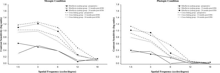
Contrast sensitivity was determined in each eye with the BSCVA at spatial frequencies of 1.5, 3, 6, 12, and 18 cycles per degree in mesopic and photopic conditions.
Endothelial Cell Count
Endothelial cell count measurements were performed at the center of the cornea. When measurements could not be performed centrally, the inferior, superior, nasal, or temporal area was measured, because of the difficulty in the keratoconic corneas. Three recorded endothelium pictures (0.1 × 0.1 mm) from each eye were taken, and the mean cell density was determined.
Ultrasound Pachymetry
The cornea was anesthetized with topical 0.5% (w/v) proxymetacaine hydrochloride. The seated patient was asked to look straight at a distant fixation target. The pachymeter probe was aligned perpendicular and central to the pupil as precisely as possible, and the mean of 3 measurements was calculated.
Corneal Cross-linking Procedure
The CXL procedure was performed by the same surgeon (M.C.) according to the standard protocol. Proxymetacaine hydrochloride 0.5% (w/v) eyedrops were applied to achieve preoperative anesthesia. The central 9 mm of the corneal epithelium was removed using a blunt knife. As a photosensitizer, riboflavin 0.1% (w/v) solution (10 mg riboflavin-5-phosphate in 10 mL dextran-T-500 20% [w/v] solution) was applied onto the cornea every 5 minutes for a total of 30 minutes before UVA irradiation. Using a slit lamp with a blue filter, the surgeon confirmed the presence of riboflavin in the anterior chamber before UVA irradiation. The cornea was exposed (using a lid speculum) to UV light emanating from a solid-state device (UV-X System; Peschke Meditrade GmbH, Hunenberg, Switzerland), which emits light at 370 ± 5 nm at an irradiance of 3 mW/cm 2 or 5.4 J/cm 2 for 30 minutes. During the irradiation time, the cornea was rinsed with riboflavin/dextran solution and fixation was achieved by instructing the patient to focus on the central light-emitting diode of the probe. Topical anesthetic was applied as needed throughout the surgery. After the treatment, a soft bandage contact lens was applied until re-epithelialization was complete. A combination of moxifloxacin 0.5% (w/v) and dexamethasone phosphate 0.1% (w/v) eyedrops (Alcon Laboratories, Fort Worth, Texas, USA) were prescribed 4 times per day for 2 weeks. Follow-up examinations were performed daily until complete re-epithelialization occurred.
Intrastromal Corneal Ring Segment Implantation
After 3 months of riboflavin eyedrops or CXL, all patients underwent insertion of the Keraring. All operations were performed by M.C. and M.F.S. The surgical decision to implant the ICRS was made according to the nomogram (version 2008) provided by the manufacturer, available at http://www.mediphacos.com.br . The Keraring is made of polymethylmethacrylate (PMMA) and is characterized by a triangular cross-section. The apical diameter is 5.0 mm, and the flat basis width is 0.6 mm with varying thickness (0.15-0.30 mm thickness with 0.5-mm steps) and arc lengths (90, 120, 160, and 210 degrees). The optical zone is 5.0 mm in diameter. To summarize, 1 or 2 segments were made according to the distribution of the ectatic area on the corneal surface, and the thickness of the segment was made according to the spherical and cylinder diopters.
ICRS insertion was performed in an operating room under sterile conditions using topical anesthetic drops. The procedure was initiated by marking a reference point for pupil centration. A 5.0-mm marker was used to locate the exact ring channel. Tunnel depth was set at 80% of the thinnest corneal thickness on the tunnel location in the femtosecond laser (IntraLase Corp, Irvine, California, USA).
An incision was made on the steepest topographic axis. A 60-kHz femtosecond laser was used to create the ring channels. Special attention was given to centralizing the disposable suction ring and marking the central point to minimize decentration. The following parameters were set: 1) channel’s inner diameter 5.0 mm; 2) channel’s outer diameter 5.9 mm; 3) entry cut thickness 1 μm (at the steepest topographic axis); 4) ring energy 1.70 μJ for channel creation; and 5) entry cut energy 1.10 μJ. Channel creation with the femtosecond laser was 15 seconds. The ICRS was implanted with the accompanying forceps immediately after channel creation and before the disappearance of the bubbles, which revealed the exact tunnel location. After surgery, a combination of moxifloxacin 0.5% (w/v) and dexamethasone phosphate 0.1% (w/v) eyedrops (Alcon Laboratories, Fort Worth, Texas, USA) were again prescribed 4 times per day for 2 weeks. The patients were instructed to avoid rubbing their eyes and to use artificial tears frequently. On the first and seventh postoperative day, slit-lamp biomicroscopic examination was performed. Wound healing and segment migration were evaluated.
Data Analysis
The statistical analysis was performed using the statistical packages Stata 11.1 (StataCorp, College Station, Texas, USA) and Statistica 8.0 (StatSoft, Inc, Tulsa, Oklahoma, USA). A minimum sample size of 17 participants in each group was needed to achieve 80% power and to detect a difference of 0.20 logMAR in BSCVA between groups, assuming a significance level of 0.05 and a standard deviation in each group of 0.20. Analysis of covariance was used to compare visual acuity, corneal curvature and thickness, endothelial cell count, refraction, intraocular pressure, and corneal biomechanical properties between the 2 groups at the 12-month and 24-month visits after ICRS insertion. The Mann-Whitney test was used to compare the change from baseline (preintervention) measurements in contrast sensitivity between the 2 groups at the 12-month and 24-month visits after ICRS insertion.
Results
All patients were followed with scheduled examinations for a total of 24 months. Of the 31 patients, 23 were women (74.19%) and 8 were men (25.80%).The mean ages of the patients were 28.30 ± 9.3 years (range, 17–55 years) in the CXL group and 30.40 ± 9.1 years (range, 22–55 years) in the riboflavin eyedrops group. Both groups were similar before treatment as assessed by corneal keratometry (flattest-K1 and steepest-K2 meridian) and spherical equivalent measurements ( Table 1 ).
| Group | Measure | N | Mean (D) | SD (D) | Minimum (D) | Median (D) | Maximum (D) | P a |
|---|---|---|---|---|---|---|---|---|
| CXL | K1 | 19 | 48.1 | 4.21 | 41.41 | 47.50 | 55.87 | .197 |
| RE | K1 | 20 | 46.5 | 3.39 | 41.66 | 46.29 | 54.87 | |
| CXL | K2 | 19 | 53.26 | 5.11 | 43.10 | 52.98 | 61.25 | .454 |
| RE | K2 | 20 | 52.17 | 3.89 | 45.06 | 51.09 | 63.92 | |
| CXL | SE | 19 | −7.41 | 3.48 | −14.0 | −6.50 | −1.75 | .090 |
| RE | SE | 20 | −5.45 | 3.53 | −13.5 | −4.50 | 0.75 |
Slit-Lamp Examination
After CXL, most patients demonstrated complete re-epithelialization in 5 days and no evidence of epithelial defects or opacities. Faint haze (cotton-like) was observed in all patients at 1 and 3 months after CXL. Post ICRS insertion, there were no epithelial defects, corneal inflammation, or anterior chamber reactions. Good positioning and a slight fluorescein staining were observed in all implants.
Visual Acuity
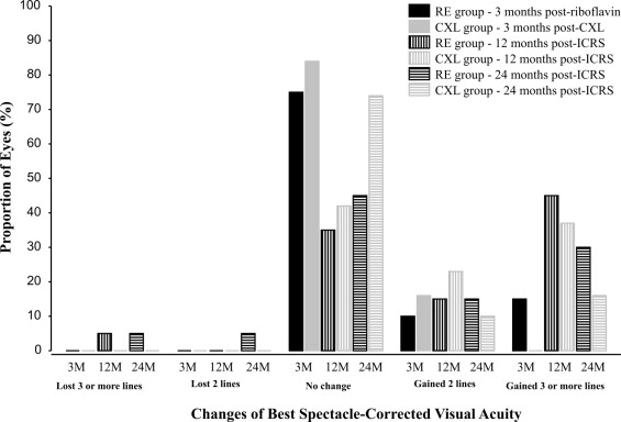
Figure 1 and Table 2 summarize the UCVA and BSCVA expressed in logMAR.
| Visual Acuity logMAR | Preintervention Mean (SD) | Postintervention | Post Intrastromal Corneal Ring Segments | |||||
|---|---|---|---|---|---|---|---|---|
| 1-Month Mean (SD) | 3-Month Mean (SD) | 1-Month Mean (SD) | 3-Month Mean (SD) | 6-Month Mean (SD) | 12-Month Mean (SD) | 24-Month Mean (SD) | ||
| UCVA | ||||||||
| Riboflavin eyedrop group | 0.84 (0.49) | 0.79 (0.52) | 0.66 (0.29) | 0.54 (0.26) | 0.51 (0.31) | 0.57 (0.28) | 0.55 (0.26) | 0.62 (0.28) |
| Cross-linking group | 1.12 (0.59) | 1.06 (0.53) | 0.94 (0.53) | 0.69 (0.40) | 0.65 (0.27) | 0.64 (0.28) | 0.73 (0.45) | 0.79 (0.50) |
| BSCVA | ||||||||
| Riboflavin eyedrop group | 0.45 (0.23) | 0.36 (0.15) | 0.35 (0.22) | 0.30 (0.21) | 0.21 (0.19) | 0.26 (0.23) | 0.23 (0.18) | 0.32 (0.21) |
| Cross-linking group | 0.68 (0.43) | 0.62 (0.41) | 0.60 (0.44) | 0.46 (0.42) | 0.44 (0.27) | 0.46 (0.48) | 0.43 (0.43) | 0.52 (0.45) |
There was no statistical difference between groups in UCVA ( P = .70), and the mean (95% confidence interval) difference between the CXL and riboflavin eyedrops groups in the BSCVA adjusted for baseline measurements was 0.02 (−0.14 to 0.18; P = .78) at 24 months.
Refractive Results
The mean (standard deviation [SD]) preoperative spherical equivalent (SE) was −5.45 (3.53) D in the riboflavin eyedrops group, with a mean sphere of −3.42 (3.78) D and a mean net cylinder of −4.10 (1.69) D. In the CXL group, the mean (SD) preoperative SE was −7.38 (3.49) D, with a mean sphere of −5.17 (3.35) D and a mean net cylinder of −4.00 (1.39) D. At 24 months postoperatively, mean (SD) SE in the riboflavin eyedrops group was −4.19 (2.89) D, with a mean sphere of −2.65 (2.82) D and a mean net cylinder of −3.21 (0.95) D. In the CXL group at 24 months, mean (SD) SE was −5.49 (3.77) D, with a mean sphere of −4.03 (3.50) D and a mean net cylinder of −3.21 (1.21) D. Table 3 shows the refractive results.
| Refraction Component (Diopters) | Preintervention Mean (SD) | Postintervention | Post Intrastromal Corneal Ring Segments | |||||
|---|---|---|---|---|---|---|---|---|
| 1-Month Mean (SD) | 3-Month Mean (SD) | 1-Month Mean (SD) | 3-Month Mean (SD) | 6-Month Mean (SD) | 12-Month Mean (SD) | 24-Month Mean (SD) | ||
| Spherical component | ||||||||
| Riboflavin eyedrop group | −3.42 (3.78) | −3.54 (4.12) | −3.76 (4.09) | −2.85 (3.36) | −2.80 (3.10) | −2.77 (2.93) | −2.66 (2.84) | −2.65 (2.82) |
| Cross-linking group | −5.17 (3.35) | −5.43 (3.49) | −5.71 (3.01) | −4.97 (4.19) | −4.51 (3.57) | −4.28 (3.85) | −4.17 (3.61) | −4.03 (3.50) |
| Cylindrical component | ||||||||
| Riboflavin eyedrop group | −4.10 (1.69) | −4.16 (1.61) | −4.30 (1.52) | −3.56 (1.24) | −3.48 (1.45) | −3.16 (0.94) | −3.17 (0.94) | −3.21 (0.95) |
| Cross-linking group | −4.00 (1.39) | −4.29 (1.26) | −4.33 (1.16) | −3.03 (1.73) | −3.07 (1.58) | −3.12 (1.44) | −3.09 (1.39) | −3.21 (1.21) |
| Spherical equivalent | ||||||||
| Riboflavin eyedrop group | −5.45 (3.53) | −5.59 (3.83) | −5.88 (3.81) | −4.59 (3.48) | −4.47 (3.24) | −4.33 (2.98) | −4.20 (2.92) | −4.19 (2.89) |
| Cross-linking group | −7.38 (3.49) | −7.55 (3.55) | −7.83 (3.05) | −6.45 (4.53) | −6.00 (3.84) | −5.78 (4.17) | −5.62 (3.96) | −5.49 (3.77) |
Adjusting for preoperative levels, there was no statistical difference between groups in terms of the spherical component ( P = .64), cylindrical component ( P = .97), and SE ( P = .94) at the 24-month follow-up.
Topographic Results
Topographic data are displayed in Tables 4 and 5 . The values correspond to 3 central millimeters of the simulated keratometry. Mean (SD) baseline flattest keratometry (K1), steepest keratometry (K2), and average keratometry (mean power) were 46.54 (3.39) D, 52.17 (3.89) D, and 51.75 (4.66) D, respectively, in the riboflavin eyedrops group and 48.15 (4.21) D, 53.26 (5.11) D, and 53.65 (5.30) D in the CXL group, respectively. At 24 months, these readings were 45.37 (3.12) D, 48.97 (3.13) D, and 50.52 (4.02) D in the riboflavin eyedrops group, and 46.82 (3.79) D, 49.62 (4.12) D, and 51.63 (4.97) D in the CXL group, respectively. There was no statistical difference between groups at the 24-month postoperative follow-up for all 3 parameters (K1 [ P = .81], K2 [ P = .68], and mean power [ P = .52]).
| Corneal Topography Index (Diopters) | Preintervention Mean (SD) | Postintervention | Post Intrastromal Corneal Ring Segments | |||||
|---|---|---|---|---|---|---|---|---|
| 1-Month Mean (SD) | 3-Month Mean (SD) | 1-Month Mean (SD) | 3-Month Mean (SD) | 6-Month Mean (SD) | 12-Month Mean (SD) | 24-Month Mean (SD) | ||
| Flattest curvature | ||||||||
| Riboflavin eyedrop group | 46.54 (3.39) | 46.67 (3.58) | 46.60 (3.66) | 45.36 (3.24) | 45.38 (3.11) | 45.23 (3.10) | 45.38 (3.15) | 45.37 (3.12) |
| Cross-linking group | 48.15 (4.21) | 48.70 (4.50) | 48.55 (4.50) | 46.88 (3.81) | 46.48 (3.63) | 46.13 (3.87) | 46.62 (3.73) | 46.82 (3.79) |
| Steepest curvature | ||||||||
| Riboflavin eyedrop group | 52.17 (3.89) | 52.34 (4.00) | 52.25 (3.56) | 49.45 (3.50) | 49.33 (3.37) | 49.07 (3.34) | 49.17 (3.17) | 48.97 (3.13) |
| Cross-linking group | 53.26 (5.11) | 54.41 (5.92) | 53.95 (5.72) | 50.08 (4.94) | 49.82 (4.37) | 49.53 (3.82) | 49.69 (4.19) | 49.62 (4.12) |
| Mean power | ||||||||
| Riboflavin eyedrop group | 51.75 (4.66) | 52.47 (5.05) | 52.01 (4.28) | 51.18 (5.41) | 51.21 (5.33) | 50.66 (4.68) | 50.37 (4.02) | 50.52 (4.02) |
| Cross-linking group | 53.65 (5.30) | 54.54 (5.34) | 53.76 (5.41) | 52.99 (6.52) | 52.72 (5.25) | 51.42 (4.92) | 51.93 (5.06) | 51.63 (4.97) |
| Topography Index | Reduction After ICRS Implantation a | ||||
|---|---|---|---|---|---|
| 1 Month Mean, Diopters (%) | 3 Months Mean, Diopters (%) | 6 Months Mean, Diopters (%) | 12 Months Mean, Diopters (%) | 24 Months Mean, Diopters (%) | |
| Flattest curvature | |||||
| Riboflavin eyedrop group | 1.24 (2.7) | 1.22 (2.6) | 1.37 (2.9) | 1.22 (2.6) | 1.23 (2.6) |
| Cross-linking group | 1.67 (3.4) | 2.07 (4.3) | 2.42 (5.0) | 1.93 (4.0) | 1.73 (3.6) |
| Steepest curvature | |||||
| Riboflavin eyedrop group | 2.79 (5.3) | 2.92 (5.6) | 3.18 (6.1) | 3.08 (5.9) | 3.27 (6.3) |
| Cross-linking group | 3.87 (7.2) | 4.13 (7.7) | 4.42 (8.2) | 4.26 (7.9) | 4.33 (8.0) |
| Mean power | |||||
| Riboflavin eyedrop group | 0.82 (1.6) | 0.79 (1.5) | 1.35 (2.6) | 1.63 (3.1) | 1.49 (2.9) |
| Cross-linking group | 0.77 (1.4) | 1.04 (1.9) | 2.34 (4.4) | 1.83 (3.4) | 2.13 (4.0) |
Contrast Sensitivity
The mesopic and photopic contrast sensitivity at spatial frequencies 1.5, 3, 6, 12, and 18 cycles per degree did not differ significantly between groups. In the mesopic condition, the values were: 1.5 cycles/degree ( P = .52); 3 cycles/degree ( P = .23); 6 cycles/degree ( P = .13); 12 cycles/degree ( P = .63); and 18 cycles/degree ( P = .51). In the photopic condition, the values were: 1.5 cycles/degree ( P = .61); 3 cycles/degree ( P = .10); 6 cycles/degree ( P = .82); 12 cycles/degree ( P = .16); and 18 cycles/degree ( P = .33; Figure 2 ) .
Endothelial Results
Mean (SD) baseline endothelial cell counts were 2780.2 (257.2) cells/mm 2 in the riboflavin eyedrops group and 2784.3 (306.8) cells/mm 2 in the CXL group. At 24 months, the endothelial cell counts were 2714.8 (230.8) cells/mm 2 in the riboflavin eyedrops group and 2687.4 (270.4) cells/mm 2 in the CXL group. The difference between baseline and 24 months was not significant ( P = .71), indicating that CXL did not induce endothelial damage in the 24-month follow-up period.
Pachymetry Results
Pachymetry results are available for the entire follow-up period with the Pentacam, except at 24 months because of technical problems with the device. Table 6 shows the pachymetry data at the central and thinnest position on Visante OCT, Pentacam, Orbscan, and ultrasound pachymetry. There was no statistical difference between groups at 24 months of follow-up in any parameter: central and thinnest Visante pachymetry ( P = .89 and P = .82), respectively; central and thinnest Pentacam pachymetry at 12 months of follow-up ( P = .30 and P = .38); central and thinnest Orbscan pachymetry ( P = .88 and P = .94); and ultrasound pachymetry ( P = .39).
| Pachymetry Device | Preintervention Mean (SD) | Postintervention | Post Intrastromal Corneal Ring Segments | |||||
|---|---|---|---|---|---|---|---|---|
| 1-Month Mean (SD) | 3-Month Mean (SD) | 1-Month Mean (SD) | 3-Month Mean (SD) | 6-Month Mean (SD) | 12-Month Mean (SD) | 24-Month Mean (SD) | ||
| Visante | ||||||||
| Central position (μm) | ||||||||
| Riboflavin eyedrop group | 453.9 (30.4) | 465.4 (34.3) | NM | 471.4 (39.6) | NM | 460.6 (34.6) | 457.3 (33.5) | 457.5 (35.9) |
| Cross-linking group | 444.9 (32.2) | 446.2 (33.6) | NM | 453.0 (34.0) | NM | 449.2 (36.4) | 445.1 (34.5) | 447.2 (37.6) |
| Thinnest position (μm) | ||||||||
| Riboflavin eyedrop group | 429.3 (32.4) | 433.5 (35.0) | NM | 442.6 (35.1) | NM | 438.2 (34.0) | 436.2 (34.0) | 433.1 (33.7) |
| Cross-linking group | 417.9 (35.0) | 420.4 (34.7) | NM | 425.5 (37.2) | NM | 422.3 (36.5) | 417.3 (44.7) | 420.6 (39.5) |
| Pentacam | ||||||||
| Central position (μm) | ||||||||
| Riboflavin eyedrop group | 464.3 (30.0) | 464.4 (31.0) | 467.1 (31.4) | 477.9 (34.2) | 478.4 (32.5) | 478.1 (34.0) | 475.4 (33.4) | NM |
| Cross-linking group | 455.2 (32.3) | 437.8 (33.6) | 447.8 (35.0) | 458.0 (32.7) | 459.7 (32.4) | 461.7 (35.9) | 460.9 (35.1) | NM |
| Thinnest position (μm) | ||||||||
| Riboflavin eyedrop group | 444.8 (30.7) | 445.1 (31.7) | 445.9 (31.5) | 461.8 (35.9) | 461.8 (34.4) | 462.3 (33.8) | 456.3 (34.2) | NM |
| Cross-linking group | 433.8 (31.7) | 417.4 (34.0) | 424.5 (34.2) | 440.6 (40.2) | 441.8 (40.3) | 442.4 (37.3) | 440.3 (38.2) | NM |
| Orbscan | ||||||||
| Central position (μm) | ||||||||
| Riboflavin eyedrop group | 399.4 (49.3) | 396.7 (48.2) | 394.4 (46.4) | 414.6 (56.6) | 417.4 (53.6) | 414.7 (56.8) | 412.3 (56.5) | 407.1 (58.4) |
| Cross-linking group | 394.8 (49.0) | 299.3 (26.0) | 337.9 (41.5) | 373.8 (65.9) | 385.4 (56.1) | 388.2 (69.4) | 400.2 (64.3) | 400.8 (61.2) |
| Thinnest position (μm) | ||||||||
| Riboflavin eyedrop group | 378.2 (47.3) | 375.2 (51.6) | 374.0 (47.8) | 394.2 (53.9) | 399.4 (54.9) | 400.5 (55.1) | 395.8 (53.3) | 388.3 (54.0) |
| Cross-linking group | 372.6 (48.1) | 273.7 (23.1) | 322.2 (45.0) | 355.4 (66.4) | 365.7 (58.0) | 370.1 (66.3) | 380.2 (61.5) | 382.8 (60.5) |
| Ultrasound (μm) a | ||||||||
| Riboflavin eyedrop group | 479.4 (36.8) | 498.8 (46.4) | 510.3 (36.1) | 517.3 (37.6) | 513.0 (41.7) | 502.9 (41.7) | 477.4 (40.3) | 481.4 (37.5) |
| Cross-linking group | 468.5 (38.8) | 482.5 (34.6) | 487.1 (40.9) | 501.6 (36.8) | 503.2 (36.3) | 476.1 (41.2) | 457.0 (47.2) | 467.9 (41.1) |
Stay updated, free articles. Join our Telegram channel

Full access? Get Clinical Tree


