Rhinoplasty: Reconstruction of the Saddle Nose Deformity Using Costal Cartilage Harvest
Dean M. Toriumi
INTRODUCTION
The saddle nose deformity is characterized by a depression of the nasal dorsum that extends from the bony dorsum to the nasal tip. Deficiencies of the nasal septum are frequently implicated in the development of this problem. There are differing degrees of deformity, which may not be related to the severity of the septal damage. Reconstruction of the saddle nose deformity can be accomplished by using either onlay cartilage grafting or reconstruction of the damaged nasal septum using cartilage grafting. Selection of the method of reconstruction will depend upon the severity of the saddle nose deformity, the patient’s expectations, and the experience of the surgeon. Materials for reconstruction include autologous cartilage, homologous cartilage, and alloplastic implants. I prefer using autologous materials including nasal septal cartilage, auricular cartilage, and costal cartilage. This chapter describes the technique of costal cartilage reconstruction of the saddle nose deformity.
HISTORY
The preoperative evaluation of the saddle nose deformity requires a thorough history. History of any nasal trauma or previous septal surgery is very important. Previous nasal trauma could have left the patient with a septal hematoma (with or without infection) that could compromise the blood supply to the septal cartilage and result in a loss of septal support. Loss of septal support of the dorsum can result in a saddle nose deformity. Immediate drainage of a septal hematoma can help prevent damage to the septal cartilage and subsequent saddling of the nasal dorsum.
Saddle nose deformity can also occur after septal surgery if there is loss of integrity of the L-shaped septal strut. Saddling of the middle nasal vault or other related deformities can occur. When performing a septoplasty, care must be taken to preserve a continuous cartilaginous support structure extending from the nasal spine to the perpendicular plate of the ethmoid bone at the keystone area (Fig. 17.1).
Patients who present with a saddle nose deformity without a history of nasal trauma or previous septal surgery must be thoroughly evaluated in order to identify the etiology. Another possible etiology for saddle nose deformity includes septal perforation. The septal perforation must extend to compromise the L-shaped septal strut. Patients who have abused vasoconstrictive agents such as cocaine may develop large septal perforations that can compromise the support of the L-shaped septal strut. Disease processes such as Wegener’s granulomatosis or sarcoidosis must be ruled out if no other diagnosis can be found. Historically, syphilis was an important etiology of the saddle nose deformity, although its incidence has declined substantially in the modern antibiotic era.
 FIGURE 17.1 L-shaped septal strut showing continuous cartilage segment extending from the nasal spine to perpendicular plate of the ethmoid. |
Patients are routinely assessed for the presence of nasal obstruction prior to any nasal surgery. Allergic symptoms are common and are identified at this time. In addition to obtaining a detailed symptom history, obstructive symptoms are quantified using a validated patient-reported quality of life instrument known as the Nasal Obstruction Symptom Evaluation (NOSE) scale. This is also used postoperatively as a tool to track outcomes.
Finally, a thorough medical history is obtained including any comorbid medical conditions, medication use, allergies, prior surgeries, and a social history. Medications and supplements that are known anticoagulants receive special attention. Patients are routinely advised to avoid aspirin and nonsteroidal anti-inflammatory (NSAID) medications for 3 weeks prior to surgery.
PHYSICAL EXAMINATION
A thorough examination of the head and neck is very important when evaluating patients with a saddle nose deformity. The examination should determine the degree of saddling and can be quantified by measuring the degree of concavity of the nasal dorsum. Palpation of the saddled middle nasal vault can determine the degree of support provided by the nasal septum. When pushing down on the middle nasal vault if robust resistance to compression is noted, then the dorsal strut may be partially or totally intact. If compression of the middle nasal vault reveals a weakness and little resistance to compression, this likely represents a severely damaged or absent dorsal septal strut.
The examination should include anterior rhinoscopy as well as endoscopic examination of the entire nasal cavity with particular attention to the septum. A thorough intranasal examination should reveal any septal perforations, mucosal irregularities, previous incisions, or nasal valve compromise.
INDICATIONS
Indications for reconstruction include patients with a saddle nose deformity or other similar depressed areas of the nasal dorsum. Typically, this is an aesthetic deformity of the nose and the patient may or may not present with nasal obstruction. If the patient does have nasal obstruction, reconstruction of the septal deformity will likely improve the nasal airway breathing. Patients may also have nasal valve obstruction that can be improved by stabilizing the dorsal septum and caudal margin of the upper lateral cartilages and internal nasal valve. Nasal obstruction associated with saddle nose deformity is also an indication for reconstruction. In patients with a large septal perforation that is impinging on the dorsal septal support, septal reconstruction may be indicated to avoid collapse of the middle nasal vault.
CONTRAINDICATIONS
Contraindications to repair of the saddle nose deformity include factors related to underlying pathologic problems. For example, if a patient with a saddle nose deformity has active Wegener’s granulomatosis or active sarcoidosis, reconstruction should be delayed until these disease processes are stabilized or inactive. This is very important to avoid progression of the disease and worsening of the patient’s condition. If the saddle nose deformity is secondary to cocaine abuse or chronic use of other vasoconstrictive agents, it is imperative that the use of these agents is discontinued. Other contraindications include medical problems prohibiting a long operation. Patients who require use of costal cartilage for repair of their deformity must have noncalcified rib cartilage. Patients older than 55 years of age will typically have some calcification of their rib cartilage, which may still be usable depending on the degree of calcification. This can be determined
by transcutaneous needle palpation of the rib in the office prior to surgery. Care must be taken to avoid perforating the chest cavity during needle palpation as this can result in pneumothorax. If the patient has calcified rib cartilage, their own costal cartilage will not be usable and other sources of cartilage will need to be identified.
by transcutaneous needle palpation of the rib in the office prior to surgery. Care must be taken to avoid perforating the chest cavity during needle palpation as this can result in pneumothorax. If the patient has calcified rib cartilage, their own costal cartilage will not be usable and other sources of cartilage will need to be identified.
PREOPERATIVE PLANNING
Preoperative planning for reconstruction of the saddle nose deformity is very important. Once the etiology of the saddle nose deformity is determined, a surgical plan can be initiated. If reconstruction is planned, material for grafting must be harvested. In some cases, septal cartilage is available and can be used for the reconstruction. However, in most cases, there will be inadequate septal cartilage for complete correction of the deformity. Therefore, auricular cartilage may be needed in addition to whatever septal cartilage is available. In some cases, there may be inadequate septal and auricular cartilage to reconstruct the deformity. In these cases, costal cartilage may be needed to reconstruct the nasal septum and correct the saddle nose deformity. Alloplastic materials can be used for reconstruction but carry the risk of infection, extrusion, and deformity. I prefer not to use alloplastic materials in the nose as the risks of infection or extrusion are always present.
Decisions must be made as to whether any other cosmetic changes will be made to the nose. Patients with a saddle nose deformity may also have retraction of the columella, ptotic nasal tip and underprojected nasal tip, acute nasolabial angle (Fig. 17.2A-E). Correction of these deformities may require more extensive use of cartilage grafting.
Preoperative computer imaging is a very effective means of communicating realistic expectations to the patient and family. Care must be taken in showing the patient realistic outcomes based on the experience of the surgeon. Preoperative computer imaging does not guarantee an outcome but provides an estimation of what is a realistic proposed outcome.
Imaging
Imaging studies are not very helpful in the diagnosis and management of the saddle nose deformity. On rare occasions a CT scan of the sinuses may be helpful to rule out chronic sinus disease. Patients with Wegener’s granulomatosis or other metabolic disorders should have a CT scan to evaluate the sinuses.
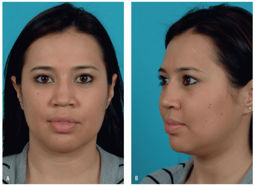 FIGURE 17.2 A-E. Patient with a saddle nose deformity demonstrating a depressed middle nasal vault, retracted columella, acute nasolabial angle, and underprojected nasal tip. This patient is the primary case used in this chapter to demonstrate the surgical technique for costal cartilage reconstruction of the nasal septum and saddle nose deformity. |
 FIGURE 17.2 (Continued) |
SURGICAL TECHNIQUE
Correction of the saddle nose deformity will usually require gaining exposure using the external rhinoplasty approach. The surgery is performed under general anesthesia, typically in an ambulatory surgery setting. The patient is placed in supine position with the head stabilized by a foam donut pillow. Perioperative antibiotics are administered prior to making incision. I typically perform a thorough endoscopic examination prior to beginning the operation. Local anesthetic (lidocaine 1% to 1:100,000 epinephrine) is injected into the nasal
septum and external nose. The patient is then widely prepped with Betadine at both surgical sites and draped in sterile fashion. The clean-contaminated nasal surgical field is kept separate from the sterile chest surgical field throughout the case, including maintaining separate instruments for each site.
septum and external nose. The patient is then widely prepped with Betadine at both surgical sites and draped in sterile fashion. The clean-contaminated nasal surgical field is kept separate from the sterile chest surgical field throughout the case, including maintaining separate instruments for each site.
The external rhinoplasty approach is carried out using a midcolumellar inverted-V incision in combination with bilateral marginal incisions. Once the lower lateral cartilages are exposed, dissection between the medial crura will permit direct exposure of the caudal margin of the nasal septum. If this approach is used, it is important to reconstitute the support of the lower lateral cartilages. Many patients will benefit from placement of an end-to-end caudal septal extension graft or caudal septal replacement graft.
Saddle nose deformities with a stable dorsal septum by palpation can be treated with an onlay dorsal graft. An onlay dorsal graft can be positioned through an endonasal approach. Bilateral intercartilaginous incisions can be made and a pocket created over the nasal dorsum. The dorsal pocket should be as tight as possible and should allow the dorsal graft to fit snugly onto the dorsum of the nose.
If it is clear that costal cartilage will be required for reconstruction of the saddle nose deformity, the rib cartilage harvest can be performed prior to beginning the saddle nose repair. I typically harvest the rib cartilage from the 6th rib, which in the female patient is located at the inferior margin of the breast (Fig. 17.3). If the patient has a large pendulous breast or has breast implants, the inferior aspect of the breast may be overlapping the 7th rib. I usually harvest the rib using a 1.1- to 1.5-cm incision placed along the inframammary crease of the right breast (Fig. 17.4). This places the incision just over the 6th rib in most patients. If a smaller incision is made, the location of the correct length and shape of the rib is very important as the size of the incision will limit access to a relatively small area of the rib. Alternatively, if a large incision (over 3 cm) is made, the positioning of the incision is less critical as a large portion of the 6th rib will be exposed and easily accessible. I do not recommend using a small incision for costal cartilage
harvest unless the surgeon has extensive experience harvesting rib cartilage in order to avoid complication such as pneumothorax.
harvest unless the surgeon has extensive experience harvesting rib cartilage in order to avoid complication such as pneumothorax.
 FIGURE 17.3 The 6th rib is typically located at the level of the inframammary crease allowing access to this rib through an inframammary incision. |
 FIGURE 17.4 Inframammary crease incision over the 6th rib. |
The 6th rib has a rather predictable curvature with a genu along the ribs contour. This is usually not a problem unless a long (>3 cm) straight segment of cartilage is needed for dorsal augmentation or long spreader grafts. In this case, the placement of the incision is important as the majority of the cartilage will need to be harvested either medial or lateral to the genu. Additionally, if the cartilage/bone junction is positioned more medial, this will leave less cartilage to harvest lateral to the genu. If the cartilage/bone junction is more lateral, then there will be a larger segment of cartilage available lateral to the genu. Most younger patients have a more laterally positioned cartilage/bone junction. As patients age, the cartilage/bone junction tends to advance medially. In patients with a prominent genu, I prefer to harvest the 7th rib even though the incision is not in the inframammary crease.
To know precisely where the cartilage/bone junction is located, a 1.5-inch long 27-gauge needle can be inserted through the skin and used to transcutaneously palpate the costal cartilage. The tip of the needle can be used to penetrate the outer 1 to 2 mm of the surface of the rib cartilage. The tip of the needle can be advanced along the rib cartilage until bone is encountered. The cartilage/bone junction is identified, and the bone is encountered with the tip of the 27-gauge needle. If a needle is used to palpate the rib cartilage, special care must be taken to avoid advancing the needle deep to or between the ribs as this will risk puncturing the parenchyma of the lung creating a small air leak that could develop into a pneumothorax.
Once the site of the incision is determined, local anesthetic (1% lidocaine with 1:100,000 epinephrine) is injected into the area around the incision. The incision is made using no. 15 blade initially cutting through skin and subcutaneous tissues. Dissection is then advanced down to the muscle layer. I try to avoid cutting the muscle as this will increase postoperative pain. The muscle fibers are separated to expose the underlying rib cartilage. Once the rib is exposed, the cartilaginous segment is assessed for curvature and contour. It is helpful to remove a large strip of perichondrium off of the surface of the 6th rib. Once the perichondrium is removed from the outside surface, the remaining perichondrium on the superior and inferior margins of the rib cartilage can be dissected away from the rib. To insure that dissection of the perichondrium is performed in the proper plane, I make a superficial incision into the rib cartilage using a Freer elevator and then continue the dissection around the superior and inferior equator points of the rib (Fig. 17.5). By making a superficial incision into the cartilage, it is less likely that the elevator will leave the proper plane and perforate the pleura. Once the perichondrium is elevated off of the superior and inferior surface of the 6th rib, the cartilage can be incised medially and laterally and then lifted from the chest. When making the medial and lateral incisions, a no. 15 blade is used to cut halfway through the rib, and then, a freer elevator is used to cut through the remainder of the rib. In most patients, a 3-cm-long segment of costal cartilage will be adequate for reconstruction as most cartilage grafts are 3 cm or less in length (Fig. 17.6).
After the rib cartilage is removed, saline solution is placed into the rib harvest site and a Valsalva maneuver is used to see if there is a pleura leak. If an obvious defect in the pleura is noted, the wound can be closed after placing a red rubber catheter into the defect. After closing the wound, the lungs can be expanded and the catheter can be pulled out. A postoperative chest radiograph can be performed postoperatively to assess the presence of pneumothorax.
Prior to closure, the edges of the remaining rib cartilage should be trimmed using a Takahashi forceps to prevent any sharp edges that may be palpable or can cause postoperative pain. I prefer to close the chest incision at the end of the operation so we can harvest additional cartilage or perichondrium if necessary. Initially, the muscle layer is closed tightly using 3-0 PDS sutures. Then, the subcutaneous tissue layer is closed with 4-0 PDS sutures. Care is taken to make sure that the breast moves freely over the deep layer closure so the breast tissue does not pucker when the patient stands. The skin can be closed using 5-0 nylon sutures. Typically, the scars for the rib cartilage harvest are small and not very noticeable (Fig. 17.7).
Stay updated, free articles. Join our Telegram channel

Full access? Get Clinical Tree


