Purpose
To evaluate the quality of the image of a grating target placed in a model eye viewed through multifocal intraocular lenses (IOLs).
Design
Laboratory investigation.
Methods
Refractive (NXG1 or PY60MV) or diffractive (ZM900 or SA60D3) multifocal IOLs were placed in a fluid-filled model eye with human corneal aberrations. A United States Air Force resolution target was placed on the posterior surface of the model eye. A flat contact lens or a wide-field contact lens was placed on the cornea. The contrasts of the gratings were evaluated under endoillumination and compared to those obtained through a monofocal IOL.
Results
The grating images were clear when viewed through the flat contact lens and through the central far-vision zone of the NXG1 and PY60MV, although those through the near-vision zone were blurred and doubled. The images observed through the central area of the ZM900 with flat contact lens were slightly defocused but the images in the periphery were very blurred. The contrast decreased significantly in low frequencies ( P < .001). The images observed through the central diffractive zone of the SA60D3 were slightly blurred, although the images in the periphery were clearer than that of the ZM900. The images were less blurred in all of the refractive and diffractive IOLs with the wide-field contact lens.
Conclusion
Refractive and diffractive multifocal IOLs blur the grating target but less with the wide-angle viewing system. The peripheral multifocal optical zone may be more influential on the quality of the images with contact lens system.
Refractive and diffractive multifocal intraocular lenses (IOLs) are widely used because of their ability to correct for both far and near vision. Recent studies have reported further patient satisfaction with multifocal IOLs with additional power for near vision. With the increase in the number of eyes implanted with multifocal IOLs, the possibility of having to perform vitreoretinal surgery in these eyes must be considered. Only a limited number of papers have reported on performing vitreoretinal surgery (eg, retinal detachment and preretinal membranes) while viewing the fundus through multifocal IOLs. Some surgeons concluded that the surgery was difficult because of the design of the optics of the multifocal lenses while others reported no difficulties.
We believe that knowledge of the retinal images observed during vitreous surgery through the different types of multifocal IOLs is important. Thus in an earlier study on pig eyes, we investigated the differences in the quality of the image viewed through implanted multifocal IOLs. However, there were limitations of these experiments including the physiological condition of the pig eyes—for example, the optical quality of the cornea and the firmness of the eye.
A comparison of the images viewed through different multifocal IOLs has been reported for a model eye system. To study an eye that was more comparable to the human eye, we constructed a model eye whose corneal lens had the average value of spherical aberration of human eyes. The aim of this study was to compare the quality of a grating target placed in our model eye and viewed through refractive and diffractive multifocal IOLs.
Materials and Methods
Model Eye and Wavefront Analyses of Cornea
A model eye was constructed based on Gullstrand’s model of the human eye ( Figure 1 , Top left). The body of the eye was made of metal, and the axial length was 24 mm ( Figure 1 , Top right). The diameter of the pupil was 7.0 mm. The distance from the corneal anterior surface to the implanted IOLs was 5 mm. The cornea was made of polymethylmethacrylate (PMMA), and the radius of curvature of the anterior surface was 7.70 mm and that of the posterior surface was 7.46 mm ( Figure 1 , Bottom left). The anterior surface was aspherical, and the cornea was constructed to have a spherical aberration of +0.220 μm. which is comparable to the mean value of human eyes with a pupil diameter of 6 mm.
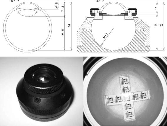
A 1951 United States Air Force (USAF) test target (Edmund Optics, Barrington, New Jersey, USA) was glued to the posterior surface of the model eye at the position of the retina ( Figure 1 , Bottom right). The target consisted of gratings of different orientations and spatial frequencies.
The model eye was filled with balanced salt solution at room temperature with care taken to remove all air bubbles. The eye was aligned so that the measuring light entered the pupil along the optical axis of the model eye. A wavefront analyzer (KR-1W; Topcon Medical System Inc, Tokyo, Japan) was used to measure the spherical aberration of the cornea without the IOLs. The measurements were performed 10 times, and the average was used for the analyses.
Images of Grating Through Multifocal Intraocular Lenses in Model Eye
The refractive multifocal IOLs, NXG1 (ReZoom; Abbott Medical Optics, Santa Ana, California, USA) and PY60MV (AF-1 iSii; HOYA Surgical Optics, Frankfurt, Germany), and the diffractive multifocal IOLs, ZM900 (Tecnis Multifocal; Abbott Medical Optics) and SA60D3 (ReSTOR; Alcon Laboratories, Fort Worth, Texas, USA), were studied. The monofocal IOL, SA60AT (AcrySof; Alcon Laboratories), was used as control ( Table 1 ). All of the IOLs had the same spherical power of +20.0 diopters (D) and were centered on the optical axis of the eye.
| IOL | Model | Material | Focusing | Optics | Spherical Aberration (μm) |
|---|---|---|---|---|---|
| ReZoom | NXG1 | Acryl | Refractive multifocal | Aspherical | −0.27 |
| AF-1 iSii | PY60MV | Acryl | Refractive multifocal | Aspherical | — |
| TECNIS multifocal | ZM900 | Silicone | Diffractive multifocal | Aspherical | −0.27 |
| ReSTOR | SA60AD | Acryl | Diffractive multifocal | Spherical | — |
| AcrySof | SA60AT | Acryl | Monofocal | Spherical | — |
Two 20-gauge wide-angled endoilluminating light pipes (Bullet Wide Angle Fiberoptic Illuminator; Alcon Laboratories) were inserted through two openings on opposite sides of the model eye at positions similar to the sclerotomy sites used during pars plana vitreous surgery ( Figure 2 ). A flat contact lens with a 30-degree prism made of quartz glass (HOYA Corporation, Tokyo, Japan) or a wide-angle viewing contact lens (Mini Quad; Volk Optical, Inc, Mentor, Ohio, USA) was placed on the cornea to view the grating target.
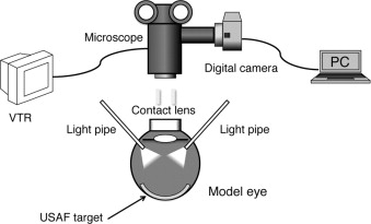
The grating target was photographed with a digital camera (EOS KISS X3; Canon Inc, Tokyo, Japan) through a surgical microscope (VISU140; Carl Zeiss Meditec, Tokyo, Japan) with a 25.5× magnification. To evaluate the differences in the quality of the images quantitatively, the contrasts of the gratings were measured using Photoshop CS4 (Adobe System Inc, San Jose, California, USA). The intensity at the center of the black stripe was set as I max and the intensity at the center of the white stripe was set at I min . The contrast was calculated as (I max – I min )/(I max + I min ) at each spatial frequency (cycle/mm). The results were compared among the different IOLs for flat or wide-angle viewing contact lenses.
Results
Wavefront Analysis of Corneal Aberrations of Model Eye
The root mean square (RMS) of the front surface of the model eye was 0.033 μm for a 4-mm-diameter pupil and 0.248 μm for a 6-mm pupil. The spherical aberration of the cornea, the value of Z4-0, was +0.032 μm for a 4-mm pupil and +0.247 μm for 6 mm. These results are in good agreement with the design of the front surface of the model eye of +0.220 μm. The corneal astigmatism was -0.06 D at axis 86 degrees.
Subjective Assessments of Grating Target Viewed Through Refractive Multifocal Intraocular Lens
The refractive multifocal NXG1 has five different optical zones: a 2.1-mm central zone for far vision and four annular zones. The refractive power of the first, and the third and fifth annular zones (the second and fourth zones), is set for near vision, and that of the second and fourth annular zones (the third and fifth zones) is set for far vision. The image viewed with the flat contact lens through the central optical zone of the NXG1 was well focused, as were the images viewed through the second annular zone. The images through the first and third annular zones were defocused with a doubling of the images ( Figure 3 , Top left), although it was difficult to identify the images through the third and fourth annular zones.
The refractive multifocal PY60MV has three optical zones with the 2.3-mm-diameter central zone and second annular zone set for far vision. The first annular zone is set for near vision. The grating target viewed with the flat contact lens through the central zone was well focused and the first annular zone surrounding the central zone was defocused with a doubling of the image ( Figure 3 , Top center). The images through the first annular zone were not as clear as those viewed through the monofocal IOL ( Figure 3 , Top right). The images from the second annular zone were as well focused as those with the NXG1.
The peripheral images observed through the prism contact lens for both the NXG1 ( Figure 3 , Middle left) and PY60MV ( Figure 3 , Middle center) were more defocused than the images of the monofocal IOL ( Figure 3 , Middle right). However, the images through the PY60MV were more in focus than those of the NXG1.
The images through both types of refractive multifocal IOLs observed through a wide-angle viewing lens were in good focus ( Figure 3 , Bottom left and center). The quality of the images was comparable to those through monofocal IOLs ( Figure 3 , Bottom right).
Contrast of Gratings Viewed Through Refractive Multifocal Intraocular Lenses
The contrasts of the gratings viewed through the flat contact lens with both refractive NXG1 and PY60MV were lower than that of monofocal SA60AT at both lower and higher frequencies ( Figure 4 , Left). These results are in agreement with our subjective assessments of the images. The contrast of the gratings viewed with NXG1 through the flat contact lens at 16 cycles/mm was significantly lower than that of the monofocal SA60AT, although the other values were not significant ( Table 2 ). The contrasts of the grating observed through the NXG1 with the wide-angle contact lens were slightly lower than those of the PY60MV and SA60AT at both lower and higher frequencies ( Figure 4 , Right), although the values were not significant.
| With Flat Contact Lens System | |||||
|---|---|---|---|---|---|
| Frequencies (Cycles/mm) | Refractive Multifocal IOL | Monofocal IOL | |||
| NXG1 | P Value a | PY60MV | P Value a | SA60AT | |
| 16.0 | 0.261 | .044 | 0.300 | .633 | 0.316 |
| 32.0 | 0.035 | .710 | 0.044 | .424 | 0.060 |
| 64.0 | 0.010 | .319 | 0.008 | .059 | 0.015 |
| With Wide-Angle Viewing Lens System | |||||
|---|---|---|---|---|---|
| Frequencies (Cycles/mm) | Refractive Multifocal IOL | Monofocal IOL | |||
| NXG1 | P Value a | PY60MV | P Value a | SA60AT | |
| 16.0 | 0.205 | .071 | 0.225 | .421 | 0.229 |
| 32.0 | 0.021 | .147 | 0.041 | .886 | 0.030 |
| 64.0 | 0.000 | .999 | 0.000 | .999 | 0.000 |
a P value was calculated by unpaired t test compared with the contrast of SA60AT.
Subjective Assessments of Grating Target Viewed Through Diffractive Multifocal Intraocular Lenses
The gratings viewed through the diffractive multifocal ZM900 were well focused with the flat contact lens ( Figure 5 , Top left) but not as clear as the images viewed through the monofocal IOL. The images in the peripheral area were doubled, and ghost images were present. The ghost images were displaced from the central optical zone.


