Objective
To evaluate the recurrence rate after upper eyelid epiblepharon repair in patients with Down syndrome.
Design
Retrospective, observational study.
Methods
Total of 578 Korean children (21 with Down syndrome patients, 557 with non-Down syndrome patients), who had undergone epiblepharon repair and were followed up for more than 2 months, were included in this study. The recurrence rate was compared between two groups at 2, 6 months after surgery. Recurrence was defined as the re-appearance of cilia touching to cornea. The recurrence rate was also analyzed according to whether patients had undergone concomitant z-medial epicanthoplasty or not.
Results
Lower eyelid epiblepharon repair was performed on 22 eyelids of Down syndrome patients, and 1072 eyelids of non-Down syndrome patients. At 3 months after surgery, the recurrence rate was not significantly different between two groups ( P = 1.00). Upper eyelid epiblepharon was repaired on 40 eyelids of Down syndrome patients, and 204 eyelids in non-Down syndrome patients. At 2 and 6 months after surgery, the recurrence rate was significantly higher in Down syndrome patients (27.5% and 29.4%) than non-Down syndrome patients (3.4% and 4.6%) ( P = 0.000, P = 0.004, respectively). The recurrence rate of upper eyelid epiblepharon repair was not affected in both groups whether Z-epicanthoplasty was combined or not ( P = 1.00 in both groups).
Conclusions
In Down syndrome patients, the recurrence rate after upper eyelid epiblepharon repair was higher than non-Down syndrome patients. The effect of combined Z-medial epicanthoplasty was limited in both groups.
Epiblepharon is rather common condition among Asian children ; in this condition, a fold of skin and the underlying orbicularis muscle press the lashes against the eyeball. In general, the lower eyelids are commonly involved as compared to the upper eyelids. Recurrence rates after surgical correction are considerably low, and recent techniques of correction include cilia rotating tarsal fixation suture and Z-medial epicanthoplasty. However, the incidence and clinical manifestations of epiblepharon that occurs in Down syndrome patients remarkably differ from those of epiblepharon that occurs in non-Down syndrome Asian children. The incidence of epiblepharon is much higher in Down syndrome patients than in non-Down syndrome patients, and characteristically, the upper eyelids are predominantly involved in the case of Down syndrome patients. Moreover, natural remission of the upper and lower eyelid epiblepharon rarely occurs in the case of Down syndrome patients; this is in contrast to that observed in the case of non-Down syndrome patients. Therefore, almost all Down syndrome patients with epiblepharon need surgical correction. To our best knowledge, surgical success rate of epiblepharon repair in the case of Down syndrome patients has not been reported. In this study, we evaluated and compared the surgical results of epiblepharon repair in the case of Asian Down syndrome patients and in that of non-Down syndrome patients.
Methods
This study was a retrospective observational study. The following patients were included: those who had undergone upper eyelid epiblepharon repair performed by 2 of the authors (H.K.C & N.J.K) at Seoul National University Hospital, Seoul Metropolitan Government Seoul National University Boramae Medical Center, Seoul National University Bundang Hospital, between June 2003 and July 2008. Epiblepharon was diagnosed when cilia touched cornea with horizontal fold of redundant skin and the underlying pretarsal orbicularis oculi muscle without inward rotation of eyelid margin. Surgical indications were severe corneal erosion, i.e., horizontal erosion, in more than one-third of the cornea or irritative symptoms such as foreign body sensation, ocular pain, or tearing that the parents thought were remarkable even after the affected children were treated with artificial tears. The following patients were excluded: those who were not followed up for more than 2 months; those who had any other disease, in which affected the eyelid position is affected, such as Stevens-Johnson syndrome; and those with ocular cicatricial pemphigoid or facial trauma. For cosmetic reasons, epiblepharon repair was performed on both the right and left sides for all the patients including those with epiblepharon on only 1 side. Lower eyelid epiblepharon was repaired using cilia rotating tarsal fixation suture and excision of the redundant skin and the pretarsal orbicularis muscle. Upper eyelid epiblepharon was repaired by an incisional double-eyelid surgery with tarsal fixation suture. We performed a combined Z-medial epicanthoplasty on patients with a prominent epicanthal fold by using modified Park’s Z-epicanthoplasty. We used the classification system described by Park to evaluate the severity of the epicanthal fold; further, this procedure was indicated in patients with epicanthal fold type II and type III ( Table ). At follow-up, the patients were evaluated for direction of the lashes and status of the cornea; recurrence was defined when the cilia touched the cornea again at each time for each eyelid. We compared the recurrence rate between the Down syndrome and non-Down syndrome patients. The recurrence rate between the 2 groups was also analyzed in both groups according to whether the patients had undergone concomitant Z-medial epicanthoplasty. In the case of simultaneous upper and lower epiblepharon repair, each eyelid was independently analyzed, because we think that the upper eyelid epiblepharon repair does not affect the lower eyelid epiblepharon repair and vice versa. Statistical analyses were performed using Statistical Package for Social Sciences (SPSS) software v. 13.0 (SPSS Inc, Chicago, IL), and two-sided P < 0.05 was considered statistically significant.
| Type | Definition |
|---|---|
| Type I | No epicanthal fold. |
| Type II | The upper eyelid skin margin covers the tarsal border as it approaches the medial canthal ligament, and the lacrimal lake is covered only partially. |
| Type III | The upper eyelid skin margin curves over the lacrimal lake, covering the entire medial angle of the palpebral fissure. |
| Type IV | The lower eyelid skin crosses over the lacrimal lake, forming a reverse epicanthal fold. |
Results
This study included 811 Korean patients who underwent epiblepharon repair during the study period; of these, 233 patients who were not followed up for more than 2 months were excluded. Thus, 578 patients fulfilled the inclusion criteria; of these, 21 were Down syndrome patients, and 557, non-Down syndrome patients. The clinical characteristics of the patients were as follows: male versus female ratio was 10:11 among the Down syndrome patients, and 274:283, among the non-Down syndrome patients ( P = 0.887, chi-square test); the mean age of the Down syndrome patients at operation was 7.33 ± 2.65 years and that of the non-Down syndrome patients was 6.19 ± 3.87 years ( P = 0.179, independent T-test); further, the mean follow-up period for the Down syndrome patients was 12.5 ± 12.9 months and that for the non-Down syndrome patients was 8.6 ± 9.7 months ( P = 0.17, independent T-test). The follow-up period of the non-Down syndrome patients was shorter than that of the Down syndrome patients although the difference was not significant.
In lower eyelid epiblepharon repair, 1 of 22 eyelids recurred (4.8%) in Down syndrome patients, and 59 of 1072 eyelids recurred (5.5%) in non-Down syndrome patients by the two month postoperative visit. ( P = 1.000, Fisher’s exact test). However, the recurrence rate did not significantly differ between the 2 groups.
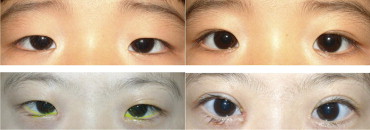
Upper eyelid epiblepharon repair was performed on 20 Down syndrome patients (40 upper eyelids) and 102 non-Down syndrome patients (204 eyelids; Figure 1 ). In upper eyelid epiblepharon repair, 11 of 40 eyelids recurred (27.5%) in Down syndrome patients, whereas 7 of 206 eyelids recurred (3.4%) in non-Down syndrome patients by the two month postoperative visit ( P = 0.000, chi-square) and 5 of 24 eyelids recurred (20.8%) versus 7 of 146 eyelids recurred (4.8%) by the six month postoperative visit ( P = 0.004, chi-square test; Figure 2 ). Kaplan–Meier survival curves for the recurrence of epiblepharon are shown in Figure 3 . The cumulative probability of survival was significantly lower among the Down syndrome patients than among the non-Down syndrome patients (Log Rank P = 0.000, Breslow P = 0.000).