Purpose
To report novel vitreous body microarchitecture findings using high-resolution spectral-domain optical coherence tomography (HR-SD-OCT).
Design
Prospective, cross-sectional study.
Methods
Horizontal and vertical retinal cross-sectional images that were 10 mm long were acquired from 17 eyes of 17 young healthy volunteers using HR-SD-OCT with enhanced vitreous imaging (EVI). Images were acquired through the fovea, upper vessel arcade, and lower vessel arcade.
Results
Three new findings on vitreous body microarchitecture were found. First, material located between the retina and posterior vitreous cortex was easily detected in 90% of upper and lower vessel arcade scans. Most scans contained hyperreflective dots and multilayered hyperreflective lines around the detached vitreous cortex. Second, a lamellar structure was observed in the vitreous body in 70%–80% of all scans, excluding vertical scans of the upper arcade vessel area. Third, tubular zones of hypodensity were detected in >80% of scans, excluding horizontal scans of the macula. Interestingly, the location of tubular zones of hypodensity seemed to correspond with the location of retinal vessels. Subject age, refractive error, and axial length were not significantly different in scans with and without material between the retina and vitreous, lamellar structures, and tubular zones of hypodensity.
Conclusions
The microarchitecture of the vitreous body can be visualized using HR-SD-OCT and EVI.
The vitreous body of the eye, which occupies 80% of the eye volume, is a gelatinous material consisting of 3-dimensionally organized collagen fibers and hyaluronan. The physiological functions of the vitreous body include maintenance of the eyeball shape, shock absorption, light transmission, and physiological management of the effects of oxygen on the lens and other surrounding structures. However, the vitreous body can also be the origin of retinal diseases and a hotbed of abnormal proliferation; consequently, it is often a therapeutic target. However, because of its gel-like structure and transparency, it was not treated as a major subject of morphologic study until the introduction of advanced research methods involving visualization of the vitreous body using fluorescein, triamcinolone, and optical coherence tomography (OCT).
Because of a remarkable development in the hardware, software, and imaging techniques, recent OCT systems have promoted noninvasive observation of the vitreous body in living humans. Although OCT imaging is limited to the posterior part of the vitreous body, recent studies have reported detailed observation of the liquefied lacunae of the vitreous body represented in the premacular bursa, Cloquet canal (CC), and vitreoretinal interface and the associated disorders. These macro-observations support the designs of vitreous structures proposed by previous studies. However, fine structures reported by Sebag, such as fibrous structures coursing from the central and posterior vitreous body through the premacular hole, as observed using dark-field slit microscopy, and a lamellar structure in the outer layer of the vitreous body, as observed by immunohistochemistry, have not been thoroughly studied using OCT.
Therefore, in the present study, we used high-resolution spectral-domain OCT (HR-SD-OCT) to visualize the microarchitecture of the vitreous body in normal subjects.
Methods
This study was designed as a prospective, cross-sectional study. Institutional Review Board/Ethics Committee approval was obtained. The study was performed in accordance with the tenets of the Declaration of Helsinki, and informed consent was obtained from all participants after they were provided with a detailed explanation of the nature and possible consequences of the study procedures.
Subjects
OCT images were acquired for 17 eyes of 17 young, healthy Japanese volunteers without a history of ocular or systemic diseases. The average age of subjects was 29.5 ± 6.1 years (range, 22–46 years), the average refractive error was −2.41 ± 2.42 diopter (range, −6.25 to +2.00 diopter), and the average axial length was 24.7 ± 0.7 mm (range, 23.5–25.8 mm).
Optical Coherence Tomography Imaging
The HS100 (Canon Inc, Tokyo, Japan) HR-SD-OCT system was used to obtain the study images. The HS100 has an extended-bandwidth, superluminescent diode light source with a full-width at half-maximum (FWHM) value of 100 nm. The light source comprises 2 chips that deliver a 3-μm optical axial resolution, whereas several commercially available OCT devices provide a maximum optical axial resolution of up to 6 μm. Although the HS100 was originally designed to acquire a maximum of 50 imaging scans to generate 1 B-scan image for multiple scan averaging, we developed system control software to acquire a maximum of 150 imaging scans to achieve more effective speckle noise reduction.
Enhanced Imaging of the Vitreous Body
An enhanced vitreous imaging (EVI) technique reported by Pang and associates was used to obtain images of the vitreous body. In brief, once the focus was auto-aligned on the retina, the focus plane was manually defocused on the vitreous body until the signal from the vitreous body could be detected on the monitor window. Ten-millimeter-long horizontal and vertical retinal section images were acquired through the fovea, upper arcade vessels, and lower arcade vessels of both eyes. All imaging procedures were performed 3 times through each area without pupil dilation. Only images that could be generated using over 100 scans for speckle noise reduction were included in our analysis. The image contrast was adjusted using ImageJ software (National Institutes of Health, Bethesda, Maryland, USA; http://rsb.info.nih.gov/ij/index.html ) to facilitate visualization of the vitreous structures.
Statistics
All values are expressed as means ± standard deviations where applicable. Differences in age, refractive error, and axial length with regard to the presence of different structures in the vitreous body were analyzed using Student t tests. A P value of <.05 was considered statistically significant. All analyses were performed using StatView statistical software (version 5.0; SAS Institute, Cary, North Carolina, USA).
Results
All types of B-scans (macular horizontal, macular vertical, upper horizontal, upper vertical, lower horizontal, and lower vertical) generated from an average of over 100 scans could be successfully acquired for all subjects. The premacular bursa was observed in all macular horizontal and macular vertical scans. Moreover, a part of CC and the septum between the premacular bursa and CC were detected in all macular horizontal scans. Channels connecting the premacular bursa and CC could be detected in 5 eyes (29.4%).
Interposed Material Between the Retina and Vitreous Body
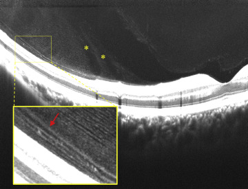
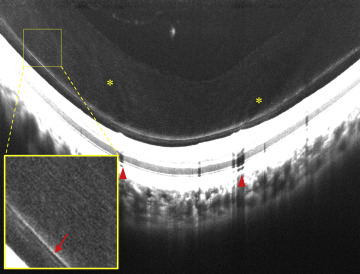
A thin hyperreflective line including the posterior wall of the premacular bursa, which may correspond to the vitreous cortex, was observed at the parafovea, but not at the fovea, on macular horizontal and vertical scans for 16 eyes (94.1%). The remaining eye showed no vitreous cortex detachment. Approximately half of the macular horizontal and vertical scans for eyes with perifoveal vitreous detachment showed moderate reflectivity in the space between the detached vitreous cortex and retinal surface ( Supplemental Figure 1 ; Supplemental Material available at AJO.com ). Several eyes showed the presence of hyperreflective dots around the detached vitreous cortex ( Figure 1 ). Moreover, multilayered hyperreflective lines of the vitreous cortex (a multilayer sign), which may be associated with vitreoschisis, were observed in some eyes. These findings were more frequently and markedly detectable on scans of the upper and lower regions ( Table 1 ). On these scans, obvious interposed material showing the same reflectivity as the vitreous body was observed between the vitreous body and retinal surface ( Figures 1 and 2 ). A distinct gap was found between the material and vitreous body, and the material itself appeared to have a lamellar structure.
| Type of Scan | Presence of Interposed Material | Presence of Hyperreflective Dots | Presence of a Multilaminar Structure |
|---|---|---|---|
| Macula horizontal | 6 (35.3%) | 1 (5.9%) | 4 (23.5%) |
| Macula vertical | 7 (41.2%) | 4 (23.5%) | 5 (29.4%) |
| Upper horizontal | 15 (88.2%) | 12 (70.6%) | 13 (76.5%) |
| Upper vertical | 15 (88.2%) | 14 (82.4%) | 15 (88.2%) |
| Lower horizontal | 16 (94.1%) | 12 (70.6%) | 13 (76.5%) |
| Lower vertical | 15 (88.2%) | 12 (70.6%) | 14 (82.4%) |
Lamellar Structure of the Vitreous Body
In addition to the multilayer sign in the interposed material outside the vitreous body, a lamellar structure was observed in the vitreous body ( Figure 3 ). This structure exhibited millefeuille-like, multilayered, thin hyperreflective lines that were parallel to the retinal surface. The interval between lines ranged from 3.4 μm to 11.9 μm. The locations of and detection rates for the lamellar structure are shown in Table 2 . The lamellar structure was detected in approximately 70%–80% of all types of scans other than the lower vertical ones; only 35.3% of these scans showed the structure. Approximately half of the scans that showed the structure exhibited good image quality that allowed counting of individual hyperreflective lines, whereas the remaining half exhibited relatively poor image quality that allowed detection of the lamellar structure but not counting of individual lines.
| Type of Scan | Presence of the Lamellar Structure | Location of the Lamellar Structure | |||
|---|---|---|---|---|---|
| Temporal | Nasal | Upper | Lower | ||
| Macula horizontal | 14 (82.4%) | 11 (64.7%) | 8 (47.1%) | – | – |
| Macula vertical | 14 (82.4%) | – | – | 14 (82.4%) | 8 (47.1%) |
| Upper horizontal | 14 (82.4%) | 13 (76.5%) | 13 (76.5%) | – | – |
| Upper vertical | 13 (76.5%) | – | – | 13 (76.5%) | 0 (0%) |
| Lower horizontal | 13 (76.5%) | 11 (64.7%) | 10 (58.8%) | – | – |
| Lower vertical | 6 (35.3%) | – | – | 0 (0%) | 6 (35.3%) |
Tubular Zones of Hypodensity
Against the background of the lamellar structure, the vitreous body exhibited long, narrow, low-intensity structures in the longitudinal direction and tubular zones of hypodensity, which could be liquefied lacunae or low-density collagen fibrils ( Figures 1, 2 , and 3 ). On poor-quality images showing an unclear lamellar structure, these tubular zones of hypodensity appeared to form the lamellar structure in the longitudinal direction ( Supplemental Figure 2 ; Supplemental Material available at AJO.com ). Interestingly, although vitreous cortex detachment and interposed material were observed, the position of these tubular zones of hypodensity appeared to approximately correspond with the position of the retinal vessels. Moreover, careful observation showed the presence of tubular zones of hypodensity, although their correspondence with the retinal vessels was not confirmed ( Figure 4 , Supplemental Figure 2 , and Supplemental Video ; Supplemental Material available at AJO.com ). The tubular zones that did not correspond with the retinal vessels were characterized by a relatively low detection rate, a closely spaced arrangement, and a uniformly thick appearance compared with those that corresponded with the retinal vessels. Although fewer tubular zones of hypodensity were observed on macular horizontal scans than on the other types of scans because of the absence of large vessels ( Table 3 ), those that did not correspond to the retinal vessels were detected in the septum between the premacular bursa and CC in several eyes. There were no significant differences in age, refractive error, and axial length with the regard to the presence of interposed material, the lamellar structure, and tubular zones of hypodensity.
| Type of Scan | Tubular Zones of Hypodensity That Correspond to the Retinal Vessels | Tubular Zones of Hypodensity That Did Not Correspond to the Retinal Vessels |
|---|---|---|
| Macula horizontal | 1 (5.9%) | 5 (29.4%) |
| Macula vertical | 15 (88.2%) | 6 (35.3%) |
| Upper horizontal | 15 (88.2%) | 4 (23.5%) |
| Upper vertical | 16 (94.1%) | 5 (29.4%) |
| Lower horizontal | 16 (94.1%) | 4 (23.5%) |
| Lower vertical | 16 (94.1%) | 5 (29.4%) |
Stay updated, free articles. Join our Telegram channel

Full access? Get Clinical Tree


