LASIK Complications and Management
Christopher J. Rapuano
▪ Introduction
The significant advantages of LASIK over photorefractive keratectomy (PRK) are somewhat tempered by the increased complexity of the surgery, which increases the type and severity of complications. While the risk of complications is low, when a complication occurs, it needs to be managed appropriately to achieve the best chance of a satisfactory result. Additionally, surgeons should continue to strive to minimize complications. This chapter will focus on intraoperative and postoperative complications and their causes, prevention, and management. It may be helpful for the reader to refer to Chapter 10 on retreatment as needed for a more detailed discussion of the indications and techniques related to re-lifting a LASIK flap. Use the intraoperative and postoperative planning index to find representative cases that demonstrate each of the complications discussed.
▪ Intraoperative Complications
SHREDDED FLAP
A shredded flap is one of the most feared complications of LASIK. It is typically only recognized after the microkeratome has made its pass and is removed from the eye. The surgeon may notice the flap to be somewhat irregular. Otherwise, it becomes obvious when the surgeon attempts to lift the flap and does not find a standard flap attached at a hinge.
Causes
Poor suction or pseudosuction. Most, if not all, modern microkeratome systems alert the surgeon when the machine senses adequate suction on the eye and will allow the microkeratome to pass only after suction is achieved. However, if the suction port or ports are occluded by conjunctiva, the machine can measure full suction without the intraocular pressure (IOP) being adequately elevated. This situation is termed pseudosuction. Patients with redundant or boggy conjunctiva, such as might occur in association with thyroid orbitopathy, after retinal surgery or after multiple attempts at achieving suction, are at greater risk for pseudosuction.
Poor blade quality. If a pass is made with an imperfect blade, the result may be a poor-quality flap. The exact condition of the blade will impact both the nature and the degree of flap damage.
Recutting a flap. Recutting a LASIK flap is a retreatment option, but a second flap may intersect with the first flap, resulting in an irregular flap and an irregular stromal bed. Because of this risk, recutting a flap is considered a less desirable approach to retreatment.
Attempting to create a thin flap. LASIK on the second eye using a microkeratome with the same blade will generally result in a thinner flap. Microkeratomes may cut thinner flaps in thin corneas. The femtosecond laser can also be set to a thickness <100 µm. In any of the above situations, flaps thinner than expected may result and the flap may be of poor quality. A shredded flap is less likely with the femtosecond laser.
Prevention
Obtain sufficient exposure. Poor suction can occur due to difficulty positioning the microkeratome onto the eye. This is more likely to occur when exposure is inadequate. The surgeon may notice a hissing sound, if suction is insufficient.
Press the ring firmly against the globe. Firm posterior pressure on the suction ring presses the redundant conjunctiva against the sclera. This may help reduce the risk of pseudosuction.
Triple check the IOP:
First: As the pressure in the eye is increasing, the surgeon should monitor the pupil, which often dilates slightly and then remains enlarged.
Second: Once the machine registers full suction, the IOP should be high enough to obstruct blood flow to the eye and the patient will usually notice the vision dimming or even blacking out.
Third: Check the IOP. This can be done with a Barraquer tonometer, pneumotonometer, TonoPen, or even with digital palpation. The pressure should be >65 mmHg to obtain a quality flap with a blade microkeratome.
Check the blade. The surgeon or an experienced technician should always check the quality of the blade prior to seating it in the microkeratome.
Avoid recutting a LASIK flap for retreatment. Other retreatment options are reliable and safer.
Be cognizant of the risk of creating a very thin flap. Consider PRK if the cornea is thin. Consider using a new microkeratome blade or a thicker plate on the second eye to avoid the risk of a shredded flap.
Management
ABORT THE EXCIMER LASER ABLATION.
Replace flap pieces as best as you can. The goal is to put all of the pieces of the shredded flap back into their original location. This is often quite difficult because there may be multiple small fragments of essentially clear cornea.
Place a bandage soft contact lens (BSCL). The BSCL is usually removed 1 to 7 days later. Topical antibiotic and topical corticosteroid eyedrops are applied in routine fashion with the lens in place and after the lens is removed.
Long-Term Management
Later options include no refractive surgery, transepithelial PRK with or without mitomycin-C, and LASIK with a femtosecond laser setting a significantly deeper flap depth.
BUTTONHOLE FLAP
Buttonhole flaps (Fig. 11.1) have been described in eyes with normal corneal curvature, but steep corneas appear to be at increased risk, when using a blade microkeratome. The surgeon must always closely examine the quality of the flap as it is being lifted and reflected. Special attention should be paid to the central and paracentral cornea, looking for a fullthickness (easy to detect) or partial-thickness (harder to detect) buttonhole. Buttonholes are easier to detect on the stromal bed appearing as an area of elevation or a smooth island of tissue, but can also be seen as an irregularity or hole in the underside of the flap as it is lifted (Fig. 11.2). Although rare, buttonholes can occur with the femtosecond laser.
Causes
Steep cornea (≥48.00 diopters [D]). The proposed mechanism for steep corneas
predisposing to buttonholes is that when suction is applied, the steepness allows a large amount of tissue to be “pushed up,” above the plane of the suction ring. As this large area of corneal tissue is deformed by the mechanical microkeratome pass, it buckles posteriorly to the plane of the blade and is left uncut, creating a buttonhole (Fig. 11.3).
Localized corneal thinning or scarring. A localized area of stromal loss or scarring may be covered by thickened epithelium. As the flap is made, there may be a full-thickness stromal defect that is covered only by tenuous epithelium.
Partial- or full-thickness buttonholes can occur during the femtosecond laser pass as gas passes through a focal area of corneal thinning or scar. Alternatively, it can result from a traumatic flap lift or from debris on the applanation lens that causes focal posterior displacement of the cornea with resultant focal superficial femtosecond laser ablation.
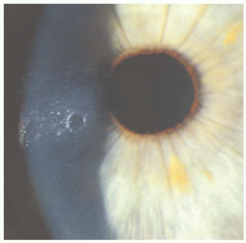 FIGURE 11.1 Flap buttonhole. (Courtesy of Christopher Rapuano, M.D.) |
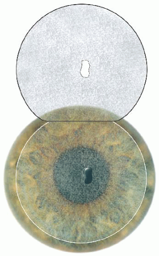 FIGURE 11.2 Smooth, elevated contour of a full-thickness buttonhole noted in the stromal bed, with a corresponding hole noted in the flap. |
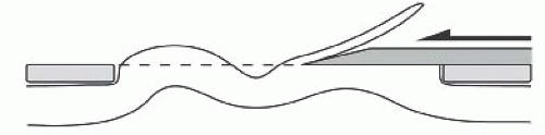 FIGURE 11.3 Proposed mechanism of the buttonhole. It is presumed that an at-risk cornea will buckle in advance of the microkeratome pass, resulting in an isolated area in which the flap is not cut. |
Prevention
Use an appropriate suction ring (e.g., use an 8.5-mm suction ring when K > 45.00 D). Most microkeratome systems are designed with a variety of suction rings. Smaller rings do not allow as much corneal tissue to protrude anteriorly. Follow the manufacturer’s recommendations regarding which suction ring size to use, based on the keratometry readings.
Femtosecond laser. The risk of a buttonhole is less because the applanating lens flattens the cornea within the suction ring and there is no mechanical pass across the cornea to induce a buckling of tissue. Consider using a thicker flap if possible when a scar or corneal facet is present within the margins of the flap. Gentle flap dissection can prevent a traumatic flap dissection. If a gas bubble is seen during the laser pass in an area of scar, extra care should be taken during flap dissection. These are very rare situations. In general, the femtosecond laser is safer than a blade microkeratome for the patient with a steep cornea.
Surface ablation. If steep keratometry readings or areas of corneal scarring/thinning are noted preoperatively, the surgeon may want to avoid the possibility of a LASIK flap complication and perform a surface ablation.
Management
DO NOT LASER.
Replace the flap as best as you can.
Place a BSCL, in the event of a full-thickness buttonhole. The BSCL is usually removed 1 to 7 days later.
Long-Term Management
Later options include no refractive surgery, transepithelial PRK (with or without mitomycin-C), and LASIK (using a microkeratome with a thicker plate or a deeper femtosecond laser ablation) (See Video 20).
SHORT OR DECENTERED FLAP WITH AN EDGE IN LASER TREATMENT ZONE
The LASIK flap needs to be properly centered and large enough to accommodate the entire centered excimer laser ablation. A 6-mm-diameter ablation does not require as large a flap as an 8-mm-diameter wavefront-guided custom ablation or a 9-mm hyperopic correction.
Causes
Poor centration of the microkeratome or femtosecond laser. The suction ring should be centered on the pupil or slightly decentered toward the planned hinge location. It may slip slightly or the globe might rotate under the ring as suction is applied to the eye.
Small flap. The flap may be too small to allow the full laser ablation size. This may be due to inadequate suction prior to the pass or due to a flat corneal contour with keratometry readings ≤40.00 D.
Incomplete flap. Loss of suction during the mechanical microkeratome pass might result in a foreshortened flap. An incomplete flap can occur with the femtosecond laser if suction is lost, if the applanation pressure is light, if fluid is present under the applanation lens, or if the meniscus is too close to the flap edge (See Video 13). Refer to Chapter 5 for a more complete discussion of the femtosecond laser.
Prevention
Re-center the suction ring. If the suction ring is decentered, suction should be released and the ring repositioned properly before the microkeratome or femtosecond laser pass. Some femtosecond lasers (e.g., IntraLase) have a toggling function that allows for minor readjustments of centration; however, the diameter of the flap decreases when this is done. Many femtosecond lasers do not have this capability.
Management
Use the reticle. If the flap has been created, use the excimer laser reticle to determine whether the treatment zone will fit entirely beneath the flap. If the hinge is slightly encroaching within a large (e.g., 8- or 9-mm diameter) ablation zone, then the laser treatment can most likely proceed safely. The hinge should be covered with a hinge protector when the treatment zone is large. Ablation of the hinge should be prevented to avoid a doubly treated area. Laser system software that can design a hingesparing ablation may exist on the particular excimer laser being used. Check with the medical director of the laser center.
DO NOT LASER if flap size is inadequate. If the flap does not cover the vast majority of the treatment zone, it is best to abort the excimer laser procedure.
Do not attempt another blade microkeratome pass during the same surgery session. The femtosecond laser is more versatile in this situation. At the first sign of a significant incomplete laser ablation, stop the femtosecond laser. Using the same applanation cone, the laser can be reset, the surface dried, and, if good applanation can be achieved, another laser pass attempted. The pocket should not be cut on the second pass of the femtosecond laser. Usually, an adequate flap can be created with this second pass; however, if the stromal bed appears very irregular, it may be best to abort the excimer laser procedure.
Do not try a lamellar dissection to increase flap diameter. Attempting to increase the flap diameter with a blade is very likely to cause irregular astigmatism and a poor visual outcome. In some cases after a femtosecond laser flap has been created, a small area of incomplete ablation can successfully be bluntly dissected.
Replace the flap. If the flap is inadequate, replace it as one would replace a routine LASIK flap.
Late Management
Later options include no refractive surgery. After waiting for 3 to 6 months, one could attempt to repeat microkeratome or femtosecond LASIK with a larger and thicker flap. Finally, surface ablation with or without mitomycin-C could be considered. Surface ablation over a previous flap increases the risk of haze.
FREE CAP
Automated lamellar keratoplasty, the predecessor to LASIK, and original LASIK techniques were performed by creating a free cap. The current hinged-flap technique improved wound alignment and the safety of the LASIK procedure. While there are reports of free caps occurring in eyes with normal corneal curvature, the risk is increased in patients with flat corneas when using a blade microkeratome.
The presence of a free cap (see Case 66, Fig. 1


Stay updated, free articles. Join our Telegram channel
Full access? Get Clinical Tree
