Purpose
To evaluate the impact of aging on conjunctivochalasis in an objective manner using anterior segment optical coherence tomography.
Design
Prospective clinical trial.
Methods
Sixty eyes of 30 healthy volunteers (15 men, 15 women; age range, 24-75 years) without any ophthalmic diseases were recruited for the study. Subjects were organized into 3 sex-matched groups, each including 10 subjects according to their ages: 20-39 years (Group 1), 40-59 years (Group 2), 60-75 years (Group 3). Cross-sectional area of conjunctivochalasis was measured at 3 locations (temporal, central, and nasal) using Fourier-domain optical coherence tomography. Tear meniscus height was also measured in all images where a typical triangular-shaped tear meniscus was obtained.
Results
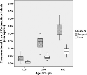
In terms of the cross-sectional area of conjunctivochalasis, there were statistically significant differences among 3 groups at temporal and nasal locations ( P < .001). At the central location, whereas there were no subjects in Groups 1 and 2, solely 3 eyes revealed conjunctivochalasis in Group 3. The severity of conjunctivochalasis affecting the temporal and nasal bulbar conjunctiva was strongly correlated with age (η 2 = 0.81, P < .001 and η 2 = 0.78, P < .001). Lower central tear meniscus height was compared among the groups and a significant increase with age was observed ( P < .001).
Conclusion
Fourier-domain optical coherence tomography provides an objective and quantitative approach for assessing the stages of conjunctivochalasis. The current study objectively confirms that conjunctivochalasis can be observed even in younger, healthy eyes; its severity increases with age; and it may alter tear distribution along the lower lid.
Conjunctivochalasis, which is characterized by a loose, redundant, nonedematous inferior bulbar conjunctiva interposed typically between the globe and the lower eyelid, has been reported to be an important cause of ocular discomfort and tear instability. The condition can be localized in the nasal, central, and temporal part of the lower eyelid and usually occurs bilaterally.
There is no consensus regarding the exact cause and pathogenesis of conjunctivochalasis. Whereas conjunctivochalasis has been reported to be associated with aging and dry eye symptoms, ocular surface inflammation has been proposed as one of the most important causative factors by some studies. However, it is not clear whether ocular surface inflammation causes conjunctivochalasis or vice versa. Coexisting aging confounds the results of studies investigating the clinical impact of conjunctivochalasis on ocular surface health and/or the etiopathogenesis of conjunctivochalasis tear dysfunction or different subjective grading schemes for conjunctivochalasis. Accordingly, the objectives of the current study were to investigate the impact of aging on the formation of conjunctivochalasis in otherwise healthy volunteers without evidence of tear film dysfunction or ocular surface disease in an objective manner using anterior segment optical coherence topography (AS-OCT), and to provide an objective method for grading conjunctivochalasis.
Methods
Subjects
Sixty eyes of 30 healthy volunteers (15 men, 15 women; range, 24-75 years) without complaints of eye irritation were evaluated. Exclusion criteria included diabetes mellitus; atopic dermatitis; rosacea; history of contact lens wear; use of any topical medications; prior ocular surgery or trauma; eyelid abnormalities such as entropion, ectropion, and trichiasis; ocular atopy; anterior blepharitis; and meibomian gland disease beyond normal age-related changes.
Subjects were organized into 3 sex-matched groups according to their ages: 20-39 years (Group 1: 10 subjects), 40-59 years (Group 2: 10 subjects), and 60-75 years (Group 3: 10 subjects).
Objective Evaluation of Conjunctivochalasis
Cross-sectional area of conjunctivochalasis was measured as mm 2 at 3 locations (temporal, central, and nasal) using the RTVue-100 Fourier-domain optical coherence tomograph (Optovue, Fremont, California, USA) using the standardized protocol described in our previous study. Lower central tear meniscus height (TMH) was also measured in microns in all images where a typical triangular-shaped tear meniscus was visualized. All measurements were performed in a standardized fashion by the same experienced examiner (K.G.) at the same time period of the day.
Statistical Analysis
The data were analyzed using SPSS 15.0 for Windows (SPSS Inc, Chicago, Illinois, USA). All data were first analyzed as to whether their distribution was normal. Lower central tear meniscus height values were transformed into logarithmic values. The differences among the 3 age groups were assessed using the Welch analysis of variance. The post hoc Tamhane test for samples without equal variances was used to determine which, if any, means were significantly different. The correlation between age groups and cross-sectional conjunctivochalasis area values was analyzed using the Eta correlation coefficients. Linear regression analysis was applied to verify the impact of age on the severity of conjunctivochalasis after adjustment for sex. A value of P < .05 was considered to be statistically significant.
Results
The age and sex distribution of the participants is shown in the Table .
| Group 1 N = 20 | Group 2 N = 20 | Group 3 N = 20 | P Value | |
|---|---|---|---|---|
| Age (years) | 24-37 29.2 ± 4.1 | 42-58 49.5 ± 5.4 | 60-75 65.9 ± 5.6 | |
| Sex (male/female), n (%) | 10 (50%)/10 (50%) | 10 (50%)/10 (50%) | 10 (50%)/10 (50%) | >.999 |
| Temporal CCha cross-sectional area (mm 2 ) a | 0.03 ± 0.02 | 0.14 ± 0.04 | 0.23 ± 0.05 | <.001 |
| Nasal CCha cross-sectional area (mm 2 ) a | 0.01 ± 0.01 | 0.04 ± 0.01 | 0.08 ± 0.02 | <.001 |
| Central CCha cross-sectional area (mm 2 ) a | None of the subjects had central Ccha | None of the subjects had central CCha | 0.01 ± 0.04 b | NA c |
| Central tear meniscus height (μm) a | 218.4 ± 94.5 | 292.4 ± 145.1 | 316.4 ± 154.0 | <.001 |
a CCha cross-sectional area and meniscus height were calculated using the mean of both eyes.
b Three subjects had central CCha.
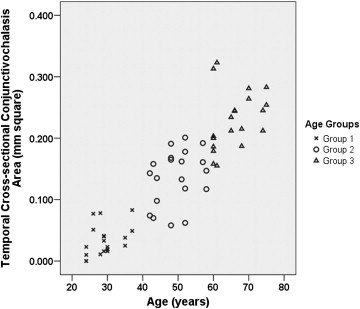
In terms of the cross-sectional area of conjunctivochalasis, there were statistically significant differences among the 3 groups at temporal and nasal locations ( P < .001). At the central location, whereas there were no subjects in Groups 1 and 2, solely 3 eyes revealed conjunctivochalasis in Group 3. Details regarding the cross-sectional area values of conjunctivochalasis and the distribution are given in the Table and Figure 1 .
The severity of conjunctivochalasis affecting the temporal and nasal bulbar conjunctiva was strongly correlated with age (η 2 = 0.81, P < .001 and η 2 = 0.78, P < .001, respectively). The correlation graphs are provided in Figures 2 and 3 . Additionally, the relationship between conjunctivochalasis and age was verified using a linear regression model with adjustment for sex. In this model, age was found strongly correlated to the severity of conjunctivochalasis both at temporal (β = -0.115, β1 = 0.005, P < .001) and nasal locations (β = -0.057, β1 = 0.002, P < .001).