Histologic Development of the Human Fovea From Midgestation to Maturity


Purpose


To describe the histologic development of the human central retina from fetal week (Fwk) 22 to 13 years.


Design


Retrospective observational case series.


Methods


Retinal layers and neuronal substructures were delineated on foveal sections of fixed tissue stained in azure II–methylene blue and on frozen sections immunolabeled for cone, rod, or glial proteins. Postmortem tissue was from 11 eyes at Fwk 20–27; 8 eyes at Fwk 28–37; 6 eyes at postnatal 1 day to 6 weeks; 3 eyes at 9 to 15 months; and 5 eyes at 28 months to 13 years.


Results


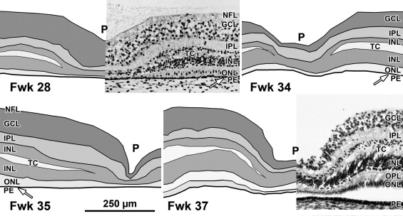
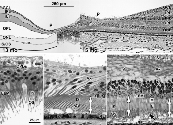
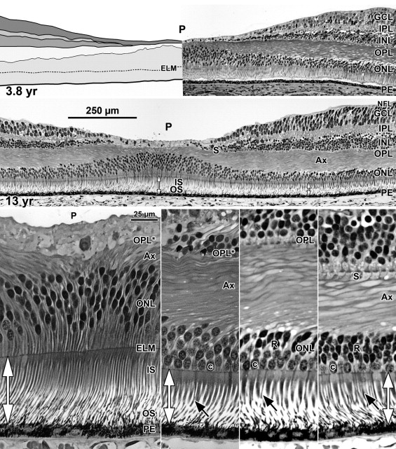
At Fwk 20–22 the fovea could be identified by the presence of a single layer of cones in the outer nuclear layer. Immunolabeling detected synaptic proteins, cone and rod opsins, and Müller glial processes separating the photoreceptors. The foveal pit appeared at Fwk 25, involving progressive peripheral displacement of ganglion cell, inner plexiform, and inner nuclear layers. The pit became wider and shallower after birth, and appeared mature by 15 months. Between Fwk 25 and Fwk 38, all photoreceptors developed more distinct inner and outer segments, but these were longer on peripheral than foveal cones. After birth the foveal outer nuclear layer became much thicker as cone packing occurred. Cone packing and neuronal migration during pit formation combined to form long central photoreceptor axons, which changed the outer plexiform layer from a thin sheet of synaptic pedicles into the thickest layer in the central retina by 15 months. Foveal inner and outer segment length matched peripheral cones by 15 months and was 4 times longer by 13 years.


Conclusions


These data are necessary to understand the marked changes in human retina from late gestation to early adulthood. They provide qualitative and quantitative morphologic information required to interpret the changes in hyper- and hyporeflexive bands in pediatric spectral-domain optical coherence tomography images at the same ages.


The fovea is the most critical portion of the primate retina for high visual acuity and color vision. Its early development begins at fetal week (Fwk) 12 with a complex expression sequence of cell-specific molecules. After midgestation an outward displacement of inner retinal layers occurs to form a foveal pit, and after birth an inward displacement of the photoreceptors occurs to raise foveal cone density 10×. Spectral-domain optical coherence tomography (SDOCT) has become a valuable tool for clinical diagnosis of normal and diseased adult human retina. Recently, Maldonado and associates used a portable research hand-held SDOCT unit to image human retinas from premature infants through adults. They documented the immaturity of the neonatal human fovea and the marked difference in SDOCT layers from the standard adult SDOCT image. However, there is still little detailed description of histologic development and almost no quantitative information about human central retina around birth. This paper provides the first detailed histologic description of human central retinal development from midgestation to early adulthood. In particular, it shows the marked change in foveal photoreceptors from a single layer of short undeveloped cones up to birth to tightly packed and highly elongated cones in the adult. Understanding these changes is essential to make the correct interpretation of neonatal SDOCT images.


Methods


Tissue sections for this study were from eyes obtained under Human Subjects Approval #010447. Eyes younger than Fwk 22 were from elective abortions; only normal-appearing fetuses were used. Older prenatal and neonatal eyes were from infants who survived days to weeks in the neonatal intensive care unit; babies with diagnosed conditions that might affect retinal development were not included in this report. Infant eyes were obtained from children who died of accidental causes, or of diseases that did not affect the retina.


Eyes were enucleated 2–8 hours after death, the cornea and lens removed, and the globe immersed in a mixture of 4% paraformaldehyde and 0.5% glutaraldehyde in pH.7.4 phosphate buffer (PB) for 1 day to months. In 1 of the pair of eyes, the horizontal meridian was embedded in glycol methacrylate and cut serially at 2–3 μm. This method gives excellent preservation and imaging of tissue. In a few eyes the entire globe was embedded in paraffin and sectioned serially at 12 μm. This method was less time-consuming, but the retina was usually detached from the pigment epithelium. In some eyes the horizontal meridian was frozen-sectioned serially at 12 μm, allowing immunolabeling at a known locus with a wide variety of antibodies. In each method, every tenth slide was stained with 1% azure II–methylene blue to locate the fovea.


Immunolabeling


Frozen sections were labeled as previously reported. The rod marker was mouse anti-rod opsin (4D2, 1/300; gift of R. Molday). Cone markers included rabbit anti-long-and-medium (L&M)-wavelength-selective cone opsin (1/2000, gift of J. Saari), rabbit anti-short (S)-wavelength-selective cone opsin (1/10 000, gift of J. Nathans), mouse anti-cone arrestin (7G6, 1/250–500; gift of P. MacLeish), and mouse anti-cone alpha transducin (A1.1, 1/250–500; gift of J. Hurley). Mouse anti-synaptophysin (1/1000; Sigma-Aldrich S5768) labels cone synaptic pedicles heavily and rod synaptic spherules lightly. Rabbit anti–cellular retinaldehyde-binding protein (CRALBP; 1/1000; gift of J. Saari) was used to label Müller glial cytoplasm.


Imaging


All photographs were from sections in which the foveal center could be reliably identified. Plastic or paraffin sections stained with azure II–methylene blue were imaged digitally using a Nikon E1000 wide-field digital microscope (Nikon Instruments, Inc, Melville, New York, USA). Immunolabeled sections were imaged using a high-sensitivity Hamamatsu camera (Hamamatsu Corp, Bridgewater, New Jersey, USA) or in an Olympus Fluoview 1000 confocal microscope (Olympus America, Center Valley, Pennsylvania, USA). All images were processed in Adobe Photoshop CS5 for size, color balance, sharpness, and contrast.


Segmentation of Light Micrographs


A low-power foveal montage was created at selected ages. One half was imaged in Photoshop and paths drawn along the inner surface of the retina and the borders between the ganglion cell (GCL) and inner plexiform layer (IPL); the IPL and inner nuclear layer (INL); the INL and outer plexiform layer (OPL); the OPL and outer nuclear layer (ONL); and the inner border of the retinal pigment epithelium (RPE). Layers were differentially shaded (( Figure 1 provides an orientation to the layers; Figures 2 through 7 show shaded layers).). In young retinas, the OPL was thin and included in ONL, but in older retinas the OPL was distinguished as a separate layer. A dotted path was drawn along the external limiting membrane (ELM), separating the proximal ONL from the distal inner segment/outer segment (IS/OS) layer.


FIGURE 1


Layers and histology of normal adult human retina. Adult human retina 2 mm nasal from the fovea center. The layers and their abbreviations used in this paper and in subsequent figures are choroid (CH); retinal pigment epithelium (PE); outer nuclear layer (ONL), which is subdivided into the photoreceptor outer segment (OS) and inner segment (IS) layers distal to the external limiting membrane (ELM); and the nuclear layer containing a single row of cone (C) cell bodies near the ELM and multiple rows of deeper rod (R) cell bodies. The thin rod IS are indicated (R-IS). The rod OS are about half the thickness of the rod IS, are longer and reach the PE. The larger tapered cone IS have a shorter OS (arrow). The ELM marks the distal edge of the retina. IS myeloid (M) and ellipsoid (E) are indicated. The outer plexiform layer (OPL) contains a distal layer made up of the fibers of Henle or photoreceptor axons (Ax) and a proximal synaptic contact layer (S) containing cone pedicles and rod spherules. The inner nuclear layer (INL) contains the cell bodies of horizontal cells (HZ) lying most distal, bipolar cells (BP) in the middle, Müller glia (MG), and amacrine cells (AM) lying most proximal. The inner plexiform (IPL), ganglion cell (GCL), and nerve fiber (NFL) layers complete the inner retina.


FIGURE 2


Layers and histology of midgestation fetal human retina. Development of the human fovea at (Top left) fetal week (Fwk) 22, (Top right) Fwk 25, and (Middle left and right) Fwk 27. Retinal layers have been drawn from digital photographs using Adobe Photoshop (see Methods) and the layers shaded as indicated. (Top left) At Fwk 22 the foveal region is composed of 5 layers with a thick GCL and a thin ONL. (Top right, Middle right) After Fwk 25 the foveal pit (P) begins to invaginate the inner retinal layers. (Bottom left) At Fwk 25 there is only a very narrow space (vertical double arrow) between the ELM (horizontal white arrowheads) and the PE in central retina. The cone marked with arrowheads is shown at higher power in the inset to the right. A short thick IS extends distal to the ELM (white arrowheads) and a small synaptic pedicle (arrow) is present. (Bottom middle) At 800 μm from the fovea there is little difference from the fovea except that 2 rods (R) are present. (Bottom right) At 2 mm from the fovea a slightly wider PE/ELM space is present (vertical double arrow) and is filled with cone and rod IS. OS are not obvious. Scale in Middle right for Top and Middle rows; in Bottom middle for Bottom row. C = cone; CH = choroid; ELM = external limiting membrane; GCL = ganglion cell layer; INL = inner nuclear layer; IPL = inner plexiform layer; IS = inner segment; NFL = nerve fiber layer; ONL = outer nuclear layer; OPL = outer plexiform layer; PE =retinal pigment epithelium.


FIGURE 3


Layers of late-gestation fetal human retina. Development of the human fovea at (Top left) Fwk 28, (Top right) Fwk 34, (Bottom left) Fwk 35, and (Bottom right) Fwk 37. A transient layer of Chievitz (TC) is present as a gap in the INL of all eyes from this age group. The foveal pit (P) has thinned the GCL, IPL, and INL compared to layers surrounding the pit (the foveal slope). Scale in Bottom left for all. GCL = ganglion cell layer; INL = inner nuclear layer; IPL = inner plexiform layer; NFL = nerve fiber layer; ONL = outer nuclear layer; OPL = outer plexiform layer; PE =retinal pigment epithelium.


FIGURE 4


Histology of late-gestation fetal human retina. A Fwk 35 retina is shown (Top left, Bottom left), at the fovea, (Top middle) at 800 μm from the fovea where there are 2-3 layers of rods, and (Top right, Bottom right) at 2 mm from the fovea. Longer axons (Ax) are appearing on cones (C) and rods (R) outside the fovea (Top middle, Top right), which tilt away from the pit center (fovea to left). Note that the space between the ELM and PE (vertical double arrows) is widest in the periphery. Short OS are present on both rods and cones in the periphery (Top right, Bottom right) but are difficult to recognize close to or in the fovea (Bottom left, Bottom middle, black arrowhead). (Bottom middle) Two Fwk 30 foveal cones shown in a low-magnification electron micrograph. The desmosomal junctions between Müller (MG) glia and cones forming the ELM can be resolved (white arrowhead). Each cone is surrounded by pale MG cytoplasm. IS myeloid (M) and ellipsoid (E) are indicated. Scale in Top left for Top row; scale in Bottom left for Bottom row. ELM = external limiting membrane; INL = inner nuclear layer; IS = inner segment; ONL = outer nuclear layer; OPL = outer plexiform layer; OS = outer segment; PE =retinal pigment epithelium.


FIGURE 5


Layers and histology of postnatal-term human retina. Development of the human fovea at (Top left) postnatal (P) 1 day (d) and (Middle left) P8d. These retinas illustrate the range in development found around birth. In both retinas the pit is wider and more shallow than before birth and has displaced most of the inner layers and extends close to the cone synapses. A TC is present in the INL. Cones on the pit slope are 2-3 cell bodies deep and have long axon (Ax), but a single layer of cones still is present over the pit center. At P1d rods (R) are within 500 μm of the foveal center. (Top right) One of the cones (asterisk) on the slope from another P1d eye is shown at higher power. The IS is longer and narrower than before birth, and a short OS is present (arrowhead). The long Ax ends in a synaptic spherule (arrow). Note the large amount of pale M cytoplasm surrounding each cone, also seen around foveal cones in Middle left. (Middle right) A P8d retina at 1 mm. Both cone (C) and rod (black arrowhead) OS are obvious. P8d photoreceptors (Bottom left) at the foveal center, (Bottom middle-left) on the foveal slope at the first rods, and (Bottom middle-right) 800 μm and (Bottom right) 2 mm from the fovea. Note that the distance between ELM and PE (vertical double arrows) has increased at the fovea, but is still narrower than in the periphery. OS are present at all locations but are much shorter in the fovea compared to the periphery. The fibrous acellular nature of the TC can be seen in Bottom middle-left and middle-right. Scale in Middle left for Top and Middle left; scale in Bottom left for all others. CH = choroid; ELM = external limiting membrane; GCL = ganglion cell layer; INL = inner nuclear layer; IPL = inner plexiform layer; IS = inner segment; OS = outer segment; NFL = nerve fiber layer; ONL = outer nuclear layer; OPL = outer plexiform layer; PE =retinal pigment epithelium; TC = transient layer of Chievitz.


FIGURE 6


Layers and histology of human retina during infancy. Postnatal maturation of the human fovea at (Top left) 13 months and (Top right) 15 months. Over the early postnatal months, the pit becomes wide and shallow with almost no neurons in the center except cone cell bodies. The drawing in Top left illustrates the large postnatal growth in thickness of the OPL, mainly attributable to the increased number and length of cone axon (Ax). (Bottom left) Note the increase in ONL cone cell bodies over the 13 months pit center attributable to postnatal cone packing. These have long thin IS and OS. At 15 months, by comparing the double vertical arrows, it can be seen that (Bottom middle-left) foveal IS/OS, and IS/OS on photoreceptors at (Bottom middle-right) 800 μm and (Bottom right) 2 mm from the fovea now are similar in length. Long thin rod OS (Bottom right, black arrow) are prominent outside of the foveal center. Cone synapses (S) are absent from the foveal center (Bottom left and middle-left) because they have been displaced onto the pit slope (Bottom right, S). Scale in Top left for Top row; scale in Bottom left for Bottom row. CH = choroid; ELM = external limiting membrane; GCL = ganglion cell layer; INL = inner nuclear layer; IPL = inner plexiform layer; IS = inner segment; OS = outer segment; ONL = outer nuclear layer; OPL = outer plexiform layer; PE =retinal pigment epithelium.


FIGURE 7


Layers and histology of human retina during childhood. The final maturation stages of the human fovea are shown at (Top left) 3.8 years (yr) and (Top right) 13 years with higher magnification of the 13 years (Middle) foveal center, (Bottom left) first rods at 300 μm, (Bottom middle-left) 500 μm, and (Bottom middle-right) 2 mm from the fovea. The foveal pit is wide and shallow. The foveal center is composed of long thin cone OS, IS, and cell bodies 8-12 cones deep. The central OPL contains a thick layer of axon (Ax). The foveal OPL contains only Ax (OPL*) out to 500 μm, where synaptic pedicles (Bottom middle-left, OPL; Bottom middle and far right, S) are first encountered. Long thin rod OS (Bottom row, black arrows) become more prominent with eccentricity. Note the marked increase in cone IS diameter from the foveal center to 300 μm with a small further increase at 2 mm. Scale in Middle for Top and Middle rows; scale in Bottom left for Bottom row. ELM = external limiting membrane; GCL = ganglion cell layer; INL = inner nuclear layer; IPL = inner plexiform layer; IS = inner segment; OS = outer segment; NFL = nerve fiber layer; ONL = outer nuclear layer; OPL = outer plexiform layer; PE =retinal pigment epithelium.


Results


Normal Adult Retina


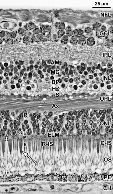
A section from adult human retina ∼2 mm nasal to the fovea is shown in Figure 1 for orientation to the layers and abbreviations used in the developing retina. A young adult fovea is shown in Figure 7 (Bottom row).


Development of the Human Fovea


Fetal week 20–27 (11 retinas examined)


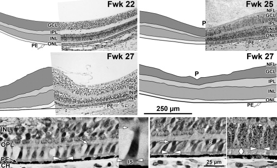
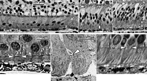
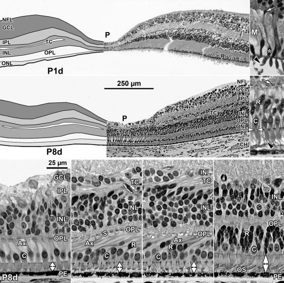
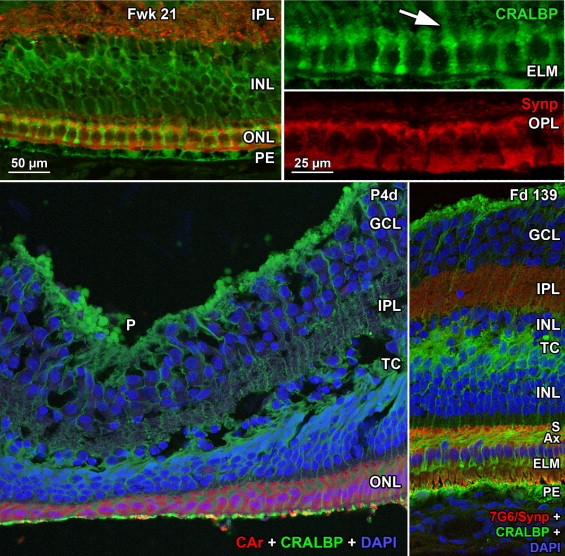
At midgestation, the future fovea had 3 distinctive differences from peripheral retina. First, the GCL is much thicker than in the periphery. Second, up to birth the ONL is formed by a single layer of cone photoreceptors ( Figure 2 , Bottom left; Figure 8 , Top left). This is in marked contrast to the thicker peripheral ONL containing both cones and rods ( Figure 2 , Bottom right; Figure 8 , Top middle-left and middle-right). Third, the OPL is a narrow band of cone synaptic pedicles ( Figure 2 , Bottom left and insert, Bottom middle; Figure 8 , Top right upper panel, and Bottom). These layers will undergo developmental changes that are critical to interpreting late prenatal and neonatal SDOCT images. In the following narrative, numbers like 800 μm or 2 mm indicate distance from the fovea center.


FIGURE 8


Immunocytochemistry of photoreceptor maturation. (Top row) Human Fwk 24-25 retina immunocytochemically labeled for rod opsin (red) and cone L&M opsin (green). The entire cone and rod membrane are labeled at this age, outlining the cell shape. (Top left) Each foveal cone has a short vertical axon ending in a row of synaptic pedicles (S) forming the OPL. A short cone OS is indicated by the white arrow. A single rod (R) with a tiny OS (black arrow) is on the edge of the fovea. (Top middle-left and middle-right) Photoreceptors at 2 mm from the fovea. Note the longer OS (arrows). (Top right) Foveal cones stained for L&M opsin (green; upper panel) and synaptophysin (red; lower panel), a synaptic vesicle marker. The synaptic pedicle (S) is labeled in all L&M cones and in 1 short-wavelength-selective cone (white arrowhead) unlabeled for L&M opsin. (Middle row) Postnatal 1 day (P1d) retina showing the change in photoreceptor morphology from the (Middle left) foveal cones to (Middle center) rods and (Middle right) cones 800 μm from the fovea. The foveal cones are short and relatively flat while the peripheral photoreceptors have axons tilted away from the foveal center. Note the difference in length between foveal and peripheral OS (arrows). (Bottom) A 37-year-old (37 yr) retina at 1 mm labeled for short-wavelength-selective cone opsin. The cone on the left can be traced from OS to synaptic pedicle (S). The striking change in central cone morphology from midgestation to adult can be appreciated by comparing Bottom to Top middle-left. Scale in Bottom for all. Ax = axon; ELM = external limiting membrane; IS = inner segment; OS = outer segment; OPL = outer plexiform layer.


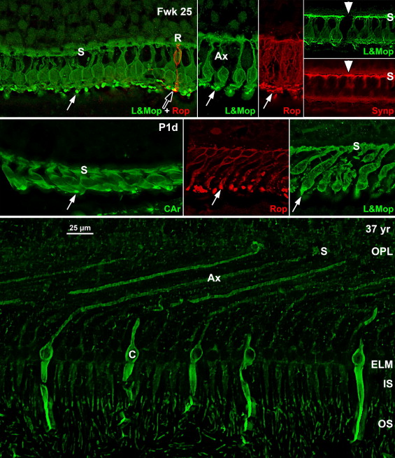
Rods are absent across the central 1500–1800 μm, forming a region called the rod-free zone underlying the thickened GCL. Central cones are 6–8 μm wide, with a large prominent nucleus ( Figure 2 , Bottom left insert; Figure 8 , Top left; Figure 9 , Top right lower panel). Each cone has a short axon (Ax), which ends below the cell body in a synaptic pedicle ( Figure 2 , Bottom left insert, arrow; Figure 8 , Top middle-left); these form the thin OPL ( Figure 8 , Top row). Foveal cones are separated by Müller cell processes filling the space between cones. These end in small bulbs forming junctions with the cone cell bodies, creating the ELM ( Figure 2 , Bottom left, white arrowhead; Figure 9 , Top row).


FIGURE 9


Immunocytochemistry of Müller cell morphology. (Top row) Fwk 21 fovea immunocytochemically labeled for the Müller cell protein CRALBP (green) and synaptophysin (red). Müller cell processes extend from the cell body deep in the INL, run between each cone (Top right upper panel, arrow), and form small clubs at the ELM. In the fovea Müller cytoplasm fills all space between foveal cones. (Bottom left) Postnatal 4 day (P4d) fovea triple labeled for cone arrestin (red), CRALBP (green), and nuclear label DAPI (blue). Cones over the pit (P) are in a thin single layer, but are longer on the slope. The TC is filled with Müller processes. (Bottom right) The foveal slope from a late prenatal Macaca monkey fetus triple-labeled for a mixture of cone transducin and synaptophysin (red), CRALBP (green), and nuclear label DAPI (blue). This retina more clearly shows the glial fiber makeup of the TC. Note how the Müller cell processes follow the angle of the cone axons (Ax) and then turn 90 degrees to run between the cones to form the ELM. Scale in Top left for Top left and Bottom row; in Top right for Top right upper and lower panels. ELM = external limiting membrane; GCL = ganglion cell layer; INL = inner nuclear layer; IPL = inner plexiform layer; NFL = nerve fiber layer; ONL = outer nuclear layer; OPL = outer plexiform layer; PE =retinal pigment epithelium; TC = transient layer of Chievitz.

Only gold members can continue reading. Log In or Register to continue

Stay updated, free articles. Join our Telegram channel

Jan 12, 2017 | Posted by in OPHTHALMOLOGY | Comments Off on Histologic Development of the Human Fovea From Midgestation to Maturity

Full access? Get Clinical Tree

Get Clinical Tree app for offline access