Purpose
To describe 4 patients who sustained facial nerve injury during temporal artery biopsy.
Design
Retrospective, observational case series.
Methods
The medical records were reviewed of 4 patients (2 men, 2 women; mean age 72.8 years, range 60 to 87), referred for evaluation of palsy of the frontal branch of the facial nerve following temporal artery biopsy. Main outcomes measured were site of incision, length of follow-up, and degree of recovery.
Results
In all cases, incisions were made in the preauricular region or on the pretrichial temple within 3 cm of the lateral canthal angle. Follow-up ranged from 1 month to over 5 years. No patient recovered completely; 2 had partial return of function, and 2 reported no improvement.
Conclusions
Branch facial nerve palsy can occur with temporal artery biopsy and is likely to result in permanent disability. In all cases the incision was placed within the known course of the frontal branch of the facial nerve. To prevent this rare complication, we advocate biopsy of the parietal, rather than the frontal, branch of the superficial temporal artery.
Giant cell arteritis (GCA) is an idiopathic vasculitis that affects medium- to large-sized arteries throughout the body. Long-term immune suppression, most often with prednisone, remains the mainstay of therapy. Particularly in elderly patients, this drug carries significant risk of complications. Therefore, prior to assigning the diagnosis of GCA, histopathologic confirmation is desirable. Biopsy of the superficial temporal artery remains the gold standard for diagnosis and is generally felt to be a low-risk procedure. However, both minor and more serious complications have been described. In this report, we describe 4 patients with injury to the facial nerve, which occurred during temporal artery biopsy.
Methods
A retrospective, observational case series was compiled by reviewing the records in the tertiary care neuro-ophthalmology practices at the University of California–San Francisco from October 1, 2004 to January 31, 2009. Four patients (2 male and 2 female, mean age 72.8 years, ranging from 60 to 87) who had been specifically referred to our department for evaluation of facial nerve injury following temporal artery biopsy were included. Medical records were reviewed with particular attention given to the site of incision, length of follow-up, and the degree of spontaneous recovery. Other causes of facial nerve palsy, including idiopathic and post-neurosurgical, were not included. Attempts were made to acquire the operative reports from the referring practices.
A PubMed search using the term “temporal artery biopsy” was performed and any reports addressing complications were assessed. Three cases of facial nerve injury were identified and were compiled with the cases from this series. Site of incision and degree of recovery were noted.
Results
The Table summarizes the results. One patient was referred from an ophthalmologist, 1 from a general plastic surgeon, and 2 from vascular surgeons. Each patient was referred from a different physician. Only 1 operative note was available for review, and this did not document difficulty with the surgery or abnormalities in the anatomy.
| Patient | Age (Years) | Sex | Site of Biopsy | Surgeon Specialty | Follow-up | Recovery |
|---|---|---|---|---|---|---|
| 1 | 78 | M | Temporal | Ophthalmology | 1 month | None |
| 2 | 60 | F | Pre-auricular | Vascular surgery | 5.5 years | 10% |
| 3 | 87 | M | Temporal | Vascular surgery | 9 months | 75% |
| 4 | 66 | F | Temporal | Plastic surgery | 9 months | None |
| Study mean | 72.8 | 1.8 years | ||||
| Slavin 1986 | 55 | F | Temporal | Not listed | 6 month | 70% |
| Bhatti 2000 | 63 | F | “Temporal scalp region” | General surgery | 1 month | None |
| Bhatti 2001 | 75 | M | Temporal | General surgery | 6 months | None |
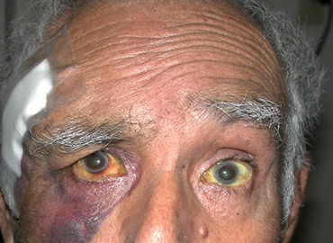
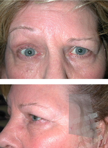
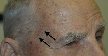
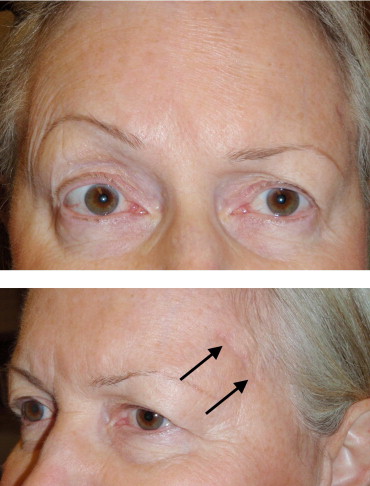
Three patients had unilateral and 1 had bilateral superficial temporal artery biopsies. In the patient with biopsies performed on both sides, only the right side was injured. On initial evaluation, complete loss of brow elevation and partial loss of eyelid closure were observed. Patients had biopsy in the pretrichial temporal (n = 3) or preauricular (n = 1) regions ( Figures 1 through 4 ). In the 3 cases with incisions performed in the pretrichial region, the incision extended to within 3 cm of the lateral orbital rim. Improvement (based on self-reporting and clinical evaluation) in frontalis function ranged from zero to 75 percent. Follow-up ranged from 1 month to 5.5 years (mean 1.8 years). No patients underwent surgery or other therapy to correct brow position. There were no other significant complications.
Three previously reported cases were identified with review of the literature and were included in the Table for comparison. Including these patients in our series gave a total of 7 patients (3 male, 4 female, mean age 69.1 years, range 55 to 87 years). Recovery of function ranged from zero to 75 percent.
Results
The Table summarizes the results. One patient was referred from an ophthalmologist, 1 from a general plastic surgeon, and 2 from vascular surgeons. Each patient was referred from a different physician. Only 1 operative note was available for review, and this did not document difficulty with the surgery or abnormalities in the anatomy.
| Patient | Age (Years) | Sex | Site of Biopsy | Surgeon Specialty | Follow-up | Recovery |
|---|---|---|---|---|---|---|
| 1 | 78 | M | Temporal | Ophthalmology | 1 month | None |
| 2 | 60 | F | Pre-auricular | Vascular surgery | 5.5 years | 10% |
| 3 | 87 | M | Temporal | Vascular surgery | 9 months | 75% |
| 4 | 66 | F | Temporal | Plastic surgery | 9 months | None |
| Study mean | 72.8 | 1.8 years | ||||
| Slavin 1986 | 55 | F | Temporal | Not listed | 6 month | 70% |
| Bhatti 2000 | 63 | F | “Temporal scalp region” | General surgery | 1 month | None |
| Bhatti 2001 | 75 | M | Temporal | General surgery | 6 months | None |
Three patients had unilateral and 1 had bilateral superficial temporal artery biopsies. In the patient with biopsies performed on both sides, only the right side was injured. On initial evaluation, complete loss of brow elevation and partial loss of eyelid closure were observed. Patients had biopsy in the pretrichial temporal (n = 3) or preauricular (n = 1) regions ( Figures 1 through 4 ). In the 3 cases with incisions performed in the pretrichial region, the incision extended to within 3 cm of the lateral orbital rim. Improvement (based on self-reporting and clinical evaluation) in frontalis function ranged from zero to 75 percent. Follow-up ranged from 1 month to 5.5 years (mean 1.8 years). No patients underwent surgery or other therapy to correct brow position. There were no other significant complications.


