Purpose
To evaluate the efficacy of perfluoro-n-octane as a postoperative short-term tamponade after vitrectomy in pediatric cases with complex retinal detachment (RD) and proliferative vitreoretinopathy (PVR).
Design
Prospective, noncomparative, interventional case series.
Methods
The medical records of 10 eyes of 9 children (6 boys and 3 girls), whose age ranged from 3 months to 11 years, with a median of 7.5 months, were reviewed. The cause of the PVR was retinopathy of prematurity (7 eyes of 6 patients); familial exudative vitreoretinopathy; or tractional RD associated with congenital optic nerve anomalies, and persistent fetal vasculature. Perfluoro-n-octane was injected into the eyes at the primary surgery in 2 eyes and at the repeat surgeries in 8 eyes. The perfluoro-n-octane was removed after 1 to 4 postoperative weeks. The patients were followed for 5 to 43 months.
Results
At the last examination, the retinas were reattached in 8 eyes (80%). In the other two eyes, a retinal attachment was not obtained. Postoperatively, the best-corrected visual acuity improved from hand motion to 0.1 in 1 eye and could not be measured in the other 9 patients because of their ages. No apparent adverse events related to the use of perfluoro-n-octane were noted.
Conclusions
Although cautions should be exercised regarding potential mechanical retinal injuries by heavy liquids in the eye, short-term perfluoro-n-octane tamponade was effective in pediatric cases with severe PVR in which retinal reattachment is considered to be difficult with conventional gas or silicone oil tamponade.
Pediatric retinal detachments (RDs) have many characteristics that are distinct from adult RDs. One of these characteristics is that they are more often associated with congenital or developmental abnormalities, such as familial exudative vitreoretinopathy (FEVR), tractional RD associated with congenital optic nerve anomalies, persistent fetal vasculature, or retinopathy of prematurity (ROP), than are adult RDs. One other characteristic is late detection, which often means that the RDs are longstanding, have macular involvement and are associated with proliferative vitreoretinopathy (PVR). These features make pediatric RDs complex and difficult to treat satisfactorily.
Long-acting gas and silicone oils are commonly used for postoperative tamponade to treat the severe rhegmatogenous retinal detachments (RRDs) associated with PVR. However, their effectiveness is limited when proliferative changes exist in the inferior retina because of their low specific gravity. Because of this, patients are requested to maintain a prone position for several days to weeks postoperatively. However, it is difficult to have an infant maintain this position for any length of time. For these cases, perfluoro-n-octane (C 8 F 18 ), which has a specific gravity of 1.75, greater than that of water, theoretically should be more effective as a tamponade.
We report the surgical results of pediatric complex RRDs with PVR that underwent vitrectomy using perfluoro-n-octane as a short-term postoperative tamponade.
Methods
The multicenter study was performed in accordance with the tenets of the Declaration of Helsinki. The Institutional Review Board of the Naha City Hospital and Sakai Hospital Kinki University Faculty of Medicine approved this prospective study before the beginning of the study, and a written informed consent was obtained from the parents of all patients. The protocol of this study was in compliance with Health Insurance Portability and Accountability Act requirements.
A perfluoro-n-octane tamponade was used in 10 eyes of 9 young patients to treat various vitreoretinal disorders at the Naha City Hospital and the Sakai Hospital Kinki University Faculty of Medicine between August 2009 and January 2013. Of those patients, 1 eye of 1 patient had FEVR, 1 eye of 1 patient had an RD associated with congenital optic nerve anomalies, 1 eye of 1 patient had persistent fetal vasculature, 6 eyes of 5 patients had stage 5 retinopathy of prematurity (ROP), and 1 eye of 1 patient had stage 4B ROP. The ages of the patients ranged from 3 months to 11 years, with a median of 7.5 months ( Table ). Of the patients, 3 were female children and 6 were male children.
| No. | Sex | Age | Disease | Number of vitrectomy before PFO tamponade | Period of PFO tamponade (weeks) | Anatomic results |
|---|---|---|---|---|---|---|
| 1 | Boy | 11 Y | FEVR, PVR | 5 | 3 | RA |
| 2 | Girl | 8 M | Stage 5 ROP | 1 | 2 | RA |
| 8 M | Stage 5 ROP | 1 | 2 | RA | ||
| 3 | Boy | 3 M | Stage 5 ROP | 1 | 2 | RA |
| 4 | Boy | 3 M | Stage 5 ROP | 1 | 2 | RA |
| 5 | Boy | 1 Y | TRD associated with congenital optic nerve anomalies | 0 | 1 | No RA (redetachment after 2 months of RA) |
| 6 | Boy | 6 M | Stage 5 ROP | 2 | 1 | RA |
| 7 | Boy | 16 M | PFV, PVR | 1 | 3 | RA |
| 8 | Girl | 4 M | Stage 4 B ROP | 0 | 4 | RA |
| 9 | Girl | 4 M | Stage 5 ROP | 1 | 1 | No RA |
Perfluoro-n-octane was used in 2 eyes at the primary operation; 1 with RD associated with congenital optic nerve anomalies and 1 with stage 4B ROP. Perfluoro-n-octane was used in the 8 other eyes at the time of the reoperation. The follow-up-period ranged from 5 to 43 months, with a median of 19.5 months.
All surgeries were performed by one surgeon (S.K.). A standard 3-port pars plana vitrectomy using 23-gauge instruments was performed. Pars plana lensectomy, membrane segmentation, delamination, and peeling were performed, depending on the condition of the retina. If the retinal detachment had a closed funnel shape in the ROP cases, a vitreous scissors and/or forceps were used to cut the anterior vitreous fibers and proliferative membranes to gain access to the midvitreous cavity and the optic disc. An encircling #240 silicone band was placed at the equator in 3 eyes: 1 eye with FEVR, 1 eye with RD associated with congenital optic nerve anomalies, and 1 eye with persistent fetal vasculature. The crystalline lens was removed in all of the cases.
During the reoperations, fibrous membranes were removed as completely as possible to relieve the traction on the retina. Then, perfluoro-n-octane was injected slowly through a 25- or 27-gauge blunt needle to fill the vitreous cavity with a single bubble. This was followed by laser ablation around the retinal breaks. The perfluoro-n-octane was left in the eye, and the sclerotomy sites were securely sutured.
The parents of the patients were instructed to try to keep their children in a supine or sitting position as much as possible for 1 to 4 weeks. Postoperatively, fundus examinations, slit-lamp examinations, and intraocular pressure measurements using iCare (Tiolat, Helsinki, Finland) or Tono-Pen XL (Reichert, Depew, NY, USA) were performed periodically. Then vitrectomy, with an exchange of the perfluoro-n-octane by balanced salt solution (BSS plus) was performed with care being taken to ensure that all of the perfluoro-n-octane was removed. To remove as much of the perfluoro-n-octane as possible, we first aspirated the perfluoro-n-octane with BSS plus irrigation and then with air irrigation. A small amount of BSS plus was then injected onto the posterior retina. This then made the residual perfluoro-n-octane visible and easy to remove. If proliferative membranes were found, they were removed, and 10% to 20% sulfur hexafluoride (SF 6 ) or 10% perfluoropropane (C 3 F 8 ) was used, according to the surgeon’s decision.
In all patients, the perfluoro-n-octane tamponade duration was 1 to 2 weeks; however, it was delayed for 1 to 2 additional weeks in 3 eyes of 3 children because of their general medical conditions. One of these cases was an 11-year-old boy. The anatomic status of the retina, visual outcomes, and complications were assessed at the final follow-up examination.
Results
Case Report 1
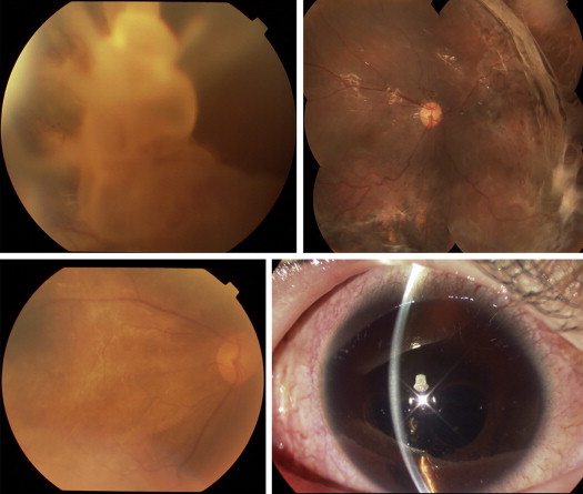
Case 1 was an 8-year-old boy who was referred to us for the treatment of a rhegmatogenous RD involving the macula of the right eye. Fundus examinations revealed a retinal break in the inferior temporal retina of the right eye and a peripheral avascular area with fibrovascular membranes in the temporal retina in both eyes. These findings suggested that the patient had FEVR in both eyes. The decimal best-corrected visual acuity (BCVA) was 0.04 in the right eye. The retina was reattached by scleral buckling; 6 months later, the BCVA had improved to 0.1 in the right eye.
Three years later, he developed a total retinal detachment with PVR in the right eye. A large tear of approximately 60 degrees was present in the upper nasal peripheral retina. The BCVA was hand motion in the right eye. Because of the repeated detachments, vitrectomy with silicone oil or C 3 F 8 gas tamponade was performed 5 times. Because all of the redetachments were due to proliferation in the inferior retina, we decided to use perfluoro-n-octane as a postoperative tamponade. During the sixth vitrectomy, perfluoro-n-octane was injected into the eye. The patient was instructed to rest in a supine or sitting position after the perfluoro-n-octane tamponade. After 2 weeks, multiple perfluoro-n-octane bubbles were noted in the inferior anterior chamber. The perfluoro-n-octane was removed 3 weeks later and replaced with 10% C 3 F 8 gas tamponade. The retina remained reattached during the 43-month follow-up. The BCVA at the last visit was 0.1 in the right eye ( Fig. 1 ).