Diabetic Macular Edema: Clinical Trials


Diabetic Macular Edema: Clinical Trials


Yoshihiro Yonekawa MD

Dean Eliott MD


I. DIABETIC MACULAR EDEMA


Early Treatment Diabetic Retinopathy Study


Introduction

In the 1960s, diabetic retinopathy was a growing public health problem and an important cause of blindness, chiefly because of proliferative diabetic retinopathy (PDR) and diabetic macular edema (DME). The Diabetic Retinopathy Study (DRS) was successfully completed in the 1970s, and it served as the foundation for additional prospective, multicenter, randomized clinical trials. The DRS (discussed in detail in the section on Proliferative Diabetic Retinopathy) conclusively demonstrated that scatter panretinal photocoagulation (PRP) was effective in the treatment of PDR, and the remarkable benefit associated with treatment had important public health implications. Whereas the DRS results offered tremendous hope for patients with PDR, DME remained a significant clinical challenge, as macular edema was the leading cause of moderate visual loss in diabetic patients. It was in this historical context that the Early Treatment Diabetic Retinopathy Study (ETDRS) was organized.


Background

The ETDRS was established to address important questions related to diabetic retinopathy. Conducted in the 1980s, the ETDRS was even larger in scope and size than the recently completed DRS.

Before the ETDRS, there was no consensus regarding the optimal management of DME. Several small trials reported encouraging results using photocoagulation; however, it was suggested that treatment benefit might be limited to certain subgroups, such as eyes with focal rather than diffuse fluorescein leakage, or eyes with intact rather than damaged perifoveal capillaries.1,2,3,4 One study involving macular photocoagulation sometimes used scatter treatment also, suggesting that scatter treatment itself might be beneficial for macular edema.2 Questions regarding the roles of focal macular photocoagulation and scatter PRP in the treatment of DME remained unanswered. The ETDRS was designed to address these questions, as well as questions involving the use of scatter PRP in the treatment of earlier stages of retinopathy (mild to severe nonproliferative diabetic retinopathy [NPDR] and early PDR) and the use of aspirin.

The ETDRS sought to determine answers to three questions: whether focal photocoagulation was effective in the treatment of DME, when scatter PRP should be initiated to be most effective in the management of diabetic retinopathy, and whether aspirin was effective in altering the course of diabetic retinopathy. Each of these study questions is addressed separately. The management of DME is addressed in the subsequent text, and the other two arms of the ETDRS are reviewed in Chapter 7C.


Study Question

Is focal photocoagulation beneficial in the management of DME?


Patients Included in the Study

A total of 3,711 patients, with or without macular edema and mild-to-severe NPDR or early PDR (less than high risk), were enrolled.
Visual acuity (VA) criteria were 20/40 or better for eyes without macular edema and 20/200 or better for those with macular edema. Eyes with macular edema were analyzed separately as one arm of the study.

Macular edema was defined as retinal thickening or hard exudates at or within one-disc diameter of the center of the macula.4,5 Clinically significant diabetic macular edema (CSDME) is defined in the following text. Definitions of mild, moderate, and severe NPDR as well as early PDR are included in the section discussing the early scatter treatment arm of the ETDRS (Chapter 7C).


Intervention and Outcome Measures

Eyes with macular edema were randomized to the immediate photocoagulation (focal and/or scatter) arm or the no treatment arm.

Specifically, eyes were divided among those without macular edema, those with macular edema and less severe retinopathy (mild or moderate NPDR), and those with macular edema and more severe retinopathy (severe NPDR or early PDR). One eye of each patient was randomized to deferral of treatment, and one eye to early photocoagulation using different combinations of scatter panretinal and macular focal photocoagulation (see Fig. 7B.1A-C). If an eye assigned to treatment deferral developed high-risk proliferative retinopathy, then scatter panretinal laser was initiated as per the DRS recommendations.

In eyes with macular edema and less severe retinopathy, those assigned to early photocoagulation received one of the four combinations: immediate focal and delayed mild scatter photocoagulation, immediate focal and delayed full scatter photocoagulation, immediate mild scatter and delayed focal photocoagulation, or immediate full scatter and delayed focal photocoagulation (Fig. 7B.1B).4 In eyes with macular edema and more severe retinopathy, those assigned to early photocoagulation received one of the four combinations: immediate mild scatter and immediate focal photocoagulation, immediate mild scatter and delayed focal photocoagulation, immediate full scatter and immediate focal photocoagulation, or immediate full scatter and delayed focal photocoagulation (Fig. 7B.1C).4,6

Focal photocoagulation (also called focal/grid) was performed using a combination of direct focal treatment to microaneurysms (Fig. 7B.2) and/or grid photocoagulation to areas of diffuse fluorescein leakage or capillary nonperfusion (Fig. 7B.3). Focal photocoagulation consisted of treatment to all focal points of leakage located between 500 µm and two disc diameters (3,000 µm) from the center of the macula. Fifty- to one hundred-micrometer spots at 0.05 to 0.1-second duration were used. Focal lesions located between 300 and 500 µm from the center were treated only if the VA was 20/40 or worse and if the treating ophthalmologist did not believe that treatment would destroy the remaining perifoveal capillary network (see Table 7B.1). Grid photocoagulation consisted of 50 to 200 µm spots at 0.05 to 0.1 second duration, placed at least 500 µm from the center of the macula and no closer than 500 µm from the edge of the optic disc. The argon blue-green wavelength was used initially, but the green wavelength was used later (Table 7B.1).4,5,6

Outcome measures included moderate visual loss, defined as a loss of 15 or more letters (three lines on the ETDRS VA chart) from baseline, which is equivalent to a doubling of the visual angle (for example, a decrease from 20/25 to 20/50 or from 20/50 to 20/100).


Major Findings

In patients with macular edema, the ETDRS identified features that were associated with a particularly high risk of visual loss, termed CSDME. CSDME was defined by the ETDRS as any one of the following: (a) retinal thickening at or within 500 µm of the center of the macula; (b) hard exudates at or within 500 µm of the center of the macula, if associated with adjacent retinal thickening; (c) a zone or zones of retinal thickening one-disc area or larger in size, any part of which is within one-disc diameter of the center of the macula (see Table 7B.2)6 CSDME was assessed by stereo-contact lens biomicroscopy and stereo photography.


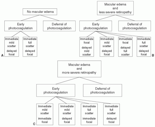
FIGURE 7B.1 (A) Early Treatment Diabetic Retinopathy Study (ETDRS) photocoagulation treatment scheme for eyes without macular edema and moderate-to-severe nonproliferative or early proliferative retinopathy. Eyes were assigned randomly to early photocoagulation or deferral of photocoagulation. Eyes assigned to early photocoagulation were further assigned randomly to either mild or full scatter (panretinal) photocoagulation. (From Early Treatment Diabetic Retinopathy Study Research Group. Early Treatment Diabetic Retinopathy Study design and baseline characteristics. ETDRS Report No 7. Ophthalmology. 1991;98:741-756.) (B) ETDRS photocoagulation treatment scheme for eyes with macular edema and less severe retinopathy (mild-to-moderate nonproliferative retinopathy). Eyes were assigned randomly to early photocoagulation or to deferral of photocoagulation. Eyes assigned to early photocoagulation were further assigned randomly to either mild or full scatter (panretinal) photocoagulation, and to either immediate focal or delayed focal treatment. For eyes assigned to immediate focal treatment, the assigned scatter treatment was not applied initially, but only if severe nonproliferative retinopathy or worse developed during follow-up. (From Early Treatment Diabetic Retinopathy Study Research Group. Early Treatment Diabetic Retinopathy Study design and baseline characteristics. ETDRS Report No 7. Ophthalmology. 1991;98:741-756.) (C) ETDRS photocoagulation treatment scheme for eyes with macular edema and more severe retinopathy. Eyes were assigned randomly to early photocoagulation or to deferral of photocoagulation. Eyes assigned to early photocoagulation were further assigned randomly to either mild or full scatter (panretinal) photocoagulation, and to either immediate focal or delayed focal treatment for at least 4 months. (From Early Treatment Diabetic Retinopathy Study Research Group. Early Treatment Diabetic Retinopathy Study design and baseline characteristics. ETDRS Report No 7. Ophthalmology. 1991;98:741-756.)


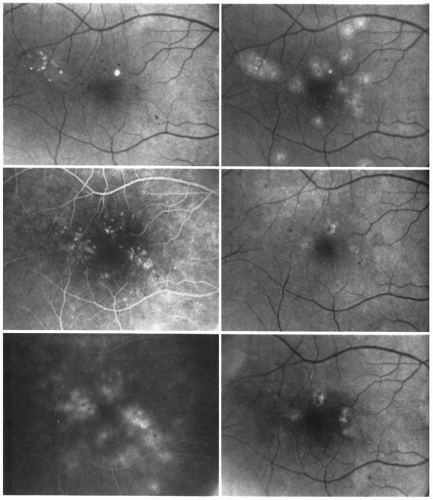
FIGURE 7B.2 Focal treatment of microaneurysms. The right eye of a 69-year-old woman with diabetes of 22 years’ duration. Top left: At baseline visit, definite retinal thickening could be seen (with stereoscopic examination) nasal to the center of the macula and above it, probably involving the center. A few small microaneurysms and hard exudates are visible in the thickened area. Visual acuity was 20/30. Center left: Mid-phase angiogram shows microaneurysms surrounding the center of the macula most within 1,000 µm of the center and some within 500 µm. Bottom left: Late-phase angiogram shows leakage from the microaneurysms. Top right: Posttreatment photograph shows mild-to-moderate intensity focal treatment of most of the microaneurysms. The microaneurysms closest to the center have not been treated. Center right: One year after treatment, the center of the macula appears flat. Hard exudates and microaneurysms have decreased. Visual acuity was 20/50. Bottom right: Between the 1- and 2-year visits, additional focal photocoagulation was applied. At the 2-year visit, the center of the macula appears flat and no microaneurysms or hard exudates can be seen. Visual acuity was 20/25. (From Early Treatment Diabetic Retinopathy Study Research Group. Treatment technique and clinical guidelines for photocoagulation of diabetic macular edema. ETDRS Report No 2. Ophthalmology. 1987;94:761-774.)


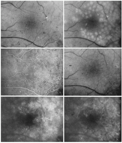
FIGURE 7B.3 Focal treatment of microaneurysms combined with a grid patter to areas of diffuse fluorescein leakage and capillary dropout. The left eye of a 49-year-old man with diabetes of 22 years’ duration. Top left: The pretreatment photograph shows extensive retinal thickening with a few scattered microaneurysms and small hard exudates temporal to the center of the macula. Retinal thickening at the center is mild. Visual acuity was 20/40. Center left: Mid-phase angiogram shows moderate capillary dilation above and temporal to the center of the macula, with mild perifoveal capillary dropout. Scattered microaneurysms are also present. Bottom left: Late-phase angiogram shows extensive small cystoid spaces above, below, and temporal to the center of the macula. Some of the large microaneurysms fill only partially with fluorescein. Top right: Posttreatment photograph shows focal burns to microaneurysms and a grid pattern of burns above, below, and temporal to the macula. Center right: Four months later, microaneurysms and hard exudates have decreased. Retinal thickening is less and no longer involves the center of the macula. Visual acuity was 20/25. Bottom right: Late-phase angiogram shows treatment scars but most of the microaneurysms and cystoid spaces have disappeared. (From Early Treatment Diabetic Retinopathy Study Research Group. Treatment technique and clinical guidelines for photocoagulation of diabetic macular edema. ETDRS Report No 2. Ophthalmology. 1987;94:761-774.)


TABLE 7B.1 Specific Techniques for Scatter (Panretinal) and Focal Photocoagulation in the Early Treatment Diabetic Retinopathy Study


Scatter parameters


Full


Mild


Burn characteristics


Size


500 µm (at retina)


500 µm (at retina)


Exposure


0.1 s


0.1 s


Intensity


Moderate


Moderate


Number


1,200-1,600


400-650


Placement


1/2 burn apart >2 disc diameters from fovea out to equator


≥1 burn apart >2 disc diameters from fovea out to equator


Number of episodes


≥2


1


Lesion treated directly


Patches of NVE <2 disc areas


Patches of NVE <2 disc areas


Indications for follow-up treatment


Recurrent or new NVE or high-risk proliferative retinopathy


Recurrent or new NVE or high-risk proliferative retinopathy


Focal parameters


Direct


Grid


Burn characteristics


Size


50-100 µm


<200 µm (at retina)


Exposure


0.05-0.1 s


0.05-0.1 s


Intensity


Sufficient to whiten or darken large microaneurysms


Mild


Number


Sufficient to satisfactorily treat all focal leaks


Sufficient to cover areas of diffuse leakage and nonperfusion


Placement


500-3,000 µm from center of fovea


Spaced greater than one burn width apart 500-3,000 µm from center of fovea


Number of episodes


1


1


Indications for follow-up treatment


Presence of CSDME and treatable lesions at ≥4 mo


Presence of CSDME and treatable lesions at ≥4 mo


CSDME, clinically significant diabetic macular edema; NVE, neovascularization elsewhere. From Early Treatment Diabetic Retinopathy Study Research Group. Early treatment diabetic retinopathy study design and baseline characteristics. ETDRS report no 7. Ophthalmology 1991;98:741-756.


TABLE 7B.2 Definition of Clinically Significant Diabetic Macular Edema


Clinically Significant Diabetic Macular Edema— any one of the following:


1.


Retinal thickening at or within 500 µm of the center of the macula


2.


Hard exudates at or within 500 µm of the center of the macula, if associated with adjacent retinal thickening


3.


A zone or zones of retinal thickening of one-disc area or larger, any part of which is within one-disc diameter of the center of the macula


Eyes with macular edema demonstrated a considerable benefit from early focal photocoagulation, as treatment reduced the risk of moderate visual loss by approximately 50% (12% risk of moderate visual loss for treated eyes vs. 24% untreated at 3 years). In eyes with CSDME, these differences were even greater. Of eyes with CSDME, a majority had central foveal involvement, and these eyes demonstrated the most benefit from treatment (13% risk of moderate visual loss for treated eyes vs. 33% untreated at 3 years). In these eyes, early focal treatment was associated with a decrease in retinal thickening at the center of the macula. In eyes with CSDME but without central
foveal involvement, treatment resulted in a lesser, but significant, benefit (6% for treated eyes vs. 16% untreated at 2 years). In contrast, in eyes with macular edema that did not meet the definition of CSDME, there was no benefit associated with treatment (see Fig. 7B.4).5,6,7

The beneficial response to early focal treatment was most apparent in eyes with CSDME and worse VA at baseline (<20/40) as compared to those with better baseline acuity (20/25 to 20/40, 20/20 or better), but a treatment effect was demonstrated even in those with good initial VA (see Fig. 7B.5).7 Despite the reduced risk of visual loss with treatment, visual improvement was rare in the ETDRS (improvement of 15 letters occurred in <3%). Therefore, the ETDRS recommendation was to consider prompt focal treatment for eyes with CSDME, regardless of VA, to prevent visual loss.6


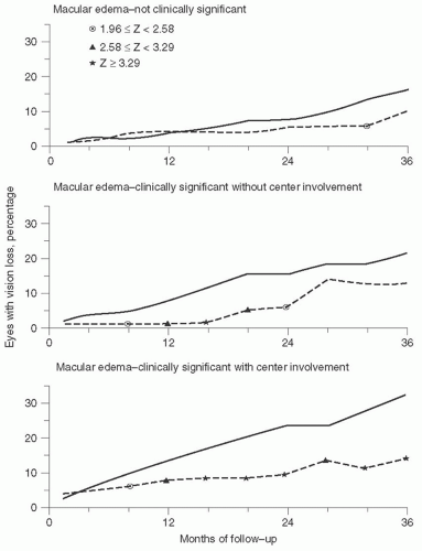
FIGURE 7B.4 Comparison of percentages of eyes with macular edema that experienced moderate visual loss classified by severity of macular edema and assigned to immediate focal treatment (broken line) or to deferral of treatment (solid line). (From Early Treatment Diabetic Retinopathy Study Research Group. Photocoagulation for diabetic macular edema. Early Treatment Diabetic Retinopathy Study Report No 4. Int Ophthalmol Clin. 1987; 27:265-272.)

The ETDRS documented treatment-related side effects, which included a small,
but not statistically significant, difference in visual field scores. Eyes assigned to focal photocoagulation demonstrated slightly more paracentral scotomata on Goldmann visual fields using the I-2 test object.6


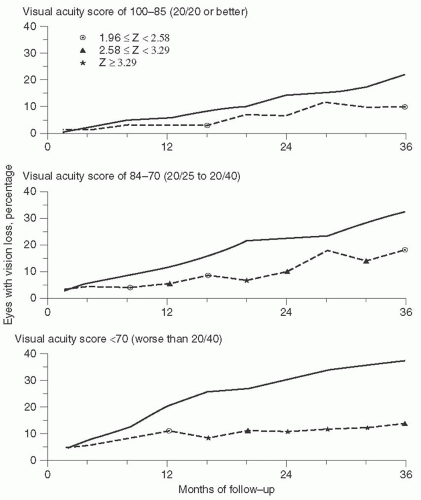
FIGURE 7B.5 Comparison of percentages of eyes with clinically significant diabetic macular edema that experienced moderate visual loss classified by baseline visual acuity and assigned to immediate focal treatment (broken line) or to deferral of treatment (solid line). (From Early Treatment Diabetic Retinopathy Study Research Group. Photocoagulation for diabetic macular edema. Early Treatment Diabetic Retinopathy Study Report No 4. Int Ophthalmol Clin. 1987; 27:265-272.)


Implications for Clinical Practice

Prompt focal photocoagulation was recommended for eyes with CSDME as defined by the ETDRS for patients who met the inclusion criteria. Treatment was recommended regardless of baseline VA, since eyes in all categories of VA (20/20 or better, 20/25-20/40, <20/40) were found to benefit from treatment. Treatment was recommended for eyes with or without thickening of the central macula, provided that they met the definition of CSDME. Treatment was most effective for those with worse VA at baseline (<20/40) and for those with central macular thickening.

Focal photocoagulation, when applied using the ETDRS treatment guidelines, resulted in a significant reduction in moderate visual loss. Since visual improvement was rare in this study, the ETDRS recommended that treatment should be considered to prevent visual loss in patients with CSDME.


Macular edema often occurs in association with severe NPDR or PDR. The DRS and the ETDRS demonstrated that scatter PRP may exacerbate macular edema and result in vision loss.8,9 If PRP can be safely delayed for a patient with CSDME and severe NPDR or early PDR, focal treatment should be applied followed by very close observation for proliferative changes. If panretinal laser cannot be safely delayed, or if a patient has CSDME and PDR with high-risk characteristics, both focal treatment and PRP should be applied, but the scatter treatment should not be given before the focal treatment.

Beginning with the ETDRS, a new VA chart was developed for use in prospective clinical research studies. This chart is still used today in clinical trials to evaluate VA in a standardized manner (see Fig. 7B.6).10,11,12


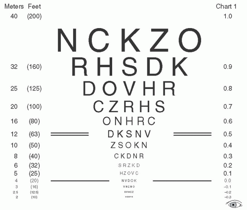
FIGURE 7B.6 One of the three Early Treatment Diabetic Retinopathy Study visual acuity charts. Fourmeter testing distance with this chart yields the following Snellen equivalent lines: 20/10, 20/12.5, 20/16, 20/20, 20/25, 20/31.5, 20/40, 20/50, 20/63, 20/80, 20/100, 20/125, 20/160, and 20/200. At 1 m, the following additional Snellen equivalent lines of visual acuity could be measured: 20/250, 20/315, 20/400, 20/500, 20/630, and 20/800. Note that every three lines is a doubling of the visual angle and that there are five letters on each line. (From Early Treatment Diabetic Retinopathy Study Research Group. Early Treatment Diabetic Retinopathy Study design and baseline characteristics. ETDRS Report No 7. Ophthalmology. 1991;98:741-756.)


Unanswered Questions

The ETDRS defined the standard of care (SOC) for the management of DME for over 20 years, and all current clinical trials continue to use ETDRS results for comparison. A direct comparison with new therapies, however, is often difficult due to the multitude of ocular and systemic variables that influence retinopathy.

Despite providing answers to critically important questions, the ETDRS results stimulated additional questions that remain unanswered. For example, the ETDRS did not evaluate eyes with VA less than 20/200. The potential effect of focal treatment in eyes with macular edema and low vision is unknown. In addition, while the ETDRS was designed to determine whether laser was effective, it was not designed to determine the best time to apply laser, and the optimal timing of
treatment remains unknown. Furthermore, the ETDRS used specific treatment guidelines for macular focal photocoagulation. Alternative laser treatment strategies have since been developed, including modified versions of the ETDRS protocol. The relative benefits of different treatment strategies are difficult to assess since a direct comparison with the ETDRS results is not possible.

Retreatment with focal photocoagulation was allowed in the ETDRS if the edema persisted or recurred. There was no clear recommendation regarding the number of laser treatments that may be beneficial in eyes requiring retreatment, and the management of refractory DME remains one of the most challenging clinical problems today, 20 years after the initial ETDRS results were reported.


II. DIABETIC MACULAR EDEMA: PHARMACOLOGIC THERAPIES


Steroids


Introduction

Intraocular corticosteroids are currently being evaluated for the treatment of DME. Triamcinolone, fluocinolone, and dexamethasone are promising pharmacologic agents that are in various phases of clinical trial development.

Triamcinolone acetonide is commonly being used off-label for diabetic and other causes of macular edema, as it is not approved by the U.S. Food and Drug Administration (FDA) for this indication (see Fig. 7B.7). Administered through an intravitreal injection, triamcinolone acetonide has been shown to be effective in improving VA and reducing macular thickness measured by optical coherence tomography (OCT) in patients with DME (see Figs. 7B.8 and 7B.9).13,14

In contrast, fluocinolone acetonide and dexamethasone are delivered via intravitreal sustained release devices, and these devices are not approved by the FDA for use in DME. These include the intravitreal fluocinolone acetonide implants (Retisert, [see Fig. 7B.10], Iluvien [see Fig. 7B.11]) and the intravitreal dexamethasone implant (Ozurdex, [see Fig. 7B.12]), which provide prolonged delivery of medication to the target tissue.


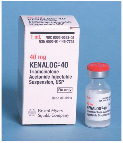
FIGURE 7B.7 Triamcinolone acetonide (Kenalog-40, Bristol-Myers Squibb). (Photograph courtesy of Ronald C. Gentile, MD, New York, NY.)

Corticosteroids act in a nonspecific manner. Although the exact mechanism of action in the treatment of DME is unknown, corticosteroids decrease the breakdown of the blood-retinal barrier, suppress inflammation, and downregulate the production of vascular endothelial growth factor (VEGF).


Diabetic Retinopathy Clinical Research Network, Protocol B


Background and Study Questions

A number of small studies had demonstrated that intravitreal injections of triamcinolone may be effective in treating DME, but sample size and follow-up were suboptimal at the time. Many practitioners began routinely treating DME with intravitreal triamcinolone, but there were no controlled clinical trials to show evidence of its efficacy. In 2002, a collaborative network of clinical practices in the United States, named the Diabetic Retinopathy Clinical Research Network (DRCR. net), was formed to facilitate multicenter
clinical trials dedicated to the investigation of diabetic retinopathy. There are currently over a hundred participating clinical sites.15 Its establishment represented a unique paradigm where multiple centers, from academic institutions to private practices, could rapidly organize large trials.


FIGURE 7B.8 The right eye of a 49-year-old man with diabetes of 18 years’ duration. Clinically significant diabetic macular edema was present despite a history of prior focal photocoagulation. Visual acuity was 20/200. (A) Fundus photograph of the macula shows cystoid retinal thickening, a few small retinal hemorrhages, and prior focal laser spots. (B) Late-phase fluorescein angiogram shows leakage in a petalloid pattern. (C) Optical coherence tomography shows cystic retinal thickening.

In order to better evaluate the use of intravitreal steroid injections for DME, the DRCR.net organized a phase III, multicenter, prospective, randomized trial to compare the efficacy and safety of 1 and 4 mg of preservative-free triamcinolone acetate (Trivaris, Allergan, Inc., Irvine, CA) to focal/grid photocoagulation. The 2-year follow-up data were published in 2008.16


Patients Included in the Study

A total of 840 eyes with DME from 693 subjects with type 1 or type 2 diabetes were examined. Inclusion criteria were ETDRS VA scores between 73 (approximately 20/40) and 24 (approximately 20/320), 250 µm or thicker central macular thicknesses on OCT,
and no expectations for requiring PRP in the subsequent 4 months. Patients with any history of intravitreal steroid treatment or pars plana vitrectomy, or those with recent periocular steroids or photocoagulation treatment were excluded. Also excluded were those with histories of open-angle glaucoma, steroid-induced ocular hypertension that required intervention, and intraocular pressure (IOP) of 25 mmHg or more. Baseline characteristics showed that the DRCR.net study patients were slightly older with better glycemic control compared to the ETDRS and PKC-DRS2 participants.17 Other variables were similar.


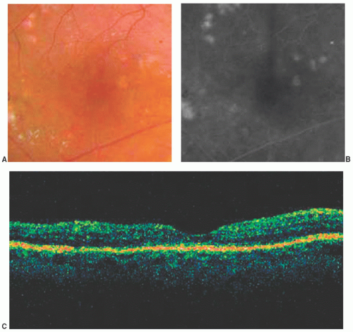
FIGURE 7B.9 Same patient as in Figure 8, 3 months after intravitreous injection of triamcinolone acetonide 4 mg. Visual acuity was 20/80. (A) Fundus photograph shows resolution of macular edema. (B) Late-phase fluorescein angiogram shows resolution of leakage. (C) Optical coherence tomography shows resolution of retinal thickening. (Three months later the patient developed recurrent edema and underwent repeat injection, with subsequent resolution of the edema.)


Intervention and Outcome Measures

Study eyes were randomized to focal/grid photocoagulation, 1 mg triamcinolone, or 4 mg triamcinolone. The laser control group allowed direct comparison to the gold standard. The photocoagulation technique was a modified ETDRS protocol, where burns were smaller and less intense (light gray, 50 µm). Subjects in the triamcinolone groups were allowed to receive laser treatment if they met
certain failure criteria as a fail-safe system. Patients were followed every 4 months, and retreated based on VA and OCT parameters. The primary endpoint was mean VA, and the secondary outcome measure was central subfield retinal thickness. At the time, this DRCR.net study was the first phase III DRS to use the OCT data.


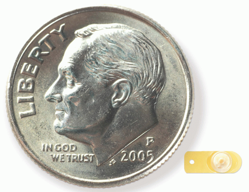
FIGURE 7B.10 Fluocinolone acetonide intravitreous sustained release implant (Retisert).


FIGURE 7B.11 Fluocinolone acetonide intravitreous sustained release insert and its injector (Iluvien).


FIGURE 7B.12 Dexamethasone intravitreous sustained release implants (Ozurdex), 350 and 700 µg doses, and the injector.


Major Findings

As we will also see with other intravitreal steroid studies, the treatment response evolved over time. At 4 months, the 4-mg triamcinolone group had the best VA, but by 1 year the difference was not statistically significant, and
starting at 16 months and persisting to 2 years the laser group developed the best mean VA (see Fig. 7B.13). At 2 years, the mean ± SD change in VA was +1 ± 17 letters in the laser group, — 2 ± 18 letters in the 1-mg group (p = 0.02), and − 3 ± 22 letters in the 4-mg group (p = 0.002). The number of treatments in each arm was similar. A subgroup analysis of pseudophakic eyes showed that the respective changes in mean visual acuities were + 2 ± 18, +2 ± 17, and −1 ± 19 letters. This analysis was performed because intravitreal steroids are strong risk factors for developing cataract.


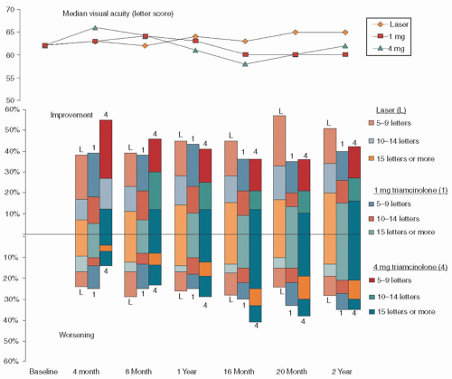
FIGURE 7B.13 Diabetic Retinopathy Clinical Research Network (DRCR.net), Protocol B. The study compared the efficacies of focal/grid photocoagulation, and 1 or 4 mg of intravitreal triamcinolone, in the treatment of diabetic macular edema. The top graph demonstrates the changes in visual acuity over 2 years. The bottom graph shows the percentages of eyes in each treatment group with the corresponding letters gained or lost at each time point. (From Diabetic Retinopathy Clinical Research Network. A randomized trial comparing intravitreal triamcinolone acetonide and focal/grid photocoagulation for diabetic macular edema. Ophthalmology. 2008;115:1447-1449.)

The effects of treatment on retinal thickness paralleled VA changes, where the 4-mg group initially performed better, but the laser group ended with the lowest central subfield thickness. At 2 years, the mean decrease in thickness was −139 ± 148 µm in the laser group, -86 ± 167 µm in the 1-mg group, and -77 ± 160 µm in the 4-mg group (all p < 0.001).

There were no cases of infectious or sterile endophthalmitis of the 1,649 intravitreal injections. An elevation of IOP more than 10 mmHg from baseline, IOP of 30 mmHg or more, and initiation of IOP-lowering medication/a new diagnosis of glaucoma were
observed at one or more visits in 40% of those in the 4-mg group, 20% in the 1-mg group, and 10% in the laser group (all p < 0.001). Cataract surgery was performed in 51% of the 4-mg group, 23% of the 1-mg group, and 13% of the laser group, during the course of the study (p < 0.001). A unique adverse event in this study was intravitreal silicone oil droplets that were found in some eyes due to the use of staked silicone syringes. The protocol was amended to use luer cone syringes instead, which eliminated the problem.

The 3-year follow-up study showed continued benefits of the laser group over both triamcinolone arms.18 The laser group gained 5 letters while the triamcinolone arms improved by 0 letters. The cumulative probabilities of cataract surgery were 31%, 46%, and 83% in the laser, 1-mg, and 4-mg groups, respectively. An IOP rise of 10 mmHg or more was noted in 4%, 18%, and 33%, respectively.

A retrospective exploratory study of the same DRCR.net cohort investigated the progression of diabetic retinopathy.19 The cumulative progression of retinopathy at 2 years was 31% in the laser group, 29% in the 1-mg group (p = 0.64 compared to laser), and 21% in the 4-mg group (p = 0.005 compared to laser). Similar results were found at the 3-year study also. However, the authors concluded that the risks of cataract and intraocular elevation did not warrant the use of intravitreal triamcinolone as a primary modality to slow or improve diabetic retinopathy.


Implications for Clinical Practice

This study demonstrated that there is potential for visual and anatomic improvement after intravitreous triamcinolone injection in the DME patients. This is a relatively cost-effective and technically easier procedure compared with the steroid implants discussed below. However, as shown in this study, VA tends to regress over time as the macular edema recurs and repeat injections are required. Furthermore, while the short-term results were superior to laser photocoagulation, the difference soon became insignificant, and the laser group developed the best outcomes after 16 months. This signifies that short-acting boluses of intravitreal steroids alone may not be sufficient to control DME long-term.

Only gold members can continue reading. Log In or Register to continue

Stay updated, free articles. Join our Telegram channel

Aug 2, 2016 | Posted by in OPHTHALMOLOGY | Comments Off on Diabetic Macular Edema: Clinical Trials

Full access? Get Clinical Tree

Get Clinical Tree app for offline access