Purpose
To evaluate the outcomes of deep anterior lamellar keratoplasty (DALK) in children.
Design
Retrospective interventional case series.
Methods
setting : Institutional, L.V. Prasad Eye Institute, a tertiary care center in south India. study population and intervention : All children less than 16 years of age undergoing DALK from January 2003 to January 2011. main outcome measure : Visual outcome and complications.
Results
Twenty-six eyes of 26 children (13 male and 13 female) with a mean age of 7.82 ± 4.64 years underwent DALK for keratoconus (8), microbial keratitis (6), corneal scar (6), corneal keloid (3), chemical injury with limbal stem cell deficiency (2), and dermoid (1). Big bubble was achieved in 5 eyes, while manual dissection was done in 21. Follow-up ranged from 1 week to 7.3 years. Seventeen patients with a minimal follow-up of 6 months were evaluated for visual outcomes. Final vision varied from counting fingers to 20/20 (mean sphere 2.32 diopters, mean cylinder -2.5 diopters). Complications encountered were suture-related graft infiltrate (3), graft dehiscence (3), and Descemet membrane detachment (2).
Conclusions
DALK is a feasible option in children with stromal corneal pathology. It offers advantages in the form of lower risk of graft rejection. However, the risk of complications such as suture-related infections and graft dehiscence persists even in these cases.
In children with corneal pathology, penetrating keratoplasty has been the standard of care until recently. However, the outcomes of primary penetrating keratoplasty in children are poor compared with adults. The active immune system in children leads to a higher incidence of graft rejection and eventual failure because of a lower reversal rate of rejection than in adults. The most common and most serious type of rejection is the endothelial rejection. A procedure that would selectively spare the host endothelium may help in reducing this event. Deep anterior lamellar keratoplasty (DALK) has gained popularity as a surgical modality for management of stromal corneal pathologies in adults. DALK, though technically challenging, offers a number of advantages over penetrating keratoplasty (PK) in terms of lower incidence of graft rejection and graft failure. DALK may offer an advantage to at least a subset of pediatric patients who undergo keratoplasty for stromal pathologies. However, the literature describing indications and outcomes of DALK in children is limited. Here, we report our experience of pediatric DALK at a tertiary care center in south India.
Methods
After Institutional Review Board approval was obtained from L.V. Prasad Eye Institute, Hyderabad Eye Research Foundation, the medical and surgical records of 26 eyes of 26 children less than 16 years of age who underwent DALK at L.V. Prasad Eye Institute, Hyderabad, India from January 1, 2003 to January 1, 2011 were retrospectively analyzed. Data collected included patient demographics, clinical and surgical details, postoperative outcomes, and complications.
Surgical Technique
Informed consent was obtained from the parents of the children prior to the surgery. All surgeries were performed under general anesthesia. The host cornea was trephined (7-11 mm), using a simple hand-held trephine, up to 50% of the corneal depth. A big bubble was attempted only in cases where the path of the needle could be clearly visible before air injection to avoid any inadvertent perforation. In the rest of the cases, a manual dissection of the stroma was done. Following trephination of the cornea, a paracentesis was made to deflate the anterior chamber. In most cases big bubble was not achieved and corneal dissection was performed layer by layer or in a spiral manner until the Descemet membrane was reached.
The donor tissue was then trephined with a disparity varying from 0.25 to 1 mm (7-12 mm) and the donor Descemet membrane was peeled off after staining with trypan blue dye. The graft was then sutured to the host bed using 16 interrupted 10-0 monofilament nylon sutures or combined interrupted and continuous 10-0 monofilament nylon sutures. All host corneal buttons were sent for histopathologic review.
Postoperative Medications
All patients were maintained on steroid eye drops administered every 2 hours (1% prednisolone; Alcon Labs, Fort Worth, Texas, USA) and antibiotic eye drops (moxifloxacin 0.3% [Vigamox]; Alcon Labs) for 2 weeks. The steroids were tapered over the next few months to achieve a maintenance dose of once or twice daily. Cycloplegic drops were used when needed. Suture removal was initiated as early as 6 weeks postoperatively in infants to 3 months postoperatively in older children. Early suture removal was done in cases where sutures were loose, broken, infiltrated, or vascularized.
All patients were followed up at 1 day, 7 days, 1 month, 3 months, 6 months, and then 1 year after surgery, and in between on an as-needed basis. Visual acuity, refraction, and corneal graft status was evaluated at each visit. Any complication in the postoperative period was noted and managed appropriately. Amblyopia therapy was instituted in all children in the form of spectacles and a patching regimen.
For the visual outcomes analysis, patients with a minimum follow-up of 6 months were included; however, for analysis of complications all the patients were included.
Results
The Supplemental Table (available at AJO.com ) summarizes the patient demographics and surgical outcomes. The mean age of the cohort was 7.82 ± 4.64 years (range 6 months to 14 years); the cohort included 13 boys and 13 girls. Eleven patients were less than 5 years of age. The mean follow-up was 1.3 years (range 1 week to 7.3 years). Thirteen patients had surgery in the right eye and 13 in the left eye. The most common indication for surgery was keratoconus (8 patients), followed by nonherpetic corneal scar (6 patients), postherpetic corneal scar (5 patients), corneal keloid (3 patients), vascularized corneal scar following limbal stem cell deficiency (2 patients), corneal dermoid (1 patient), and active Acanthamoeba keratitis not responding to therapy (1 patient). Most of the patients with keratoconus (5 of 8 cases) had corneal scarring.
Surgical Techniques and Peculiarities
A big bubble was attempted in 7 cases but was successful in only 3 of these cases. In the other 4 cases, a manual dissection was performed to reach the plane of the Descemet membrane. In 19 cases, the big bubble was not attempted and the Descemet membrane was bared using manual dissection of the stroma. Graft and host were trephined to the same size in 2 cases; a 0.25-mm disparity was used in 7 cases; a 0.5-mm disparity was used in 16 cases; and a 1-mm disparity was used in 1 case of limbal stem cell deficiency where “tuck in” lamellar keratoplasty was performed. Additional surgical procedures in the form of pannus resection, limbal stem cell transplantation, and amniotic membrane transplantation were performed in the same patient.
Visual Outcomes
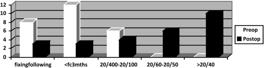
The preoperative and postoperative visual acuities have been summarized in Figure 1 . For the analysis of visual outcome, patients with a minimum follow-up of 6 months were included (n = 21). The final visual outcome in this group ranged from unsteady fixation to light to 20/20. Thirteen patients (61.90%) had visual acuity better than 20/80.
Refractive Outcome
Of the 21 patients included for the visual outcome analysis, refraction was available for 17 patients at the final follow-up. The mean spherical error was 2.32 ± 4.58 diopters and mean cylinder was −2.51 ± 4.02 diopters.
Graft Clarity
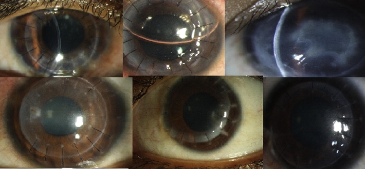
At the final follow-up, 18 of the 26 grafts (69.23%) were clear; 8 grafts (30.76%) had scarring in the graft ( Figure 2 ).
Complications
Complications were divided into early (occurring within 1 month) and late (occurring after 1 month). During the early postoperative period, 2 of the 26 patients (7.69%) had detachment of the Descemet membrane leading to a double anterior chamber. This was managed by air injection into the anterior chamber in 1 case, following which the Descemet membrane adhered well to the donor stroma. The second case had a central Descemet detachment with peripheral attachment. The Descemet membrane attached spontaneously after 1 week.
Late complications included posttraumatic graft dehiscence in 3 cases (11.53%). Two of these patients were managed with resuturing alone, but 1 patient required simultaneous lens aspiration for a traumatic cataract. Both patients had good visual outcome after resuturing. One patient had lens extrusion and required a penetrating keratoplasty and anterior vitrectomy. This patient later developed retinal detachment and the eye eventually became phthisical. Five patients had suture-related bacterial graft infection and were managed with appropriate intensive antibiotic therapy. All the patients healed with scarring. Three patients had recurrent herpes simplex keratitis in the graft, 2 had stromal keratitis, and 1 had endotheliitis. These patients were managed with topical acyclovir and topical corticosteroids. Cases with stromal keratitis resolved with a vascularized scar, whereas the patient with endotheliitis had a clear graft at final follow-up. None of the patients experienced any episodes of graft rejection during the follow-up.
Results
The Supplemental Table (available at AJO.com ) summarizes the patient demographics and surgical outcomes. The mean age of the cohort was 7.82 ± 4.64 years (range 6 months to 14 years); the cohort included 13 boys and 13 girls. Eleven patients were less than 5 years of age. The mean follow-up was 1.3 years (range 1 week to 7.3 years). Thirteen patients had surgery in the right eye and 13 in the left eye. The most common indication for surgery was keratoconus (8 patients), followed by nonherpetic corneal scar (6 patients), postherpetic corneal scar (5 patients), corneal keloid (3 patients), vascularized corneal scar following limbal stem cell deficiency (2 patients), corneal dermoid (1 patient), and active Acanthamoeba keratitis not responding to therapy (1 patient). Most of the patients with keratoconus (5 of 8 cases) had corneal scarring.
Surgical Techniques and Peculiarities
A big bubble was attempted in 7 cases but was successful in only 3 of these cases. In the other 4 cases, a manual dissection was performed to reach the plane of the Descemet membrane. In 19 cases, the big bubble was not attempted and the Descemet membrane was bared using manual dissection of the stroma. Graft and host were trephined to the same size in 2 cases; a 0.25-mm disparity was used in 7 cases; a 0.5-mm disparity was used in 16 cases; and a 1-mm disparity was used in 1 case of limbal stem cell deficiency where “tuck in” lamellar keratoplasty was performed. Additional surgical procedures in the form of pannus resection, limbal stem cell transplantation, and amniotic membrane transplantation were performed in the same patient.
Visual Outcomes
The preoperative and postoperative visual acuities have been summarized in Figure 1 . For the analysis of visual outcome, patients with a minimum follow-up of 6 months were included (n = 21). The final visual outcome in this group ranged from unsteady fixation to light to 20/20. Thirteen patients (61.90%) had visual acuity better than 20/80.


