Purpose
To evaluate corneal biomechanical deformation response using Ocular Response Analyzer (ORA) and Corvis ST data.
Design
Prospective observational case-control study.
Methods
A total of 1262 eyes of 795 patients were enrolled. Three groups were established, according to the corneal compensated intraocular pressure (IOPcc): Group I (10-13 mm Hg), Group II (14-17 mm Hg), and Group III (18-21 mm Hg). Each group included 3 subgroups, based on central corneal thickness (CCT): Subgroups 1 (465-510 μm), 2 (510-555 μm), and 3 (555-600 μm). In addition, similar groups of CCT were divided into subgroups of IOPcc. Corneal hysteresis (CH) and corneal resistance factor (CRF) were derived from ORA. The parameters of highest concavity with the parameters of first and second applanation were recorded from Corvis ST.
Results
CH and CRF, applanation 1 time, and radius of curvature at highest concavity showed significant differences between CCT subgroups for each IOPcc group ( P < .0001). CH, applanation 1 and 2 time, and applanation 2 velocity, as well as deformation amplitude (DA), showed significant differences by IOP subgroups for all CCT groups. IOPcc is correlated negatively with CH ( r = −0.38, P < .0001). There are positive correlations of IOPcc with applanation 1 time, applanation 2 velocity, and radius and negative correlations with applanation 2 time ( r = −0.54, P < .0001), applanation 1 velocity ( r = −0.118, P < .0001), and DA ( r = −0.362, P < .0001).
Conclusion
ORA and Corvis ST parameters are informative in the evaluation of corneal biomechanics. IOP is important in deformation response evaluation and must be taken into consideration.
The cornea is a complex biomechanical composite whose behavior depends on its structural subcomponents and their organizational motifs. The microstructure of the corneal stroma is composed of 300-500 lamellar sheets. Each of these sheets consists of thin, unbranched collagen fibrils that stretch from limbus to limbus. The fibrils in each sheet are arranged parallel to one another and are evenly spaced. A gel-like material, known as ground substance, fills the spaces between the fibrils and lamellae. The biomechanical properties of corneal tissue determine how it will respond and deform when placed under stress. This process depends on the viscoelastic properties of the cornea. Knowledge of biomechanical properties is important in the fields of intraocular pressure (IOP) measurement, glaucoma, corneal pathology such as keratoconus, and corneal refractive surgery. We will describe 2 devices that evaluate the biomechanical response of the cornea to an air puff–induced deformation: the Ocular Response Analyzer (ORA; Reichert Ophthalmic Instrument, Inc., Buffalo, New York. USA) and Corvis ST (Oculus, Wetzlar, Germany).
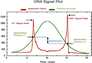
The Ocular Response Analyzer was the first device commercially available to provide an in vivo measurement of corneal biomechanical response using dynamic infrared signal analysis. ORA is a noncontact tonometer developed to provide a more accurate assessment of IOP than Goldmann applanation tonometry (GAT), and through this assessment also provides a measure of biomechanical features of the cornea through monitoring and analyzing corneal response during an air pulse. There are 4 main parameters of the ORA. During the measurement, 2 applanation pressures (P1, P2) are obtained by an electro-optic system. The difference between these 2 pressure values is termed corneal hysteresis (CH) ( Figure 1 ). CH provides information not on elastic properties—that is, how stiff or soft the cornea is—but rather on the rate-dependent viscoelastic response. Corneal resistance factor (CRF) is strongly associated with central corneal thickness by design as well as being uncorrelated to corneal compensated intraocular pressure (Luce DA. IOVS 2006;47:ARVO E-Abstract 2266). Thus, CRF may correlate with elastic properties even though it is viscoelastic by nature. Both CH and CRF are the analyzed responses of the cornea with applied air jet–induced deformation. ORA also reports 2 IOP values: the Goldmann-correlated IOPg, derived from the mean of P1 and P2; and the corneal compensated IOP (IOPcc). The IOPcc was designed to be less sensitive to corneal properties than traditional applanation tonometry and was calibrated empirically to be relatively unaffected by laser in situ keratomileusis (LASIK) (Luce DA. IOVS 2006;47:ARVO E-Abstract 2266).
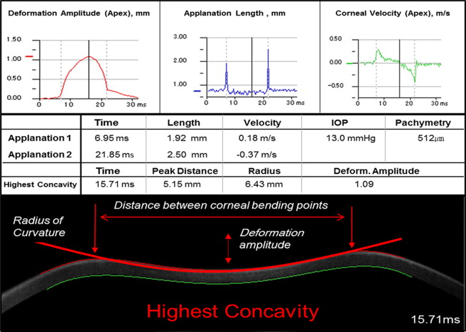
The dynamic Scheimpflug imaging analysis (Corvis ST) uses a high-speed camera at a rate of 4300 frames per second to capture a series of horizontal Scheimpflug images during corneal deformation with an air puff jet. It is a noncontact tonometer (NCT) and imaging device that measures not only intraocular pressure but also corneal thickness and provides additional information about biomechanical responses of the cornea using dynamic Scheimpflug imaging analysis. The biomechanical response of the cornea is characterized by the following parameters: time (time to reach applanation), length (the length of the flattened segment in a Scheimpflug image), and velocity (corneal velocity of movement during applanation), all at the moment of both the first and second applanation events, as well as the following characteristics at the point of highest concavity: time, deformation amplitude (DA), distance between bending points of the cornea (Dist), and concave radius of curvature (RadCurv) ( Figure 2 ). The approach taken in this study was to evaluate corneal biomechanical parameters by ORA and Corvis ST, and to investigate their relationship with IOPcc and central corneal thickness (CCT).
Patients and Methods
This was a prospective observational case-control study. The study conformed to the ethics codes established by the Ethical Board Committee of Japan. The study was carried out with approval from the Institutional Review Board (Matsumoto Clinic, Tokyo, Japan), and all subjects consented. Because of the study population, the study was conducted on Asian eyes. The study was performed to evaluate the biomechanical parameters of the cornea using the ORA and Corvis ST in normal eyes and to determine the influence of both IOPcc and CCT on corneal biomechanical response to an air puff, defined by the parameters produced. Exclusion criteria included corneal astigmatism >4.0 diopters (D), CCT >600 μm, IOP >21 mm Hg or IOP <10 mm Hg, ocular pathology, or previous ocular surgery. A total of 1262 eyes of 795 pre-LASIK patients were included. Mean age was 34 ± 8.5 years (range 18-67). Mean manifest refraction spherical equivalent was −4.75 ± 2.54 D (range 4.00 to −16.75 D). To study the influence of compensated IOP as measured by the ORA (IOPcc) on the corneal biomechanical parameters, we established 3 IOPcc groups with 3 mm Hg IOPcc ranges: IOPcc Group I (10-13 mm Hg), IOPcc Group II (14-17 mm Hg), and IOPcc Group III (18-21 mm Hg). Each IOPcc group was subdivided into another 3 subgroups based on ultrasound pachymetry measurements of the central cornea. Pachymetry measurements from Corvis ST were also recorded for comparison. Pachymetry subgroups were referred to as CCT Subgroup 1 (range 465-510 μm), CCT Subgroup 2 (range 510-555 μm), and CCT Subgroup 3 (range 555-600 μm) ( Table 1 ). Additionally, similar groups of pachymetry with IOPcc subgroups were also divided, with the same range of CCT and IOPcc.
| Groups/Subgroups | |||
|---|---|---|---|
| IOPcc/CCT (Total n = 795, 1262 eyes) | CCT 1 Range 465-510 μm | CCT 2 Range 510-555 μm | CCT 3 Range 555-600 μm |
| I | |||
| Range 10-13 mm Hg (n = 267, 435 eyes) | n = 46, 80 eyes | n = 149, 234 eyes | n = 72, 121 eyes |
| II | |||
| Range 14-17 mm Hg (n = 391, 632 eyes) | n = 61, 99 eyes | n = 205, 334 eyes | n = 125, 199 eyes |
| III | |||
| Range 18-21 mm Hg (n = 137, 197 eyes) | n = 21, 30 eyes | n = 71, 106 eyes | n = 45, 58 eyes |
All patients underwent a complete ophthalmologic examination. CCT was measured using an ultrasound pachymeter (Contact-pachymeter SP-3000; Tomey Corp) over an undilated pupil and the mean of 3 readings was recorded. The corneal biomechanical parameters were tested with ORA (Version 3.01; Reichert Ophthalmic Instrument, Inc., Buffalo, New York. USA) and Corvis ST (Oculus Corvis ST; Oculus, Wetzlar, Germany). Average measurements were used for statistical analysis. All measurements were performed at the same time of day to decrease the effect of diurnal fluctuation. The relationship between patient IOPcc and corneal biomechanical response was investigated. Corneal biomechanical parameters from both ORA and Corvis ST were compared between groups/subgroups of IOPcc and CCT.
Statistical Analysis
The results are expressed as mean ± standard deviation (SD). Normality of data samples was evaluated using Shapiro-Wilk test. The means of the groups were compared using 1-way analysis of variance (ANOVA) with Tukey honestly significantly different (HSD) post hoc test. In case of non-normally distributed data, the Kruskal-Wallis 1-way ANOVA with multiple comparisons and χ 2 test were used. The level of statistical significance was set at P < .05. Statistical analysis was carried out using JMP software version 9.0.3 (SAS Institute, Inc, Cary, North Carolina, USA). In addition, Pearson correlation coefficient ( r ) was used to investigate relationships.
Results
The results showed that eyes with similar CCT may display variable biomechanical parameters of the cornea with different ranges of IOPcc. Average parameters for each subgroup of the 3 corneal compensated intraocular pressure groups from ORA and Corvis ST are shown in Tables 2, 3, and 4 .
| IOPcc Group | CCT Subgroup | Applanation 1, Mean ± SD (Range) | Applanation 2, Mean ± SD (Range) | ||||
|---|---|---|---|---|---|---|---|
| Time, ms | Length, mm | Velocity, m/s | Time, ms | Length, mm | Velocity, m/s | ||
| Group I | CCT 1 | 7.0 ± 0.16 (6.5-7.5) | 1.7 ± 0.31 (1.1-2.4) | 0.17 ± 0.03 (0.07-0.28) | 22.1 ± 0.35 (21.2-22.8) | 1.6 ± 0.51 (0.7-2.7) | −0.45 ± 0.07 (−0.29 to 0.74) |
| CCT 2 | 7.0 ± 0.16 (6.6-7.6) | 1.8 ± 0.28 (1.2-2.4) | 0.17 ± 0.02 (0.10-0.25) | 22.0 ± 0.47 (18.6-22.8) | 1.8 ± 0.49 (0.8-2.8) | −0.41 ± 0.07 (−0.11 to −0.67) | |
| CCT 3 | 7.1 ± 0.16 (6.5-7.6) | 1.7 ± 0.29 (1.3-2.4) | 0.16 ± 0.02 (0.10-0.26) | 22.0 ± 0.39 (19.0-22.8) | 1.8 ± 0.51 (0.4-2.8) | −0.41 ± 0.07 (−0.25 to −0.75) | |
| Group II | CCT 1 | 7.1 ± 0.160 (6.8-7.8) | 1.8 ± 0.32 (1.2-2.4) | 0.17 ± 0.06 (0.10-0.27) | 21.8 ± 0.29 (21.1-22.4) | 1.8 ± 0.49 (0.9-2.7) | −0.43 ± 0.07 (−0.23 to −0.64) |
| CCT 2 | 7.2 ± 0.18 (6.1-8.1) | 1.8 ± 0.29 (1.2-2.4) | 0.16 ± 0.03 (0.06-0.25) | 21.7 ± 0.30 (20.8-22.6) | 1.8 ± 0.47 (0.8-2.8) | −0.41 ± 0.07 (−0.20 to −0.64) | |
| CCT 3 | 7.2 ± 0.19 (6.7-8.5) | 1.8 ± 0.26 (1.3-2.3) | 0.16 ± 0.02 (0.07-0.22) | 21.7 ± 0.31 (21.0-22.5) | 1.9 ± 0.47 (0.7-2.8) | −0.39 ± 0.07 (−0.23 to −0.64) | |
| Group III | CCT 1 | 7.2 ± 0.17 (6.8-7.7) | 1.8 ± 0.32 (1.2-2.4) | 0.16 ± 0.03 (0.09-0.29) | 21.5 ± 0.34 (20.9-22.5) | 1.7 ± 0.50 (1.1-2.7) | −0.40 ± 0.08 (−0.25 to −0.64) |
| CCT 2 | 7.3 ± 0.16 (6.6-7.6) | 1.7 ± 0.27 (1.3-2.3) | 0.16 ± 0.02 (0.01-0.25) | 21.5 ± 0.29 (20.8-22.3) | 1.8 ± 0.48 (1.0-2.7) | −0.38 ± 0.08 (−0.15 to −0.65) | |
| CCT 3 | 7.4 ± 0.18 (7.1-7.9) | 1.8 ± 0.26 (1.3-2.2) | 0.15 ± 0.02 (0.10-0.23) | 21.4 ± 0.37 (19.8-22.2) | 1.9 ± 0.49 (0.8-2.7) | −0.36 ± 0.07 (−0.14 to −0.56) | |
| IOPcc Group | CCT Subgroup | Highest Concavity, Mean ± SD (Range) | |||
|---|---|---|---|---|---|
| Time, ms | DA, mm | Dist, mm | RadCurv, mm | ||
| Group I | CCT 1 | 16.5 ± 0.53 (15.3-18.0) | 1.2 ± 0.08 (1.0-1.4) | 4.4 ± 1.25 (2.3-5.7) | 6.3 ± 0.59 (5.1-8.1) |
| CCT 2 | 16.5 ± 0.53 (14.8-18.0) | 1.2 ± 0.08 (0.9-1.4) | 4.4 ± 1.19 (2.3-5.8) | 6.7 ± 0.71 (5.2-8.9) | |
| CCT 3 | 16.6 ± 0.46 (15.5-17.8) | 1.1 ± 0.07 (0.9-1.4) | 4.5 ± 1.1 (0.9-1.4) | 6.8 ± 0.72 (3.8-8.5) | |
| Group II | CCT 1 | 16.3 ± 0.46 (15.3-17.3) | 1.1 ± 0.07 (1.0-1.3) | 4.3 ± 1.24 (2.3-5.7) | 6.5 ± 0.70 (4.6-9.0) |
| CCT 2 | 16.4 ± 0.56 (14.8-18.9) | 1.1 ± 0.18 (0.9-1.4) | 4.2 ± 1.23 (2.1-5.6) | 6.7 ± 0.79 (1.3-9.5) | |
| CCT 3 | 16.5 ± 0.51 (15.5-18.9) | 1.1 ± 0.08 (0.9-1.4) | 4.4 ± 1.16 (2.2-6.1) | 7.0 ± 0.72 (5.4-9.3) | |
| Group III | CCT 1 | 16.4 ± 0.97 (14.8-19.9) | 1.1 ± 0.07 (0.9-1.2) | 4.2 ± 1.25 (1.9-5.6) | 6.44 ± 0.76 (3.8-8.0) |
| CCT 2 | 16.3 ± 0.57 (15.0-18.9) | 1.0 ± 0.07 (0.9-1.3) | 4.4 ± 1.06 (2.2-5.4) | 6.8 ± 0.67 (5.3-9.1) | |
| CCT 3 | 16.3 ± 0.53 (15.0-17.3) | 1.0 ± 0.08 (0.9-1.3) | 4.2 ± 1.26 (2.0-6.0) | 7.2 ± 0.82 (4.1-9.0) | |
| IOPcc Group | CCT Subgroup | CH (mm Hg), Mean ± SD (Range) | CRF (mm Hg), Mean ± SD (Range) |
|---|---|---|---|
| Group I | CCT 1 | 10.1 ± 1.06 (10.0-13.9) | 8.9 ± 1.16 (6.2-11.6) |
| CCT 2 | 10.8 ± 1.36 (7.3-14.3) | 9.7 ± 1.39 (6.7-14.2 ) | |
| CCT 3 | 11.8 ± 1.36 (8.4-16.5) | 11.1 ± 3.35 (7.3-15.2) | |
| Group II | CCT 1 | 9.1 ± 0.85 (7.0-10.9) | 8.8 ± 1.08 (6.4-11.3) |
| CCT 2 | 10.1 ± 1.16 (7.3-14.5) | 9.9 ± 1.54 (6.5-15.0) | |
| CCT 3 | 11.0 ± 1.25 (7.5-14.9) | 11.0 ± 1.51 (7.1-15.6) | |
| Group III | CCT 1 | 8.1 ± 1.08 (5.0-9.9) | 8.8 ± 1.36 (5.0-9.9) |
| CCT 2 | 9.4 ± 1.21 (6.7-12.8) | 10.4 ± 1.42 (7.6-14.5) | |
| CCT 3 | 10.3 ± 1.27 (6.6-13.2) | 11.4 ± 1.49 (7.6-14.7) |
No significant difference in corneal thickness parameters between ultrasound pachymetry and Corvis ST ( P = .36) was found.
Analysis of the ORA parameters between CCT subgroups of each IOPcc group revealed that both CH and CRF showed significant differences by CCT in all IOPcc groups ( Table 5 ). In analysis of the Corvis ST parameters between CCT subgroups, significant differences by CCT were found in all 3 IOPcc groups only for applanation 1 time and radius of curvature at highest concavity ( Table 6 ). There was a significant negative correlation between IOPcc and CH (Pearson correlation coefficient, r = −0.38, P < .0001) ( Figure 3 , Top left) and significant positive correlation with CRF ( r = 0.075, P = .008), although quite weak with a low correlation ( Figure 3 , Bottom left). Also, we compared corneal biomechanical parameters of IOPcc subgroups for all similar CCT groups. Results of this comparison for Corvis ST are shown in Table 7 and show significant differences by IOPcc in all 3 CCT groups only for applanation 1 time, applanation 2 time, applanation 2 velocity, and DA. Results for ORA are shown in Table 8 and show significant differences by IOPcc for CH in all CCT groups, but not for CRF.
| IOPcc Groups With Subgroups of CCT (1, 2, 3) | CH | CRF |
|---|---|---|
| IOPcc I | <.0001 | <.0001 |
| IOPcc II | <.0001 | <.0001 |
| IOPcc III | <.0001 | <.0001 |
| IOPcc Groups With Subgroups of CCT (1, 2, 3) | A1 | A2 | Highest Concavity | |||||||
|---|---|---|---|---|---|---|---|---|---|---|
| Time | Length | Velocity | Time | Length | Velocity | Time | DA | Dist | RadCurv | |
| IOPcc I | <.0001 | .16 | .04 | .11 | .003 | <.0001 | .41 | <.0001 | .74 | <.0001 |
| IOPcc II | <.0001 | .32 | .37 | .39 | .21 | <.0001 | .03 | .07 | .16 | <.0001 |
| IOPcc III | <.0001 | .31 | .23 | .28 | .30 | .076 | .81 | .04 | .39 | .0008 |
Stay updated, free articles. Join our Telegram channel

Full access? Get Clinical Tree


