This article presents an overview of atypical facial pain for the practicing otolaryngologist. Discussion includes the definition of persistent idiopathic facial pain and its pathophysiology, clinical features, demographics, lack of findings on physical examination, use of imaging modalities and consultations, differential diagnosis, treatment, and prognosis within the framework of an holistic approach.
Key points
- •
Atypical facial pain (AFP) is unilateral facial pain that lasts most of the day, is poorly localized and deep, is not associated with other physical signs or loss of sensation, and does not have an obvious anatomic or structural cause.
- •
The mainstay of medical treatment of persistent idiopathic facial pain is counseling.
- •
A key aspect of the integrative approach is to consider the patient’s diet.
- •
Methods to engage the mind may play a useful role in crafting an individualized plan for pain management in patients with chronic facial pain.
- •
The role of religion and spirituality should be explored, as this may be a significant source of support for many patients with chronic pain.
- •
The best approach uses a multidisciplinary team.
Overview
In 1924, 2 neurosurgeons, Frazier and Russell, described a syndrome of pain along the territory of the trigeminal nerve that did not fit into the classically known cranial neuralgias ( Fig. 1 ). This syndrome consisted of unilateral facial pain that lasted most of the day, was described as a severe ache, burning, or crushing sensation, and no autonomic abnormalities were noted. In fact, no abnormalities on examination or tests could be found. Today, this constellation of symptoms would fit into the diagnosis of persistent idiopathic facial pain (PIFP), or as it is more commonly called, atypical facial pain (AFP). AFP has an incidence of 1 in 100,000, affects mainly adults, and is equally distributed between men and women, although women present more commonly for treatment, with postmenopausal women being more frequent.
The etiology of PIFP remains unknown. At one point, it was believed to be psychogenic. Early reports of AFP suggested that more than two-thirds of patients had a comorbid psychiatric disorder. A role for female hormones was postulated, as more women present with this condition. Today, different neuropathic mechanisms are hypothesized to be responsible.
Physiology and anatomy
Sensation to the face is mediated by the trigeminal (Greek, “3 twins”) nerve. It provides somatic sensory afferents from the skin of the face, forehead, anterior scalp, and tympanic membrane, as well as the mucous membranes of the nasal cavity, paranasal sinuses, and oral cavity, including the anterior two-thirds of the tongue, conjunctivae, and the dura of the anterior and middle cranial fossae. In addition, it also contains special visceral efferents that provide motor function to the muscles of mastication (temporalis, masseter, medial and lateral pterygoids), tensor tympani, tensor veli palatini, mylohyoid, and anterior belly of the digastric.
The nerve arises from the midsurface of the pons in the brainstem and travels to Meckel cave, a cerebrospinal fluid–filled space over the petrous portion of the temporal bone. Here, sensory neuron cell bodies are found that comprise the trigeminal or semilunar/Gasserian ganglion on the floor of the middle cranial fossa. It is posterior, inferior, and lateral to the cavernous sinus. The ganglion contains fibers from all divisions of the nerve, the ophthalmic (V1), maxillary (V2), and mandibular (V3) roots. From there, the sensory fibers of the ganglion enter the pons to terminate into 3 major nuclear complexes within the brainstem. These nuclei are the main sensory nucleus of the trigeminal nerve, the mesencephalic nucleus of the trigeminal, and the spinal nucleus of the trigeminal, which is the largest. The spinal nucleus contains 3 subnuclei, 2 of which are important in pain processing: the subnucleus interpolaris, which is associated with the transmission of tactile sense and dental pain, and the subnucleus caudalis, which is associated with nociception and thermal sensation from the head. There is no parasympathetic nucleus of the nerve, or parasympathetic ganglia, although the trigeminal nerve is associated with the parasympathetic ganglia of other cranial nerves (oculomotor, facial, and glossopharyngeal).
The trigeminal system also includes 3 tracts. The spinal tract of the trigeminal nerve carries first-order sensory fibers responsible for pain, touch, and temperature from the orofacial area to the spinal nucleus of the trigeminal. The tract also contains first-order sensory afferents from the facial, glossopharyngeal, and vagus nerves, which terminate in the spinal trigeminal nucleus. By way of the reticular formation, the thalamus receives indirect trigeminal nociceptive input (dull, aching).
Half of the sensory fibers in the nerve are similar to the A (delta) and C (nociceptive) fibers found in the spinal nerves. Pain input is modulated in the nucleus by interneurons, and further processing occurs in the postcentral gyrus of the somatosensory cortex in the brain. Electrical stimulation of the midline periaqueductal gray matter, reticular nuclei, or medullary raphe nuclei, has an inhibitory effect on the nociceptive neurons. Substance P (a peptide found in the axon terminals of neurons involved in pain transmission) receptors and opiate receptors have been localized to the subnucleus caudalis, suggesting an endogenous opioid-mediated pain processing pathway, before integration into higher brain centers.
The sensations of light touch and pain/temperature are mediated by the trigeminal nerve. Lesions proximal or at the trigeminal ganglion cause sensory dysfunction over the entire ipsilateral face and forehead. Those distal to the ganglion result in sensory loss, pain, and dysesthesia/paresthesia confined to a single division. Notably, if there is a dissociation between touch and pain/temperature, this differentiates causes affecting the principal sensory nucleus from lesions affecting the solitary tract and nucleus of the trigeminal nerve. Some injuries may not produce complete loss of sensation, but decreased sensation (hypesthesia), altered sensation (dysesthesia), abnormal sensation (paresthesia), or pain in the affected division.
Transduction of painful stimuli occurs as a result of the interaction between molecules released after tissue injury (substance P, neuropeptide Y, histamine, bradykinin, glutamate, prostaglandins) and pain receptors located on free nerve endings. Peripheral sensitization of these free nerve endings produces increased excitability of the nerve, with spontaneous activity, a reduced activation threshold, and increased sensitivity to repeated stimulation. This may partly explain the findings of hyperalgesia, spontaneous pain, and allodynia in patients with chronic pain conditions. The altered activity of peripheral receptors may in turn cause functional alterations in central pain processing that further lead to chronic pain (“central sensitization”). Interestingly, recent research has uncovered gender differences in the peripheral molecular mechanisms of pain transduction, suggesting one reason why women may be predominantly affected in chronic pain conditions.
Symptoms
According to the International Headache Society’s 2004 classification, PIFP is “Persistent facial pain that does not have the characteristics of the cranial neuralgias described above ( Table 1 ) and is not attributed to another disorder.” The diagnosis therefore, is one of exclusion.
| IHS ICHD-II Code | WHO ICD-10NA Code | Diagnosis [and Etiological ICD-10 Code for Secondary Headache Disorders] |
|---|---|---|
| 13. | [G44.847, G44.848 or G44.85] | Cranial neuralgias and central causes of facial pain |
| 13.1 | [G44.847] | Trigeminal neuralgia |
| 13.1.1 | [G44.847] | Classical trigeminal neuralgia [G50.00] |
| 13.1.2 | [G44.847] | Symptomatic trigeminal neuralgia [G53.80] + + See The Code |
| 13.2 | [G44.847] | Glossopharyngeal neuralgia |
| 13.2.1 | [G44.847] | Classical glossopharyngeal neuralgia [G52.10] |
| 13.2.2 | [G44.847] | Symptomatic glossopharyngeal neuralgia [G53.830] + + See The Code |
| 13.3 | [G44.847] | Nervus intermedius neuralgia [G51.80] |
| 13.4 | [G44.847] | Superior laryngeal neuralgia [G52.20] |
| 13.5 | [G44.847] | Nasociliary neuralgia [G52.80] |
| 13.6 | [G44.847] | Supraorbital neuralgia [G52.80] |
| 13.7 | [G44.847] | Other terminal branch neuralgias [G52.80] |
| 13.8 | [G44.847] | Occipital neuralgia [G52.80] |
| 13.9 | [G44.851] | Neck-tongue syndrome |
| 13.10 | [G44.801] | External compression headache |
| 13.11 | [G44.802] | Cold-stimulus headache |
| 13.11.1 | [G44.8020] | Headache attributed to external application of a cold stimulus |
| 13.11.2 | [G44.8021] | Headache attributed to ingestion or inhalation of a cold stimulus |
| 13.12 | [G44.848] | Constant pain caused by compression, irritation or distortion of cranial nerves or upper cervical roots by structural lesions [G53.8] + + See The Code |
| 13.13 | [G44.848] | Optic neuritis [H46] |
| 13.14 | [G44.848] | Ocular diabetic neuropathy [E10-E14] |
| 13.15 | [G44.881 or G44.847] | Head or facial pain attributed to herpes zoster |
| 13.15.1 | [G44.881] | Head or facial pain attributed to acute herpes zoster [B02.2] |
| 13.15.2 | [G44.847] | Post-herpetic neuralgia [B02.2] |
| 13.16 | [G44.850] | Tolosa-Hunt syndrome |
| 13.17 | [G43.80] | Ophthalmoplegic “migraine” |
| 13.18 | [G44.810 or G44.847] | Central causes of facial pain |
| 13.18.1 | [G44.847] | Anesthesia dolorosa [G52.800] + + See The Code |
| 13.18.2 | [G44.810] | Central post-stroke pain [G46.21] |
| 13.18.3 | [G44.847] | Facial pain attributed to multiple sclerosis [G35] |
| 13.18.4 | [G44.847] | Persistent idiopathic facial pain [G50.1] |
| 13.18.5 | [G44.847] | Burning mouth syndrome + See The Code |
| 13.19 | [G44.847] | Other cranial neuralgia or other centrally mediated facial pain + See The Code |
There are 4 criteria to be met for the diagnosis:
- 1.
There is pain in the face present for most of the day or all day, and occurring daily.
- 2.
Initially, the pain may be confined to a portion of the face, but is poorly localized and deep.
- 3.
It is not associated with other physical signs or loss of sensation.
- 4.
Workup in terms of imaging is unrevealing of an obvious anatomic or structural cause.
It may follow an operation or trauma to the face, teeth, or gums, but persists without a known local factor. Pain may start in the nasolabial crease or chin, and then spread to a wider region in the face and neck. One hypothesis for the etiology of the pain is a demyelination injury to the trigeminal nerve following trauma. In some cases, PIFP may be a precursor to trigeminal neuralgia, or the 2 may coexist and be difficult to differentiate. In a large population study out of Germany, the lifetime prevalence of PIFP was estimated to be 0.03%.
The key differential diagnoses are other causes of chronic orofacial pain, which may affect up to 10% of the adult population. This category includes temporomandibular joint disorders, atypical odontalgias, oral dysesthesias (including burning mouth syndrome), and atypical facial pain. The cranial neuralgias, listed in Table 1 , need to be considered, as does Eagle syndrome or stylohyoid complex syndrome, first bite syndrome, postherpetic neuralgia (pain that persists 1–6 months following a herpes zoster infection), headache/migraine, and rare neurologic entities, such as Raeder syndrome (trigeminal ophthalmic branch distribution of a unilateral burning pain that may be associated with miosis, ptosis, and hyperesthesia and can be caused by a middle fossa tumor, sinusitis, or syphilis), and thalamic pain syndrome (severe burning, aching unilateral facial pain that may be associated with dysethesias caused by a contralateral ventral-medial thalamic nuclei lesion). Neuralgia-inducing cavitational osteonecrosis has been hypothesized as a cause of chronic facial pain. Lung cancer may also lead to referred facial pain, and sometimes the onset of facial pain precedes the onset of symptoms and signs due to lung cancer by several months. Compression or invasion of the vagus nerve is implicated, and pain is usually temporal, along the ear or jaw, and described as a severe ache, either intermittent or continuous. Multiple sclerosis, a demyelinating disease, should also be considered.
In patients with trigeminal neuralgia, the pain is excruciating but short lasting, and localized to a branch of the trigeminal nerve. Patients are typically older. There may be triggers as well. The cause is believed to be ephaptic transmission of the nerve, or “short-circuiting,” from demyelination caused by nerve compression, such as from a vascular loop that envelops a nerve root. This is in contrast to trigeminal neuropathy, wherein injury to the trigeminal nerve, such as from facial trauma, dental trauma (most commonly), sinus trauma, or after destructive procedures (rhizotomies) used for treatment of trigeminal neuralgia is the root cause. Following injury, numbness may become associated with bothersome sensations or pain, sometimes called phantom pain or deafferentation pain. Pain is usually constant, aching, or burning, but may be worsened by exposure to triggers, such as wind and cold, and can start immediately or days to years following injury. The most extreme form, anesthesia dolorosa, presents with continuous severe pain in areas of complete numbness.
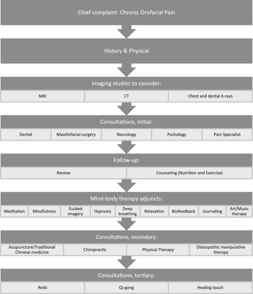
Medical treatment approaches and outcomes
The mainstay of medical treatment of PIFP is counseling. Patients should be counseled about the chronic nature of the illness, and its nonmalignant nature. Their pain should be acknowledged, and they should feel that their concerns are being heard, and a plan for addressing them is forthcoming. Toward this end, a comprehensive multidisciplinary team approach is most helpful. A short screening questionnaire for anxiety and/or depression (such as the Hospital Anxiety and Depression Scale ) or other surveys for common comorbidities in chronic pain may be administered, with appropriate referral to a psychologist/psychiatrist as necessary. A pain specialist may be able to initiate minimally invasive interventions, such as nerve blocks. Neurologists may be better able to manage the nuances of antiepileptic therapy trials.
Pharmacologic treatment
Tricyclic Antidepressants
The next line of therapy is pharmacologic. The tricyclic antidepressants have shown a moderate effect in several trials. They are postulated to work by altering the sensory discriminatory component of pain, in effect increasing the pain threshold. This effect is independent of their antidepressant action, as it is quicker in onset and observed even in nondepressed individuals, at lower dosages than those used for psychiatric use. Amitriptyline, one member of this class, has been shown to reduce the nociceptive discharges originating from myofascial tissues. Other drugs in this class are nortriptyline and duloxetine. Typically, dosages of between 25 and 100 mg daily are used.
Because one mechanism of action of the tricyclic antidepressants is to inhibit serotonin reuptake, other drugs with similar mechanisms have been tried. Fluoxetine (Prozac 10–20 mg/day), a selective serotonin reuptake inhibitor, and venlafaxine (Effexor 50–75 mg/day), which inhibits serotonin, dopamine, and norepinephrine reuptake, have both been found to have modest results when used for alleviation of chronic facial pain. Smaller doses initiated at nighttime and then titrated upward are recommended. In chronic neuropathic pain syndromes, the number of patients needed to treat (NNT) for antidepressants has been found to be 3, with NNT being an average of the number of patients who need to be treated for one to benefit compared with a control.
Anticonvulsant Drugs
The anticonvulsant drugs, including gabapentin (Neurontin), topiramax (Topamax), carbamazepine (Tegretol), and pregabalin (Lyrica), are often tried in chronic neuropathic pain, and are thought to be indicated especially when the pain is lancinating or burning. Their molecular mechanisms are varied, but typically involve action at the level of cell membrane ion channels. They have narrow therapeutic indices and are therefore best applied by knowledgeable practitioners. A 2005 Cochrane Database review concluded that, “Although anticonvulsants are used widely in chronic pain surprisingly few trials show analgesic effectiveness…There is no evidence that anticonvulsants are effective for acute pain. In chronic pain syndromes other than trigeminal neuralgia, anticonvulsants should be withheld until other interventions have been tried.” Their use in chronic orofacial pain has been systematically reviewed, with the finding that a limited to modest benefit may be derived for some patients.
Opioids
Interestingly, very few trials exist on the use of opioids for chronic facial pain. The opioids are thought to be less effective in neuropathic pain conditions, and carry with them serious adverse effect profiles including respiratory depression, hypotension, seizures, paralytic ileus, and dependency. Thus, their use is limited and requires careful monitoring.
Topical Anesthetic Agents
Topical anesthetic agents can be tested. Lidocaine patch (Lidoderm 5%) has been used in trigeminal neuralgia to decrease pain. Capsaicin, a naturally occurring compound responsible for the spiciness of chili peppers, which induces a neurogenic inflammation via release of neuropeptides, has also been studied. Amelioration of oral neuropathic pain symptoms was found in 60% of 24 patients in a small study from Vancouver, Canada. As capsaicin used around the face and eyes may be extremely irritating, a preemptive application of EMLA (eutectic mixture of local anesthetics) cream may be given first.
Botox
Botulinum toxin A (Botox) has been suggested in cases in which masticatory hyperactivity is present. This functional disorder may be commonly associated with chronic facial pain and headache. A group of researchers in Germany reported encouraging results in a randomized, blinded, placebo-controlled study of 90 patients. Ninety percent of patients who received the botox injection improved by a mean of 3.2 on a visual analog scale (VAS), a significant difference compared with placebo. In a similar vein, Italian researchers measured the electromyogram of the masseter and temporalis muscles in a group of patients with PIFP, at rest, during activation, and under kinesiography following transcutaneous electrical nerve stimulation (TENS). They used neuromuscular orthoses to correct identified discrepancies, which were found in all of their 21 patients. They found a net decrease in VAS pain score with a mean shift from 9.5 to 3.1. In a review by the European Federation of Neurologic Societies Task Force on neurostimulation therapy for neuropathic pain, TENS was found to be better than placebo, but its level of evidence was weaker than that for electroacupuncture.
Cannabis
Finally, cannabis, which is legal for medical purposes in several states including California but prohibited by federal law, has been studied for chronic pain. In a study from Canada, where medical marijuana use is allowed, a team from the department of anesthesia found that in patients with posttraumatic or postsurgical neuropathic pain, a single inhalation of 25 mg of 9.4% tetrahydrocannabinol herbal cannabis 3 times daily for 5 days reduced the intensity of pain, improved sleep, and was well tolerated. Abrams and colleagues at the University of California, San Francisco, studied vaporized cannabis for chronic pain. They reported that “vaporized cannabis augments the analgesic effects of opioids without significantly altering plasma opioid levels. The combination may allow for opioid treatment at lower doses with fewer side effects.” Vaporized treatments are used to minimize the risks of smoke to the lungs.
Stay updated, free articles. Join our Telegram channel

Full access? Get Clinical Tree


