Purpose
To describe the clinical and imaging characteristics of choroidal neovascularization (CNV) accompanied by focal choroidal excavation.
Design
Retrospective, interventional case series.
Methods
The medical records of 16 patients (16 eyes) were reviewed. Imaging findings including fluorescein angiography (FA), indocyanine green angiography, and spectral-domain optical coherence tomography (SD OCT) were analyzed.
Results
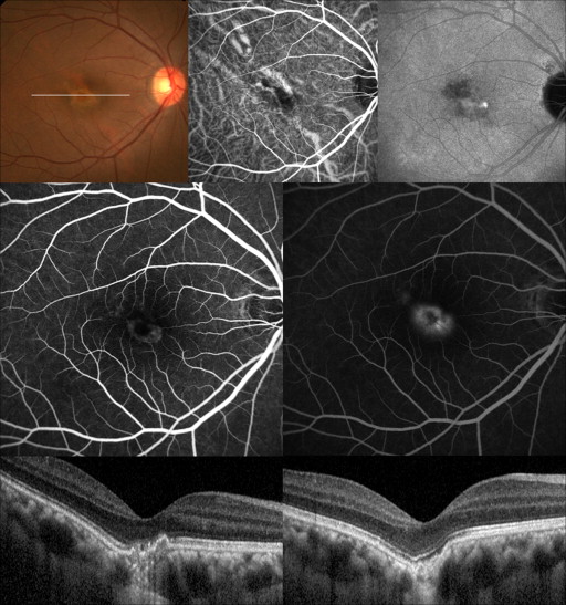
CNV complexes were primarily located beneath the retinal pigment epithelium (type 1 CNV) in 9 eyes and in the subneurosensory retinal space (type 2 CNV) in 7 eyes, as assessed by SD OCT. Seven of 8 patients over 50 years old had type 1 CNV, and 6 of 8 patients under 50 had type 2 lesions. All 7 eyes with type 2 CNV exhibited classic CNV on FA. Additionally, 7 of 9 eyes with type 1 CNV had the classic pattern, and in these eyes, the CNV complexes were confined to the concavity of choroidal excavation. In 15 patients treated by anti–vascular endothelial growth factor (anti-VEGF) injections, the mean best-corrected visual acuity improved from 20/44 to 20/26 with a mean of 3.7 injections during a mean follow-up period of 14.5 months.
Conclusions
The CNV growth pattern and extent seem to be determined by the degree of damage to the retinal pigment epithelium/Bruch membrane complex resulting from choroidal excavation, as well as age. Neovascular complexes tend to be located within the boundary of choroidal excavation and are revealed as classic patterns on FA, even in type 1 CNV. Anti-VEGF was notably effective for treating these lesions, with a low rate of recurrence.
Choroidal excavation was first reported as an unusual finding on time-domain optical coherence tomography (OCT) by Jampol and associates. After a few reports of similar cases detected by spectral-domain optical coherence tomography (SD OCT), Margolis and associates named this entity focal choroidal excavation. In 2 recent reports, most focal choroidal excavation lesions were shown to remain stable over a 1- or 3-year follow-up period. In a portion of patients, there was a history of central serous chorioretinopathy (CSC) or the concomitant presence of CSC. Cicatrized subretinal neovascularization was also noted at initial presentation in certain patients, while in others, secondary choroidal neovascularization (CNV) developed during the follow-up. The authors suggested a possible association between abnormal choroidal circulation in focal choroidal excavation and pathologic lesions. However, it has not been determined whether CNV develops as a clinical consequence of focal choroidal excavation or if they are 2 unrelated diseases that were coincidentally detected simultaneously. The paucity of cases has made it difficult to make conclusions regarding the role of focal choroidal excavation in CNV development.
There are several characteristic features of focal choroidal excavation that indicate that it may provide the appropriate milieu for CNV development. Previous studies showed that focal choroidal thinning and hypoperfusion was associated with focal choroidal excavation. Additionally, varying degrees of pigmentary disturbances and retinal pigment epithelium (RPE) alterations were noted. It is also possible that the stretching of the RPE/Bruch membrane in the area of choroidal excavation can result in a focal break in the Bruch membrane, which induces neovascular proliferation from the choroid, as in Gass type 2 CNV.
Gass classified the patterns of choroidal neovascular growth into the 2 following pathologic types: type 1, the growth of new vessels beneath the RPE; and type 2, the growth of new vessels in the subsensory retinal space. Elderly patients with age-related macular degeneration (AMD), which is associated with diffuse loosening of the firm attachment of the RPE to the Bruch membrane, are prone to developing type 1 CNV. This form of neovascularization is less permeable and proliferates slowly. Fluorescein angiography (FA) reveals a pattern that is “poorly defined” with minimal leakage, which is also described as occult CNV. In contrast, type 2 CNV occurs primarily in patients younger than 50 years and is associated with focally destructive lesions affecting the RPE/Bruch membrane. The firm attachment between the surrounding RPE and Bruch membrane in young individuals was postulated to explain the type 2 growth pattern. Type 2 CNV tends to proliferate rapidly, and FA typically exhibits a “well-defined” pattern with intense leakage, also referred to as classic CNV. SD OCT enables us to observe the details of retinal structures and the level of retinal involvement in CNV. Studies using SD OCT demonstrated a relatively good correlation between the angiographic classification (occult vs classic) and the anatomic classification of CNV (type 1 vs type 2).
The purpose of this study was to evaluate the detailed clinical and imaging characteristics in patients with CNV accompanied by focal choroidal excavation, focusing particularly on the association between the angiographic pattern and the anatomic pattern on OCT. We also suggest a hypothetical angiogenic process in this newly described disease entity.
Methods
We analyzed the medical records and imaging studies of 689 eyes from 653 consecutive patients with CNV or polypoidal choroidal vasculopathy who were referred to the ophthalmology department of Seoul St. Mary’s Hospital, The Catholic University of Korea, between the beginning of January 2009 and the end of March 2013. Sixteen patients were diagnosed as having CNV accompanied by focal choroidal excavation. Detailed clinical and imaging characteristics were evaluated in these patients. This retrospective study was approved by the Institutional Review Board of the Catholic Medical Center and conducted in accordance with the tenets of the Declaration of Helsinki.
All patients received a complete ocular examination, including best-corrected visual acuity (BCVA) measurement using a Snellen visual acuity chart, slit-lamp biomicroscopy with a noncontact or contact lens, and OCT at baseline and at each follow-up visit. Spectralis OCT (Heidelberg Engineering, Heidelberg, Germany) was used for SD OCT examinations after November 2011. The raster scan of Spectralis OCT was performed on each eye centered at the fovea using 2 scan protocols: a conventional protocol and an enhanced depth imaging (EDI) protocol. Before that time, Cirrus OCT (Carl Zeiss Meditec, Dublin, California, USA) with macular cube program consisting of 128 horizontal lines of 512 A-scans was used. All 16 patients underwent EDI OCT using Spectralis OCT at least once during their follow-up. FA and indocyanine green angiography (Heidelberg Retina Angiograph; Heidelberg Engineering, Heidelberg, Germany) were performed at baseline and on a patient-by-patient basis thereafter. However, indocyanine green angiography (ICGA) was not performed in 4 eyes at baseline. The Topcon IMAGEnet Digital Imaging System (Topcon, Tokyo, Japan) was used for FA examination in some patients.
The diagnosis of focal choroidal excavation was based on SD OCT findings of local excavation of the RPE and Bruch membrane line. Staphylomatous excavation of the sclerochoroidal junction was ruled out with EDI OCT. EDI OCT was used to determine choroidal thickness at the base of the focal choroidal excavation and the unaffected sites near the excavation by measuring the distance from the outer portion of the hyperreflective RPE line to the inner surface of the sclera with the digital calipers included in the review software. The ratio of the choroidal thicknesses of the 2 sites was calculated.
The diagnosis and classification of CNV were based on the FA and SD OCT findings. According to FA, CNVs were labeled classic or occult based on the Treatment of Age-Related Macular Degeneration With Photodynamic Therapy (TAP) protocol. The anatomic classification of CNV was made based on the location of the CNV membrane appearing as a highly reflective lesion on SD OCT with respect to the RPE band. CNV was classified as type 1 when the highly reflective lesion was primarily identified in the sub-RPE space and as type 2 when it was predominantly localized in the subretinal space. The topographic association between choroidal excavation and the hyperreflective lesion was also evaluated. The ICGA images were analyzed to identify aberrant vessels or polypoidal lesions and any abnormal hypofluorescent or hyperfluoresecnt areas.
All patients were treated with intravitreal anti–vascular endothelial growth factor (anti-VEGF) injection with either bevacizumab (Avastin; Genentech, Inc, San Francisco, California, USA) or ranibizumab (Lucentis; Genentech Inc), except for 1 patient who was lost to follow-up. Anti-VEGF treatment was performed on an as-needed basis, after either an initial 2 or 3 consecutive injections or a single injection. Repeated injections were given whenever either intraretinal or subretinal fluid involving the fovea was detected on follow-up OCT. The patients were followed up regularly at 1- to 6-month intervals, depending on lesion activity, but the intervals did not exceed 3 months in patients over 50 years old.
Results
The patient characteristics are summarized in the Table . We studied 16 eyes in 16 Korean patients, 9 of whom were women. The mean age was 49.6 years (range, 28-86 years). Eight patients were under 50 years old; 2 among them had a history of CSC in the affected eyes and 1 patient had high myopia. Soft drusen was only noted in 1 of 8 patients over 50. None of the patients had a history of medical illness, medication use, or a family history of retinal disease. None of the cases were bilaterally affected, while 2 cases showed type 1 CNV in their contralateral eye. The spherical equivalent of the refractive error ranged from −12.50 diopters (D) to 1.125 D (mean, −3.48 D).
| Case No. | Age/Sex | Eye | SE | OCT | FA | ICGA | Ratio of Choroidal Thickness a | F/U (Months) | Anti-VEGF (N b ) | Additional Injection c | BCVA | Others | |||
|---|---|---|---|---|---|---|---|---|---|---|---|---|---|---|---|
| CNV Pattern | Extent of CNV | CNV Pattern | Focal Hypofluoresence | Late Hyperfluoresence | Initial | Final | |||||||||
| 1 | 39/M | Left | −12.5 | Subsensory | Within the concavity | Classic | NA | NA | 0.50 (114/228) | 19 | Bevacizumab (1) | − | 20/32 | 20/25 | Subsequent development of CNV |
| 2 | 71/M | Left | −1.125 | Sub-RPE | Within the concavity | Classic | + | + | 0.25 (54/216) | 35 | Ranibizumab (7) | + | 20/200 | 20/25 | |
| 3 | 86/F | Left | 0.625 | Sub-RPE | Over the margin | Occult | + | + | 0.60 (168/282) | 32 | Bevacizumab (8) | + | 20/63 | 20/32 | CNV in the other eye |
| 4 | 28/M | Right | −4.5 | Subsensory | Over the margin | Classic | + | − | 0.56 (120/214) | − | F/U loss | 20/200 | Subsequent development of CNV | ||
| 5 | 69/F | Left | 1.125 | Sub-RPE | Within the concavity | Classic | NA | NA | 0.41 (83/203) | 19 | Ranibizumab (5) | + | 20/200 | 20/100 | |
| 6 | 45/M | Right | −2.125 | Subsensory | Within the concavity | Classic | + | + | 0.29 (62/217) | 17 | Bevacizumab (3) | − | 20/25 | 20/20 | History of CSC |
| 7 | 47/F | Left | −6.5 | Subsensory | Within the concavity | Classic | NA | NA | 0.69 (154/224) | 17 | Bevacizumab (3) | − | 20/40 | 20/20 | History of CSC |
| 8 | 55/M | Right | −0.5 | Sub-RPE | Within the concavity | Classic | + | − | 0.35 (134/383) | 14 | Ranibizumab (5) | + | 20/32 | 20/20 | |
| 9 | 40/F | Right | −2.75 | Sub RPE | Within the concavity | Classic | + | + | 0.46 (148/324) | 13 | Bevacizumab (2) | − | 20/50 | 20/32 | |
| 10 | 53/F | Right | −3.125 | Subsensory | Within the concavity | Classic | + | + | 0.74 (265/356) | 13 | Ranibizumab (3) | − | 20/25 | 20/20 | |
| 11 | 29/F | Right | −7.125 | Subsensory | Over the margin | Classic | NA | NA | 0.44 (118/259) | 7 | Bevacizumab (2) | − | 20/32 | 20/25 | Subsequent development of CNV |
| 12 | 53/M | Left | −0.5 | Sub-RPE | Within the concavity | Classic | + | − | 0.34 (76/221) | 8 | Bevacizumab (5) | + | 20/80 | 20/63 | |
| 13 | 66/M | Right | 0.875 | Sub-RPE | Over the margin | Occult | + | + | 0.46 (124/272) | 8 | Bevacizumab (3) | − | 20/100 | 20/32 | CNV in the other eye |
| 14 | 54/F | Right | −4.0 | Sub-RPE | Within the concavity | Classic | + | + | 0.76 (297/389) | 7 | Bevacizumab (4) | + | 20/40 | 20/20 | |
| 15 | 28/F | Left | −4.0 | Sub-RPE | Within the concavity | Classic | + | + | 0.85 (353/413) | 6 | Bevacizumab (3) | − | 20/25 | 20/25 | |
| 16 | 30/F | Right | −9.5 | Subsensory | Within the concavity | Classic | − | − | 0.78 (197/250) | 3 | Bevacizumab (1) | − | 20/100 | 20/25 | |
a Ratio of choroidal thickness at the base of the focal choroidal excavation and unaffected sites near the excavation.
At initial presentation, CNV was accompanied by focal choroidal excavation in 13 of 16 patients. CNV had developed in 3 patients at 2, 11, and 21 months of follow-up, and 2 of them showed a nonconforming pattern. Fifteen CNVs involved the subfoveal area and 1 CNV was juxtafoveal (within 200 μm from the center of the fovea). The mean baseline BCVA was 20/46 (range, 20/200-20/25). The focal choroidal excavation lesions were located subfoveally in 14 of 16 eyes (87.5%) and juxtafoveally in 2 eyes (12.5%). The mean choroidal thickness was 160 μm (range, 54-353 μm) at the base of the excavation and 272 μm (range, 203-413 μm) at the unaffected sites near the excavation. The ratio of the choroidal thicknesses of the 2 sites varied from 0.25-0.85 (mean, 0.53).
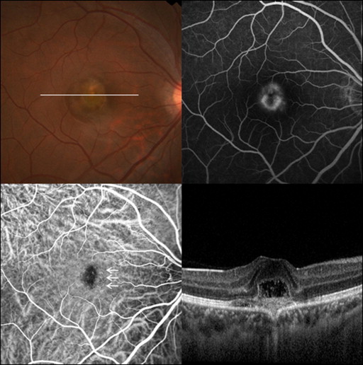
According to FA, 14 eyes exhibited a classic appearance with profuse leakage through the mid and late phase ( Figures 1-4 ). The other 2 CNVs were purely occult ( Figure 5 ). All 8 patients under 50 years had classic CNV. Six of 8 patients over 50 years old showed a classic pattern, and the other 2 had occult pattern CNV. ICGA revealed abnormal vasculature consistent with CNV, but no polypoidal lesions. Eleven of 12 eyes (91.7%) showed focal hypofluorescence at the corresponding focal choroidal excavation area, and 8 of 12 eyes (66.7%) showed choroidal venous dilation and diffuse hyperfluorescence in the late phase.
Stay updated, free articles. Join our Telegram channel

Full access? Get Clinical Tree


