Radical Neck Dissection
Jesus Medina
INTRODUCTION
The radical neck dissection is a comprehensive cancer operation that removes the lymph node-bearing tissues of one side of the neck, from the inferior border of the mandible to the clavicle and from the midline to the anterior border of the trapezius muscle. Included in the resected specimen are lymph node groups I through V, the spinal accessory nerve, the internal jugular vein, and the sternocleidomastoid muscle (Fig. 6.1).
The first description of this operation was published by Crile in 1906. It was Martin et al. in the 1950s who championed the concept that a cervical lymphadenectomy for cancer was inadequate unless all the lymph node-bearing tissues of one side of the neck were removed and that this was not possible without resecting the sternocleidomastoid muscle, internal jugular vein, and spinal accessory nerve because of the close association of the lymphatics with these structures.
As a result of Martin’s influence, the radical neck dissection was for many years the only dissection of the lymph nodes performed in patients with cancer of the head and neck.
HISTORY
The clinician should record:
The presence and duration of symptoms related to the primary tumor, such as pain, otalgia, odynophagia, hoarseness, dysphagia, cough, and hemoptysis
The occurrence and extent of weight loss and all other comorbidities
History of risk factors such as the quantity of tobacco and alcohol consumed each day
History of previous treatment to the head and neck with radiation (total dose and portals) or surgery
PHYSICAL EXAMINATION
Physical examination should include:
Examination of all the areas of the oral cavity, pharynx, indirect laryngoscopy, or fiberoptic examination of the larynx if necessary
Palpation of the neck bilaterally, recording the location (levels I-VI), size, mobility, and relationship of the node(s) to adjacent structures. This should include bimanual palpation of the submandibular area.
Documentation of the presence or absence of trismus and of actual or potential airway compromise, which may have bearing on the management of the airway during induction of anesthesia
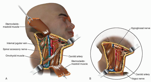 FIGURE 6.1 Radical neck dissection. A: Intraoperative appearance. B: Postoperative appearance. |
INDICATIONS
The radical neck dissection is indicated in the following situations:
Patients with multiple clinically obvious cervical lymph node metastases, particularly when they involve the lymph nodes of the posterior triangle of the neck and are found to involve or to be closely related to the spinal accessory nerve
Patients with a bulky metastatic tumor mass or with multiple matted nodes in the superior aspect of the neck
When a neck dissection is performed to remove residual disease in the neck following an ill-advised incisional biopsy of a cervical lymph node containing metastatic cancer. In such cases, extensive undermining during the biopsy procedure, postoperative inflammation, and ecchymosis often obscure the relationship of the tumor to the sternocleidomastoid muscle, spinal accessory nerve, or internal jugular vein, making their preservation problematic.
CONTRAINDICATIONS
A radical neck dissection is not indicated in the following circumstances:
In the absence of palpable cervical lymph node metastases (i.e., in the elective surgical treatment of the N0 neck)
When the diagnostic evaluation of the patient reveals (a) frank involvement of the wall of the carotid artery in patients whose preoperative evaluation indicates intolerance to carotid ligation and the location and extent of the cancer in the neck, that is, near the skull base, precludes reconstruction of the carotid and (b) involvement of the base of the skull, paraspinal muscles, transverse processes of the cervical vertebrae, and the brachial plexus
PREOPERATIVE PLANNING
In patients with advanced metastases to the neck requiring a radical neck dissection, the preoperative evaluation must include comprehensive imaging studies that address:
Resectability. In this regard, CT and MRI imaging are used to define the relationship of the metastatic cancer to critical structures such as the common and the internal carotid artery, the cervical spine, the vertebral
artery, and the brachial plexus. If the common or the internal carotid artery is suspected of involvement by cancer, a systematic preoperative evaluation should include four-vessel cerebral angiography to determine the status of the contralateral carotid artery and to assess intracerebral collateral circulation. In addition, an attempt should be made during the angiography to measure carotid back pressure and to assess dynamically the collateral circulation by using balloon occlusion techniques while monitoring the patient for evidence of neurologic deficits under normotensive and hypotensive conditions.
The presence of metastases in lymph nodes that are not routinely removed with a radical neck dissection such as the retropharyngeal, paratracheal, and upper mediastinal nodes
The possibility of distant metastases. A positron emission tomography-computed tomography (PET-CT) scan is a useful study in this regard and in staging the disease.
SURGICAL TECHNIQUE
The patient is positioned on the operating table with the neck extended, if necessary, with a rolled up towel or blanket under the shoulders and the head turned toward the opposite side and stabilized with a foam doughnut.
The incisions most commonly used to perform a radical neck dissection are outlined in Figure 6.2. Skin flaps are elevated in a subplatysmal plane. However, depending upon the size and extent of the tumor in the neck, the platysma may be left over the area involved by tumor as the skin flaps are elevated in a supraplatysmal plane. Skin that is infiltrated with cancer or the scar of a previous open biopsy should be left on the specimen.
Dissection of the Submandibular Triangle
As the superior cervical flap is elevated, it is important to keep the plane of dissection superficial to the fascia that covers the submandibular gland; this facilitates identification of the ramus mandibularis of the facial nerve. This nerve lies deep to the fascia but superficial to the facial vessels, which are exposed and divided. The submandibular prevascular and retrovascular lymph nodes, which are usually immediately below or medial to the nerve, are likewise exposed (Fig. 6.3). When these nodes are involved by tumor, it is preferable to leave the platysma attached to them. In such cases, it may not possible, nor desirable, to expose and preserve the ramus mandibularis.
The next step is to incise the fascia and adipose tissue along and medial to the inferior border of the mandible. As this is done, it is usually necessary to divide the submental artery at the angle between the anterior belly of the digastric and the inferior border of the mandible and the nerve to the mylohyoid.
The fascia of the anterior belly of the digastric muscle is incised and the fibroadipose tissue lateral to the mylohyoid muscle is dissected. A “clean” dissection of this area is important because it contains the preglandular lymph node(s), which can be the first echelon of lymphatic drainage for cancer of the floor of the mouth and oral tongue. When the dissection reaches the posterior border of the mylohyoid, this muscle is retracted forward with a Richardson retractor. This exposes three structures that are somewhat parallel but in different planes; from lateral to medial and from superior to inferior, they are the lingual nerve, Whartons duct, and the hypoglossal nerve. The submandibular ganglion located inferior to the lingual nerve and Whartons duct is divided.
Stay updated, free articles. Join our Telegram channel

Full access? Get Clinical Tree


